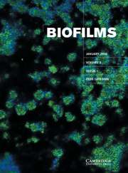
Ultrastructure of Enterococcus faecalis biofilms
Published online by Cambridge University Press: 01 September 2004
Abstract
Enterococcus faecalis is known to produce biofilms on biomaterials, but the manner in which this occurs is unknown. Herein we report that adhesion of E. faecalis in biofilms appeared to be mediated by cell wall surface projections attaching cells to the substratum. Biofilm formation was observed on the polystyrene surface of 96-well plates and also on the surface of cellulose kidney dialysis tubing used as a model for biofilm formation on catheters. Qualitative differences involved the packing of E. faecalis cells in biofilms, with greater intercellular spacing detected in the 96-well plate, whereas bacteria were tightly packed on the surface of cellulose catheters. Distribution of adherent bacterial cells accumulating on the two surfaces revealed obvious differences, with most of the bacteria attaching to the polystyrene surface as single cells or diplococci separated from neighboring organisms by intervals of uncolonized surface. In contrast, enterococci on the cellulose surface were found as multi-layer cellular aggregates or microcolonies, even when much of the total surface was free from attached bacteria. Microcolonies stained intensely for neutral hexose sugars using the periodic acid–Schiff (PAS) stain. Surface projections, presumably exopolysaccharide, anchored bacteria to the substratum and appeared to elevate the cells above the surface. These slender surface projections could be seen over the entire enterococcal cell wall, with the exception of areas adjacent to septal regions where new cell wall formation was occurring. Rod-like interconnections were also observed between adjacent diplococci. These results suggested that biofilm formation varies on different substrates and that enterococcal surface projections may be involved in E. faecalis colonization and adhesion within biofilms.
- Type
- Research Articles
- Information
- Copyright
- © 2004 Cambridge University Press
- 5
- Cited by


