


CASE
A 54-year-old woman prepares dinner around 8:00 pm that includes mushrooms that she picked from her yard. The next morning, around 8:00 am, the woman (patient), her daughter, and son-in-law all develop abdominal cramps, violent vomiting, and diarrhea. They present to the emergency department and are admitted for dehydration and intractable vomiting with a presumed diagnosis of food poisoning. Twenty-four hours later, they appear well with stable vital signs and improved symptoms. Four hours later, 36 hours post-ingestion, the patient becomes lethargic. A venous blood gas reveals pH, 7.1; PCO2, 16 mmHg; and her AST was 3140 units/L with an ALT of 4260 units/L and an INR of 3.7.
KEY CLINICAL QUESTIONS
- 1.
What is the epidemiology and pathophysiology of cyclopeptide mushroom poisoning?
Answer: Cyclopeptide-containing mushrooms are found worldwide in four genera of fungi, Amanita, Galerina, Lepiota, and Conocybe. Amanita phalloides (Figure 1) is an invasive species and, along with Galerina and Lepiota (Figure 2) species, has a range that includes space around urban trees.Reference Berch, Kroeger and Finston1 These mushrooms vary significantly in appearance and can be mistaken for edible mushrooms, especially at early stages of development when Amanita resemble the commonly edible puffballs (Figure 1). Cyclopeptide mushrooms are thought to be responsible for 90% of all mushroom-related fatalities. Children with unintentional, exploratory ingestions rarely develop severe toxicity. Case fatality rates following intentional ingestions are extremely variable, ranging from 5–40%.Reference Jander, Bischoff and Woodcock2

Figure 1. Amanita phalloides through multiple stages of maturity. At younger stages, they may resemble edible puffballs. (Photo courtesy of Justin Pierce via www.MushroomObserver.org)
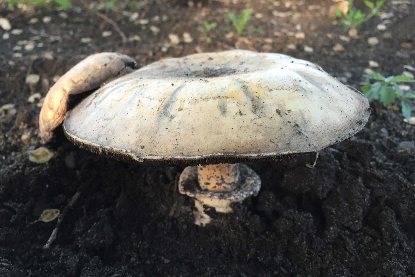
Figure 2. Lepiota sp.
There are several cyclopeptides in each of these mushrooms that can be problematic, but amatoxins are the principal toxin. Amatoxin is heat stable and acts as an irreversible inhibitor of RNA polymerase II, therefore preventing mRNA synthesis and causing a disruption of cellular protein production. The main organ of injury is the liver, but kidneys are also affected.
- 2.
What are the clinical characteristics of cyclopeptide-containing mushroom toxicity, and how can they be differentiated from other mushroom toxicity syndromes?
Answer: Toxicity is characterized by gastrointestinal distress with profuse nausea and vomiting that is delayed at least 5 hours after ingestion. When vomiting occurs less than 5 hours post-ingestion, cyclopeptide-containing mushroom toxicity is extremely unlikely unless there is a possibility of mixed mushroom ingestion.
Gastrointestinal symptoms usually abate around 24 hours post-ingestion, but signs of acute liver injury are detectable with rising aminotransferase concentrations. Over the following hours to days, acute liver failure and multisystem organ failure develop and can result in cardiovascular collapse and death. Consultation with a regional poison centre can be extremely helpful with clinical syndromes as can the involvement of a mycologist to identify photos or uneaten mushrooms when available for examination.
- 3.
Is gastrointestinal decontamination indicated?
Answer: Yes, activated charcoal is believed to be helpful in reducing cyclopeptide toxicity.Reference Ye and Liu3 Since data from charcoal hemoperfusion studies demonstrate removal of amatoxin, activated charcoal is assumed to be beneficial. In a patient who presents early after ingestion (within 2 hours) of an unidentifiable mushroom, it is reasonable to give a single dose of activated charcoal (1 g/kg) orally. With ingestions of confirmed or highly suspected features of cyclopeptide-containing mushrooms, activated charcoal can also be used to enhance elimination at a dose of 0.5–1 g/kg by NGT/OGT every 4–6 hours for the first 24–48 hours.
- 4.
What treatments and medications can be used?
Answer: Supportive care with adequate intravenous fluid and electrolyte replenishment is the mainstay of treatment given the significant gastrointestinal losses that patients experience. The administration of N-acetylcysteine is beneficial in liver injury of any etiology and appears efficacious in cyclopeptide mushroom toxicity.Reference Poucheret, Fons, Doré, Michelot and Rapior4 Amatoxin is transported from the liver sinusoids into hepatocytes. It is not metabolized and is then excreted in bile to be reabsorbed from the intestinal tract into the hepatocytes. Experimentally, silibinin decreases hepatocyte uptake of amatoxin from liver sinusoids by inhibiting the organic anion transporter protein (OATP1B3), interrupting the enterohepatic recirculation characteristic of this toxin.Reference Garcia, Costa and Carvalho5 Clinically, silibinin may be helpful, but, because interpretation of supporting data is complicated by uncontrolled administration of multimodal therapies, it is unclear whether this treatment is independently useful.Reference Poucheret, Fons, Doré, Michelot and Rapior4 Similarly, ceftazidime inhibits amatoxin uptake, but its use is only described with combined use of silibinin. Treatment with high-dose penicillin G yields no significant improvement in mortality and its clinical use is no longer recommended due to possible allergic reactions, seizures, and electrolyte disturbances.Reference Ye and Liu3
- 5.
Are extracorporeal modalities clinically useful?
Answer: Hemodialysis is generally widely available in many countries, whereas charcoal hemoperfusion, fractionated plasma separation and absorption (FPSA), and molecular adsorbent recirculating system (MARS) are only available at selected tertiary care centres. No extracorporeal modality has been proven to improve outcomes in cyclopeptide mushroom toxicity. Hemoperfusion with activated charcoal clears amatoxin significantly.Reference Mydlík, Derzsiová, Klán and Zíma6 FPSA and MARS show promise but require more supporting data prior to widespread acceptance. Hemodialysis alone or with hemoperfusion is a rational strategy and may be a useful modality to provide renal replacement in patients who develop acute kidney injury even though there may be no toxin-specific benefits. The Extracorporeal Treatments in Poisoning Workgroup (EXTRIP) is reviewing data for amatoxin, and we await their recommendations.
CASE RESOLUTION
The patient was intubated for respiratory failure and transferred to the intensive care unit. A mycologist was provided images from the family's dinner and suggested Amanita ingestion. Treatment with aggressive intravenous fluid and N-acetylcysteine was administered. Her aminotransferase and INR continued to rise and she required vasopressors for shock. Unfortunately, she expired 4 days after ingestion.
KEY POINTS
1. Cyclopeptide-containing mushroom ingestions occur worldwide and are responsible for the vast majority of mushroom-related mortality.
2. The organ of injury is the liver, and delayed gastrointestinal symptoms predominate.
3. Involvement of your regional poison centre and a mycologist are helpful to determine the species of mushroom.
4. Treatment consists of aggressive fluid replacement, multidose-activated charcoal, and N-acetylcysteine.
5. Extracorporeal therapies may be used to remove toxin or for supportive care in those with acute kidney injury.
Competing interests
None declared.


