


Introduction
Compared with other Gondwanan landmasses, particularly South America and Africa, Australia has a scant record of Cretaceous crocodyliforms. Whereas African Cretaceous crocodyliforms comprise over 35 different taxa, and South America boasts over 50, only four are currently recognized from Australia (Molnar, Reference Molnar1980; Molnar and Willis, Reference Molnar, Willis, Grigg, Seebacher and Franklin2001; Salisbury et al., Reference Salisbury, Molnar, Frey and Willis2006; Hart et al., Reference Hart, Bell, Smith and Salisbury2019; Hart, Reference Hart2020). These comprise three species of Isisfordia: I. duncani Salisbury et al., Reference Salisbury, Molnar, Frey and Willis2006, I. selaslophensis Etheridge, Reference Etheridge1917, and I. molnari Hart et al., Reference Hart, Bell, Smith and Salisbury2019, and a fourth, unnamed, mesoeucrocodylian (Molnar and Willis, Reference Molnar, Willis, Grigg, Seebacher and Franklin2001). Of the Australian representatives, only one species, Isisfordia duncani from the Albian–Turonian Winton Formation of Queensland, has been described from multiple associated and articulated skeletons (Salisbury et al., Reference Salisbury, Molnar, Frey and Willis2006; Syme and Salisbury, Reference Syme and Salisbury2018). Upon its initial description, I. duncani was hypothesized to be the basal-most eusuchian on the basis of several synapomorphies, including weakly procoelous vertebrae and secondary choanae enclosed anteromedially by ventral laminae of the pterygoids (Salisbury et al., Reference Salisbury, Molnar, Frey and Willis2006). More recent studies have postulated alternative phylogenetic positions for I. duncani as the sister taxon to Susisuchus within Susisuchidae, with this clade located either outside of Eusuchia (e.g., Turner and Pritchard, Reference Turner and Pritchard2015; Montefeltro et al., Reference Montefeltro, Bronzati, Langer and Anelli2020) or within a more inclusive Eusuchia (e.g., Leite and Fortier, Reference Leite and Fortier2018; Martin et al., Reference Martin, Smith, Salaviale, Adrien and Delfino2020). Despite the lack of consensus on the phylogenetic position of I. duncani, the taxon, and its transitional morphology, remains at the focus of discussions on the origins of Eusuchia since its description (Salisbury et al., Reference Salisbury, Molnar, Frey and Willis2006; Turner and Pritchard, Reference Turner and Pritchard2015; Leite and Fortier, Reference Leite and Fortier2018; Martin et al., Reference Martin, Smith, Salaviale, Adrien and Delfino2020; Montefeltro et al., Reference Montefeltro, Bronzati, Langer and Anelli2020).
The other two crocodyliform species from the Australian Cretaceous are Isisfordia selaslophensis and I. molnari from the Cenomanian Griman Creek Formation at Lightning Ridge, New South Wales. Etheridge (Reference Etheridge1917) described what was interpreted as a dentary fragment with six in situ teeth as the holotype of a new species, ‘Crocodylus (Bottosaurus) selaslophensis.’ Subsequently, Molnar (Reference Molnar1980) proposed that this fragment belonged to neither Crocodylus nor Bottosaurus and most likely represented an indeterminate eusuchian. Molnar (Reference Molnar1980) also referred several unassociated postcranial elements (a femur, tibiae, and vertebral remains) to ‘C. selaslophensis.’ Hart et al. (Reference Hart, Bell, Smith and Salisbury2019) reappraised the holotype of ‘C. selaslophensis,’ which was reidentified as a partial maxilla, and a more recently discovered partial braincase, both of which showed similarities to the same elements in I. duncani. The maxilla and braincase were assigned to a new species, Isisfordia molnari, with the braincase as the holotype. However, the assignment of the maxilla to I. molnari was problematic as it was a name-bearing specimen; even though ‘C. selaslophensis’ had been considered a nomen dubium (Mannion et al., Reference Mannion, Benson, Carrano, Tennant, Judd and Butler2015; supplementary information), the specific epithet retains taxonomic seniority. As a consequence, Hart (Reference Hart2020) erected the combination Isisfordia selaslophensis to represent this material. Isisfordia molnari remains a valid taxon as there is currently no material that overlaps with the holotypic braincase (which displays at least one unambiguous autapomorphy, see Hart et al., Reference Hart, Bell, Smith and Salisbury2019). However, Hart (Reference Hart2020) suggested that I. molnari is likely a junior subjective synonym of I. selaslophensis, a hypothesis that can be tested only with the discovery of more complete material that overlaps with the holotype specimens.
Additional crocodyliform material from the Griman Creek Formation (the aforementioned ‘unnamed mesoeucrocodylian’), represented by associated maxillary and dentary remains, was described by Molnar and Willis (Reference Molnar, Willis, Grigg, Seebacher and Franklin2001), who suggested that it represented a taxon that was distinct from ‘C. selaslophensis’ (now I. selaslophensis). Their conclusions were based on the apparently ziphodont dentition exhibited by some of the specimens. An alternative hypothesis is proposed herein. Hart et al. (Reference Hart, Bell, Smith and Salisbury2019) speculated that these remains are likely to pertain to Isisfordia.
The only other crocodyliform material from the Mesozoic of Australia comes from the Aptian–Albian Otway Formation of Victoria and includes isolated osteoderms, vertebrae, and a quadratojugal (see Poropat et al., Reference Poropat, Martin, Tosolini, Wagstaff, Bean, Kear, Vickers-Rich and Rich2018). Salisbury et al. (Reference Salisbury, Frey, Martill and Buchy2003) suggested this material might pertain to a susisuchid, indicating a close relationship to Isisfordia (Turner and Pritchard, Reference Turner and Pritchard2015; Montefeltro et al., Reference Montefeltro, Bronzati, Langer and Anelli2020).
Although some postcranial remains of crocodyliforms have been reported from the Griman Creek Formation (Molnar, Reference Molnar1980; Smith, Reference Smith1999), to date only cranial remains have been referred to a named taxon. This study aims to reassess previously described postcranial remains and describe new material from the Griman Creek Formation at Lightning Ridge, which includes the first partial Australian Cretaceous crocodyliform skeleton outside of the Winton Formation. This will clarify the taxonomy of Australian Mesozoic crocodyliforms and provide additional morphological information on these taxa.
Geological setting
The Griman Creek Formation, part of the Surat Basin, crops out (in part) near the outback opal mining town of Lighting Ridge in northern New South Wales (Fig. 1). Fossil material from this locality, including that discussed herein, has been excavated from underground opal mines over the past century. All Cretaceous-aged vertebrate fossils from this locality are preserved as opal (SiO2.nH2O; see Bell et al., Reference Bell, Fanti, Hart, Milan, Craven, Brougham and Smith2019b). The specimens come from claystone lenses (informally referred to as the ‘Finch Clay facies’; Scheibner and Basden, Reference Scheibner and Basden1998) within the Wallangulla Sandstone Member of the Griman Creek Formation (Bell et al., Reference Bell, Fanti, Hart, Milan, Craven, Brougham and Smith2019b). The Griman Creek Formation is considered Cenomanian in age (c. 96–100 Ma) on the basis of U–Pb dating of detrital zircons (Bell et al., Reference Bell, Fanti, Hart, Milan, Craven, Brougham and Smith2019b). Opalized fossils are extracted during opal mining activities, and in many cases accurate provenance data are missing. However, where this information has been disclosed, it is noted in Figure 1 and Table 1.
Table 1. Crocodyliform material from the Griman Creek Formation at Lightning Ridge. All of these localities except Holden's are within the ‘Coocoran’ opal field, and the cranial material housed at the Australian Museum (AM) derives from fields close to the Lightning Ridge Township.


Figure 1. Map of Australia showing the location of the Griman Creek Formation at Lightning Ridge (red circle). Mining fields in which the crocodyliform fossils were recovered are detailed in the inset. The Eromanga and Surat basins are represented by the gray area. The maximum extent of the Eromanga Sea is represented by the blue area. Australia coastline uses data taken from GEODATA COAST 100 K 2004 provided by Geoscience Australia (http://pid.geoscience.gov.au/dataset/ga/61395). Basin extents use data taken from Stewart et al. (Reference Stewart, Raymond, Totterdell, Zhang and Gallagher2013).
The terrestrial faunal assemblage at Lightning Ridge is one of the most diverse from the Mesozoic of Australia. It includes aspidorhynchids (Bell et al., Reference Bell, Fanti, Hart, Milan, Craven, Brougham and Smith2019b), lamniforms (Bell et al., Reference Bell, Fanti, Hart, Milan, Craven, Brougham and Smith2019b), dipnoans (Kemp Reference Kemp1997a, Reference Kempb), meiolaniform and chelid testudines (Smith Reference Smith2009, Reference Smith2010; Smith and Kear, Reference Smith, Kear, Brinkman, Holroyd and Gardener2013), leptocleidid plesiosaurs (Kear, Reference Kear2006), anhanguerian pterosaurs (Brougham et al., Reference Brougham, Smith and Bell2017), crocodyliforms (Etheridge, Reference Etheridge1917; Molnar, Reference Molnar1980; Molnar and Willis, Reference Molnar, Willis, Grigg, Seebacher and Franklin2001; Hart et al., Reference Hart, Bell, Smith and Salisbury2019), ankylosaurians (Bell et al., Reference Bell, Burns and Smith2017), small- and large-bodied ornithopods (Molnar and Galton, Reference Molnar and Galton1986; Molnar, Reference Molnar1996; Bell et al., Reference Bell, Herne, Brougham and Smith2018, Reference Bell, Brougham, Herne, Frauenfelder and Smith2019a, Reference Bell, Fanti, Hart, Milan, Craven, Brougham and Smithb; Kitchener et al., Reference Kitchener, Campione, Smith and Bell2019), titanosauriform sauropods (Molnar and Salisbury, Reference Molnar, Salisbury, Tidwell and Carpenter2005), megaraptorid theropods (Bell et al., Reference Bell, Cau, Fanti and Smith2015; Brougham et al., Reference Brougham, Smith and Bell2019), noasaurids (Brougham et al., Reference Brougham, Smith and Bell2020), enantiornithes (Bell et al., Reference Bell, Fanti, Hart, Milan, Craven, Brougham and Smith2019b), and mammals (Archer et al., Reference Archer, Flannery, Ritchie and Molnar1985; Flannery et al., Reference Flannery, Archer, Rich and Jones1995; Clemens et al., Reference Clemens, Wilson and Molnar2003; Pian et al., Reference Pian, Archer, Hand, Beck and Cody2016; T. Rich in Poropat et al., Reference Poropat, Martin, Tosolini, Wagstaff, Bean, Kear, Vickers-Rich and Rich2018).
Materials and methods
All the original fossil material from Lighting Ridge described herein was observed, analyzed, and photographed, with the exception of LRF 3378 (a tibia) and LRF 3379 (a calcaneum). These two specimens are replicas of originals currently held in a private collection. Before molding, the original specimen represented by LRF 3378 had been incorrectly reassembled from two pieces. A cast was made from the mold of the original specimen in its incorrect reconstruction, then divided into the original proximal and distal parts in which the fossil was initially discovered. The surfaces of these parts were scanned using a white-light scanning system at 45 μm resolution (smartSCAN3D C-5, Breuckmann) to create three-dimensional (3D) polygonal surface models. Processing of the scan data was carried out using the optoCAT scanning software (Breuckmann). The tibia was then reconstructed by reattaching these two parts in the correct alignment using Geomagic Studio v. 2014. A high-resolution 3D print was created and accessioned as LRFR 3378 into the Australian Opal Centre collection, along with the cast LRFR 3379. The analyses of all of the Lightning Ridge material were supplemented with observations of original (holotype, paratype, and referred specimens) and cast material of Isisfordia duncani at the Queensland Museum (QM) and University of Queensland (UQ). All measurement data collected are presented in Table 1.
Repositories and institutional abbreviations
Types, figured, and other specimens examined in this study are deposited in the following institutions: The Australian Opal Centre (LRF), Lightning Ridge, New South Wales, Australia; The Australian Museum (AM), Sydney, New South Wales, Australia; The Queensland Museum (QM), Brisbane, Queensland, Australia; The University of Queensland (UQ), Brisbane, Queensland, Australia.
Systematic paleontology
Crocodyliformes Hay, Reference Hay1930
Mesoeucrocodylia Whetstone and Whybrow, Reference Whetstone and Whybrow1983
Neosuchia Benton and Clark, Reference Benton, Clark and Benton1988
Eusuchia Huxley, Reference Huxley1875
Genus Isisfordia Salisbury et al., Reference Salisbury, Molnar, Frey and Willis2006
Type species
Isisfordia duncani Salisbury et al., Reference Salisbury, Molnar, Frey and Willis2006 (QM F36211) from the ‘lower’ Winton Formation, uppermost Albian–Cenomanian, Queensland.
Other species
Isisfordia molnari Hart et al., Reference Hart, Bell, Smith and Salisbury2019; Isisfordia selaslophensis Etheridge, Reference Etheridge1917.
Diagnosis
As per Hart (Reference Hart2020); broad exposure of the prootic within the supratemporal foramen anterior to the rostral aperture of the posttemporal canal; posterior maxillary alveolar groove; maximum diameter of the posterior aperture of the cranioquadrate siphonium approximately one-third the mediolateral width of the foramen magnum, with the lateral wall of the siphonium formed exclusively by the quadrate; maximum mediolateral width of the secondary choanae exceeds the minimum mediolateral width of the palatines; naris with a distinctly pear-shaped outline; posterior dentary teeth confluent and set in a shallow alveolar groove (shared with some alligatoroids); dentary and maxillary teeth flattened labiolingually at the base of the crown but become conical toward the apex; cervical, thoracic, and anterior-most caudal vertebrae weakly procoelous at maturity; caudal vertebrae weakly procoelous; sacral vertebra II with a low posterior condyle; distal extremity of ulna expanded transversely with respect to the long axis of the bone (shared with Susisuchus spp. and Theriosuchus pusillus Owen, Reference Owen1879).
Isisfordia cf. I. selaslophensis Etheridge, Reference Etheridge1917
- Reference Etheridge1917
Crocodilus (?Botosaurus) selaslophensis Etheridge (Hart et al., Reference Hart, Bell, Smith and Salisbury2019, figs. 4, 5).
- Reference Molnar1980
Crocodylus (Bottosaurus) selaslophensis Molnar (Hart et al., Reference Hart, Bell, Smith and Salisbury2019, figs. 4, 5).
- Reference Hart, Bell, Smith and Salisbury2019
Isisfordia molnari Hart et al., figs. 4, 5.
- Reference Hart2020
Isisfordia selaslophensis Etheridge (Hart et al., Reference Hart, Bell, Smith and Salisbury2019, figs. 4, 5).
Holotype
Maxillary fragment (AM F15818) from the Griman Creek Formation, Lightning Ridge, New South Wales, Australia (Hart et al., Reference Hart, Bell, Smith and Salisbury2019, figs. 4, 5).
Description of associated vertebrae
Thoracic vertebrae
LRF 750 contains six vertebrae of uncertain position within the thoracic–lumbar series, herein labeled A–F for the purpose of description, ordered from anterior- to posteriormost (Fig. 2). LRF 750.A is interpreted as more anterior due to the presence of a hypapophysis and lack of parapophyses. LRF 750.B–D are likely to be from the mid-thoracic region due to the lack of hypapophyses and the dorsally elevated transverse processes, and LRF 750.E, F are interpreted as lumbar vertebrae due to the ovoid (dorsoventrally compressed) articular surfaces of the centra (see Hoffstetter and Gasc, Reference Hoffstetter, Gasc, Gans and Parsons1969). The overall sizes of LRF 750.A–D are similar but are smaller than LRF 750.E and LRF 750.F. The difference in size between LRF 750.A–D and LRF 750.E, F is peculiar given that all crocodylomorphs have centra that are consistent in size along the vertebral series (Salisbury and Frey, Reference Salisbsury, Frey, Grigg, Seebacher and Franklin2001). Because of this difference, it is possible that LRF 750.A–D and LRF 750.E, F represent separate individuals; however, in the absence of overlapping material and the close association of all specimens, they are treated here as belonging to a single individual. It is also possible that the centra have been subject to erosion over time. All centra are roughly cylindrical in shape, with variations discussed in the following.

Figure 2. Isisfordia cf. I. selaslophensis thoracic vertebrae (LRF 750A–F) in (1, 7, 13, 19, 25, 31) anterior, (2, 8, 14, 20, 26, 32) posterior, (3, 9, 15, 21, 27, 33) left lateral, (4, 10, 16, 22, 28, 34) right lateral, (5, 11, 17, 23, 29, 35) dorsal, and (6, 12, 18, 24, 30, 36) ventral views. h = hypopophysis; na = neural arch; nc = neural canal; ncs = neurocentral suture; ns = neural spine; poz = postzygapophyses; prz = prezygapophysis; tp = transverse process. Scale bar = 5 mm. Photo credits: Tom Brougham.
LRF 750.A (Fig. 2.1–2.6) is incomplete, missing the anterior end of the centrum and lacking most of the neural arch. In lateral view (Fig. 2.3, 2.4), the ventral surface is concave, with the anteriormost point extended more ventrally than at the posterior end. This anteriormost point is most likely the remnant of a hypapophysis. The right lateral surface (Fig. 2.4) is more completely preserved than the left (Fig. 2.3) and shows no evidence of a parapophysis, indicating that LRF 750.A is from an anterior position in the trunk. The posterior articular surface of the centrum is shallowly concave (Fig. 2.2), with this concavity measuring ~5 mm in diameter. The preserved section of the neural arch is firmly fused to the centrum (Fig. 2.1, 2.2).
LRF 750.B (Fig. 2.7–2.12) has an incomplete neural arch and missing anterior end of the centrum (Fig. 2.7). Unlike LRF 750.A, the ventral surface of the centrum in lateral view is straight (Fig. 2.9, 2.10). The posterior end of the centrum is circular in posterior view with a small, shallow articular fossa with a diameter of ~5 mm (Fig. 2.8). In ventral view, the posterior end of the centrum is slightly broader and more posteriorly rounded than the remainder of the centrum (Fig. 2.12). The neural canal is large, roughly the same diameter as the posterior end of the centrum (Fig. 2.7, 2.8). The base of the neural arch contains remnants of the neural spine, left postzygapophysis, and the right transverse process (Fig. 2.7–2.10). The neurocentral suture can be observed in right lateral view (Fig. 2.10).
LRF 750.C is an incomplete centrum and partial neural arch (Fig. 2.13–2.18). In lateral view (Fig. 2.16, 2.17), the ventral surface of the centrum is slightly concave, with the anterior end extended further ventrally than the posterior end. The anterior and posterior ends of the centrum are both broken; however, in ventral view (Fig. 2.16) a slight widening can be observed at the anterior end. Unlike LRF 750.B, the neural canal of LRF 750.C is approximately half the diameter of the posterior end of the centrum (Fig. 2.13, 2.14). The partially preserved bases of the right transverse process and neural spine are visible on the neural arch (Fig. 2.13–2.15, 2.17, 2.18).
LRF 750.D (Fig. 2.19–2.24) is the anterior end of a centrum. The anterior articular surface is circular and dominated by a shallow fossa that occupies nearly the entire articular surface (Fig. 2.19). The dorsal surface of the centrum has a crenulated texture, presumably part of the sutural surface with the neural arch (Fig. 2.23).
LRF 750.E is an incomplete centrum and neural arch (Fig. 2.25–2.30). The centrum is incipiently procoelous (with the anterior end bearing a deep, broad cotyle and the posterior end a thickened rim and a small circular cavity in the center), although the anterior end shows some breakage (Fig. 2.25). The articular ends of the centrum are dorsoventrally compressed and ovoid (Fig. 2.25, 2.26). In ventral view, both the anterior and posterior ends of the centrum widen (Fig. 2.30). The neural canal is subcircular and approximately the same width (~7 mm) as the fossa on the posterior end (Fig. 2.25, 2.26). The bases of both transverse processes are preserved, with the right more complete than the left (Fig. 2.25, 2.26). The left postzygapophysis is nearly complete and extends to a point in line with the posterior edge of the centrum (Fig. 2.27, 2.28). The base of the neural spine is also present and suggests that it may have been angled dorsoposteriorly (Fig. 2.26, 2.27). This could indicate that LRF 750.E is from a more anterior location in the thoracic series than indicated previously.
LRF 750.F is the most complete vertebra of the series, reassembled from two parts (Fig. 2.31–2.36). It consists of a nearly complete centrum and partial neural arch, missing the neural spine and lateral parts of the transverse processes. The centrum appears to be incipiently procoelous; however, the posterior end shows some signs of breakage (Fig. 2.32). As in LRF 750.E, the articular ends of the centrum are ovoid (dorsoventrally compressed; Fig. 2.31, 2.32). The neural canal and articular ends have roughly the same diameter (~8mm; Fig. 2.31, 2.32). The base of the left transverse process is present as is the base of the neural spine (Fig. 2.31, 2.34). The base of the transverse process is horizontal in lateral view and does not extend the full length of the centrum. The transverse process is elevated above the centrum to a point approximately halfway up the neural arch in anterior and posterior views. The right postzygapophysis is partially preserved and extends over the centrum to near the posteriormost point (Fig. 2.33, 2.34). The left prezygapophysis is nearly complete and bears a ridge, which extends anterolaterally away from the body of the centrum (Fig. 2.33).
Caudal vertebrae
LRF 751 (Fig. 3) contains seven vertebrae from the caudal series found in association with the six thoracic vertebrae of LRF 750 (Fig. 2). The caudal vertebrae of LRF 751 are herein labeled A–G for the purpose of description. As with LRF 750, labels are in anteriormost to posteriormost order, but exact locations in the caudal series cannot be determined with any certainty although, on the basis of overall size, LRF 751.A–D are from the anterior end of the caudal series (Fig. 3.1–3.24) and LRF 751.E–G are from the posterior end of the series (Fig. 3.25–3.42).

Figure 3. Isisfordia cf. I. selaslophensis caudal vertebrae (LRF 751A–G) in (1, 7, 13, 19, 25, 31, 37) anterior, (2, 8, 14, 20, 26, 32, 38) posterior, (3, 9, 15, 21, 27, 33, 39) left lateral, (4, 10, 16, 22, 28, 34, 40) right lateral, (5, 11, 17, 23, 29, 35, 41) dorsal, and (6, 12, 18, 24, 30, 36, 42) ventral views. lg = lateral groove; lp = lateral projection; na = neural arch; prz = prezygapophysis; tp = transverse process; vg = ventral groove. Scale bar = 5 mm. Photo credits: Tom Brougham.
LRF 751.A (Fig. 3.1–3.6) is a mostly complete centrum, missing some of the posterior end (Fig. 3.2). The bases of the prezygapophyses (Fig. 3.3, 3.4) and the base of the neural arch are also preserved (Fig. 3.1). The anterior articular surface is round and slightly concave (Fig. 3.1), while the preserved portion of the posterior end is flat to slightly convex. There is a longitudinal groove on the lateral side of the centrum (Fig 3.3–3.4) and a deep median groove ventrally (Fig. 3.6). The centrum is constricted at the middle in ventral view (Fig. 3.6), giving it an ‘hourglass’ shape. The floor of the neural canal is subdivided by a low median ridge ~6 mm in length. This ridge is intersected near its midpoint by transverse ridge ~3.5 mm long (Fig. 3.5). In lateral view, both anterior and posterior ends of the centrum are slightly ventrally inclined (Fig. 3.3, 3.4).
LRF 751.B–D are complete centra with the bases of the prezygapophyses and the base of the neural arch preserved (Fig. 3.7–3.12). In general, these do not differ significantly from one another, and the following description is applicable to all three specimens. The centrum is incipiently procoelous, and the articular ends are circular (Fig. 3.7, 3.8). The lateral sides of the centrum have a weak anteroposterior groove (Fig. 3.9, 3.10). Between the articular ends, the centrum is mediolaterally constricted, creating an hourglass shape in ventral view. A deep, well-defined ventral groove is also present (~4 mm wide at its widest point), spanning nearly the entire length of the centrum (Fig. 3.12). A thin median ridge is present within the neural canal, ~5 mm long. In lateral view, the centrum is slightly ventrally downturned on both sides (Fig. 3.9, 3.10).
LRF 751.E–G are all fragmentary centra. The anterior end of LRF751.G is concave (Fig. 3.37), but all other articular surfaces of these three centra are either missing or incompletely preserved. In LRF 751.F, G, the centrum tapers mediolaterally, posterior to the anterior articular surface (Fig. 3.35, 3.42). A small lateral projection midway along LRF 751.F may represent the base of the right transverse process (Fig. 3.34).
Description of associated cranial bones
Maxilla
Two fragments, AM F103588 (Fig. 4.1–4.5) and AM F103589 (Fig. 4.6–4.10) derive from the right and left sides of the maxilla, respectively. These specimens, along with the partial mandible (AM F103587), were previously described by Molnar and Willis (Reference Molnar, Willis, Grigg, Seebacher and Franklin2001). AM F103588 retains a small portion of the maxillary palate medially, which is divided from the tooth row by a distinct groove, not dissimilar to the alveolar groove of I. selaslophensis (Hart et al., Reference Hart, Bell, Smith and Salisbury2019; Hart, Reference Hart2020). The fragment contains four complete and two partial ovoid alveoli. The third alveolus (from anterior) includes a partially erupted replacement tooth, which is subconical and bears weak apicobasal striations. Three (perhaps four) dentary tooth reception pits are present medial to the alveoli, as are three foramina on the lateral (external) side. AM F103589 is slightly larger than AM F103588, bearing five complete and two partial alveoli. The sixth alveolus (counting posteriorly) contains a partial, semi-erupted replacement tooth, which appears to be subconical and bears faint striations. The apex of the crown is missing. The five (perhaps six) preserved reception pits gradually reduce in size from anterior to posterior. Like AM F103588, AM F103589 also includes some small sections of the palate attached to the maxilla. As in AM F103588, these elements are offset from the maxilla by a distinct groove, which extends the anterior two-thirds of the entire specimen. The lateral exterior surface is adorned with at least five foramina.

Figure 4. Isisfordia cf. I. selaslophensis associated cranial material. (1–5) Right maxillary fragment (AM F103588) in (1) dorsal, (2) ventral (palatal), (3) medial, and (4) lateral views; (5) line drawing in ventral (palatal) view. (6–10) Left maxillary fragment (AM F103589) in (6) dorsal, (7) ventral (palatal), (8) medial, and (9) lateral views; (10) line drawing in ventral (palatal) view. (11–15) Reconstructed left mandibular fragments (AM F103587) in (11) ventral, (12) dorsal, (13) medial (lingual), and (14) lateral views; (15) line drawing in dorsal view. f = foramen; Mc = Meckelian canal; ms = mandibular symphysis; pp = palatal process; rp = reception pit; sp = splenial; t = tooth. Scale bar = 10 mm. Photo credits: Lachlan Hart; line drawings (5, 10, 15) modified from Molnar and Willis (Reference Molnar, Willis, Grigg, Seebacher and Franklin2001, figs. 2, 4, 6, 8).
Mandible
AM F103587 (Fig. 4.11–4.15) is the anterior portion of a left mandible, represented by two adjoining fragments. The anterior piece contains five laterally compressed, ovoid alveoli, all containing remnants of teeth, and the posterior piece contains three alveoli, two with tooth remnants. The orientation of the anteriormost alveolus indicates the corresponding tooth was procumbent. The mandibular symphysis extends from the anteriormost point on the lingual surface of the dentary to a point roughly in line with the third alveolus. From the point where the symphysis terminates on the lingual surface, the Meckelian canal extends the remaining length of the entire reconstructed specimen. The Meckelian canal is obscured posteriorly (from the position of the sixth alveolus) on the dentary by what appears to be a plate-like fragment of the splenial. The sixth and eighth alveoli contain a small portion of tooth. Faint striations are present on the surface of the sixth tooth. A series of foramina run parallel and medial to the alveoli on the oral (dorsal) surface of AM F103587. These foramina appear to be set within their own groove in the dentary. The exterior surface of AM F103587 is ornamented by pits and anteroposteriorly aligned grooves, typical of crocodyliforms.
Description of unassociated cranial bones
Basioccipital
The basioccipital (AM F125524; Fig. 5.1, 5.2) is a robust element. The basioccipitobasisphenoidal sutural surface is anteroventrally sloping and coarsely striated (Fig. 5.2). In dorsal view, this surface forms a trapezoid (broadest posteriorly) pierced by the medial eustachian foramen. Posterior to the basioccipitobasisphenoidal suture, the dorsal surface forms a shallow longitudinal trough to accommodate the path of the hindbrain. On either side of this trough, anterolaterally, is a large lateral eustachian foramen. In posterior view, the occipital condyle is reniform and occupies (in ventral aspect) nearly one-quarter the length of the entire element. Between the occipital condyle and the basal tuber, the ventral surface of the basioccipital is convex mediolaterally and concave anteroposteriorly. There is no median ridge, although a hemispherical distal tuberosity likely formed an attachment point for part of the m. longissimus colli (Frey, Reference Frey1988).

Figure 5. Isisfordia cf. I. selaslophensis cranial and postcranial remains. (1, 2) Basiocciptial (AM F125524) in (1) ventral and (2) dorsal views. (3–5) Calcaneum (LRFR 3379) in (3) medial, (4) posterior, and (5) ventral views. (6, 7) Quadrate (AM F66766) in (6) dorsal and (7) ventral views. (8–10) Osteoderms (8) AM F12893, (9) AM F121672, (10) AM F128119 in dorsal view. bt = basal tuber; cs = calcaneal socket; ct = calcaneal tuber; dg = distal groove; dt = distal tuber; El = lateral eustachian foramina; Em = medial eustachian foramen; lc = lateral occipital condyle; mc = medial lateral condyle; oc = occipital condyle; sg = sustentacular groove; sk = sagittal keel. (1, 2, 6–10) Scale bars = 10 mm; (3–5) scale bar = 5 mm. Photo credits: LRFR 3379 photo by Phil Bell; AM F121672 photo by Matthew McCurry; all other photos by Lachlan Hart.
Quadrate
AM F66766 is the posterior portion, including the articular condyles, of a right quadrate (Fig. 5.6, 5.7). The element is dorsoventrally flattened and subdivided ventrally by a posterolaterally directed ridge (Fig. 5.7) not dissimilar to features of the ventral crest system of the quadrate as per Iordansky (Reference Iordansky, Gans and Parsons1973). The lateral section of the ventral surface is adorned with striations that extend anteromedially from the lateral edge. The medial section of the ventral surface is generally smooth and depressed relative to the lateral section. The articular condyles are well preserved, with the medial articular surface approximately three times as wide mediolaterally as the lateral articular surface. The two condyles meet at a relatively sharp angle (~95°) compared with those of modern crocodiles (see Iordansky, Reference Iordansky, Gans and Parsons1973) and I. duncani (see Salisbury et al., Reference Salisbury, Molnar, Frey and Willis2006), where the division between medial and lateral condyles is less defined. The dorsal surface (Fig 5.6) is unequally divided by a crest that is laterally offset. The surface lateral to this crest is deeper and less striated than that medial to it. The medial side of this crest is also heavily rugose anteriorly, presumably due to the sutural contact with the exoccipital.
Maxilla
AM F18628 is a fragment of a right maxilla (Fig. 6.1, 6.2) preserving three circular alveoli. The first alveolus (as preserved) retains an in situ tooth crown, whereas only the root is intact in the second alveolus (Fig. 6.2). The third alveolus is vacant. The tooth crown is broken, missing the tip and some of the lingual surface (Fig. 6.1). It is roughly conical, gently curved lingually, and finely striated. A small portion of the palatal shelf is preserved medially (Fig. 6.2), which Molnar (Reference Molnar1980) suggested indicated a “low, broad snout” (p. 66). The dorsal and lateral surfaces are ornamented with dermal sculpting typical of crocodyliforms (see Iordansky, Reference Iordansky, Gans and Parsons1973), although these surfaces are quite worn. Similarly, the preservation of the palatal surface of AM F18628 is not clear enough to identify reception pits.
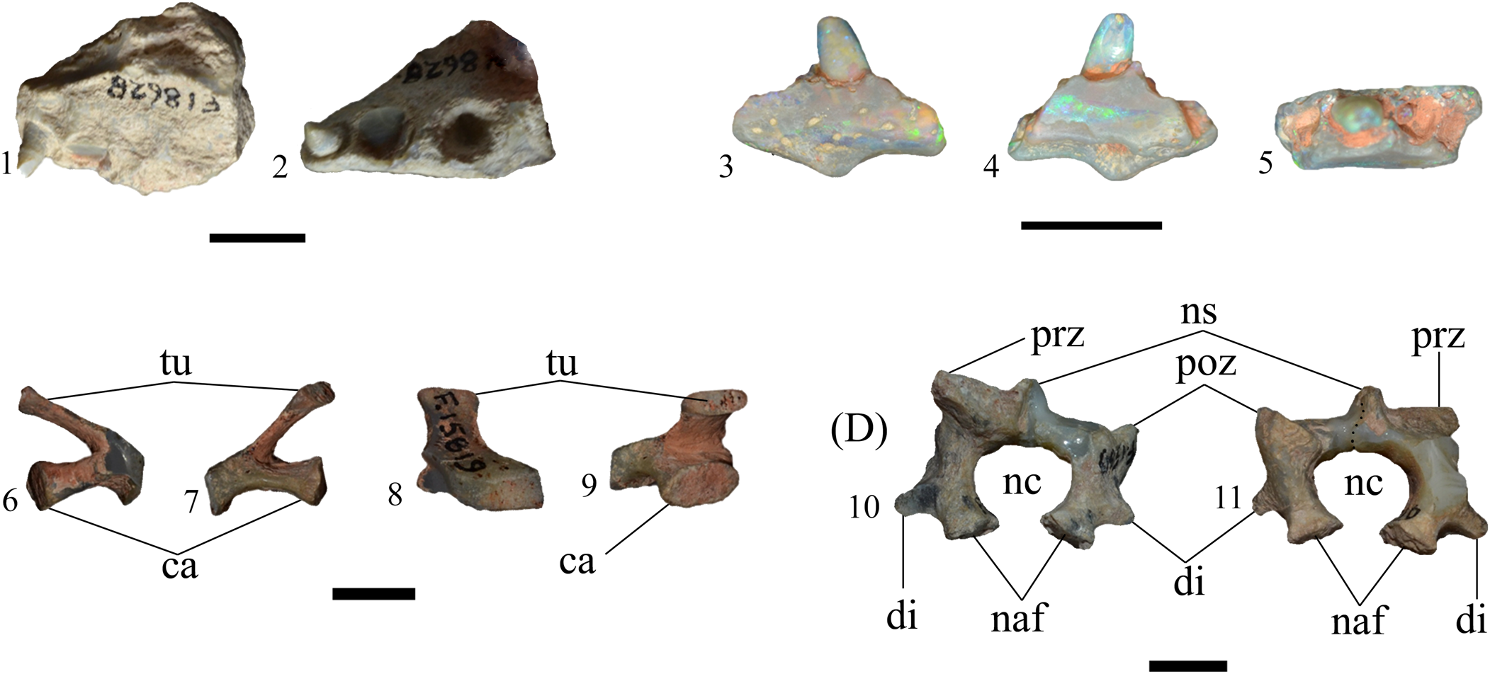
Figure 6. Isisfordia cf. I. selaslophensis cranial and postcranial remains. (1, 2) Maxillary fragment (AM F18628) in (1) medial and (2) ventral (composite image) views. (3–5) Dentary fragment (AM F68339) in (3) lateral (labial), (4) medial (lingual), and (5) dorsal views. (6–9) Cervical rib (AM F137556) in (6) anterior, (7) posterior, (8) lateral, and (9) medial views. (10, 11) Neural arch (AM F137555) in (10) anterior and (11) posterior views. ca = capitulum; di = diapophysis; naf = neurocentral articular facet; nc = neural canal; ns = neural spine; poz = postzygapophysis; prz = prezygapophysis; tu = tuberculum. Scale bars = 10 mm. Photo credits: Lachlan Hart.
Dentary
AM F68339 is a small fragment of right dentary bearing one tooth (Fig. 6.3–6.5) and parts of three (or perhaps four) other alveoli (Fig. 6.5). The medial and lateral surfaces are straight (showing very little concavity and convexity, respectively), indicating this fragment is probably from a more posterior region of the mandible. The ventral edge is uneven, with a distinct triangular ventral process at roughly the midline. This projection is likely a taphonomic artefact and not indicative of natural morphology. The lateral surface of the dentary is ornamented with unevenly dispersed neurovascular foramina (Fig. 6.3), typical of crocodyliforms (see Iordansky, 1976). The lingual surface is comparatively smooth and morphologically uninformative, with the most striking feature being a flash of green and blue opal color extending nearly the entire anteroposterior length (Fig. 6.4). The tooth (extending ~3 mm dorsoventrally from the dentary bone) is inclined lingually from the crown base. The apex of the crown is missing (perhaps worn), and no carinae or striations can be observed although this may be due to preservation and not indicative of natural morphology. In lateral views, the tooth is slightly posteriorly oriented.
Description of unassociated postcranial bones
Cervical rib
AM F60082 is a cervical rib (Fig. 6.6–6.9), previously described (but not figured) by Molnar (Reference Molnar1980) as the “proximal portion … from the left side” (p. 67), a diagnosis that we accept here. Both the anterior and posterior lateral processes of the rib are missing. The tuberculum and capitulum form an acute angle (~30°) in anterior and posterior views (Fig. 6.6, 6.7). The tuberculum is approximately 20% mediolaterally longer and less robust than the capitulum. The stout capitulum extends laterally, expanding dorsoventrally and anteroposteriorly at the articular end. The ventral surface (between the capitulum and the rib) is concave in anteroposterior view. The articular surfaces of both the tuberculum and the capitulum are ovoid (anteroposteriorly longer than tall) and adorned with crenulate sutural scars (Fig. 6.9). The articular surface of the tuberculum is roughly half the height of that of the capitulum. In lateral view, the lateralmost surface of the rib is rectangular and smooth (Fig. 6.8).
Neural arch
AM F60081 (Fig. 6.10, 6.11) was originally described and figured by Molnar (Reference Molnar1980) as “two portions of a cervical neural arch” (p. 66), and it was emphasized that, although the pieces do not share a contact, they have been reconstructed together as a single piece, as if they had originally constituted a single vertebra. The size and preservation of both pieces are very similar, but whether they formed a single element is unclear. The neurocentral articular facets are elliptical, ventromedially oriented, and retain the typical crenulated sutural texture (Fig. 6.10, 6.11). The bases of both diapophyses are preserved as short, laterally rounded subconical projections, beginning ~5 mm from the base of the neural arch (Fig. 6.10, 6.11). The breakage of the diapophyses in lateral view reveals a semicircular cross section. The neural canal is broad and subcircular, ~12 mm in diameter (Fig. 6.10, 6.11). The left postzygapophysis is preserved as the base only, whereas the right is more complete. The postzygapophysis extends dorsally and posteriorly parallel to the base of the neural arch in lateral view, creating a ‘C’-shaped concavity. The base of the neural spine is preserved as an ~5 mm tall process at the center of the neural arch (Fig. 6.10, 6.11). The left prezygapophyseal facet is also preserved, creating a medially oriented right angle with the right ascending process of the neural arch, visible in posterior view (6.11). The prezygapophyseal facet extends ~4 mm from the dorsalmost point of the neural arch body.
Thoracic vertebra
AM F112894 is probably from the mid-thoracic region due to the lack of a hypopophysis (Fig. 7.1–7.6). The hourglass-shaped centrum (in ventral view) is complete, as is much of the neural arch. The articular ends of the centrum are incipiently procoelous, (Fig. 7.1, 7.2), as in LRF 750.A, E, F. The neural canal (~5 mm diameter) is best observed in anterior view (Fig. 7.1), where it has a circular opening. In posterior view, the neural canal is dorsoventrally compressed. The left prezygapophysis is partially preserved and has a dorsomedially oriented articular facet. The lateral extension of the prezygapophysis begins approximately at the mid-height of the neural canal in anterior view (Fig. 7.1). The preserved section extends ~1 mm beyond the anterior edge of the centrum in lateral view (Fig. 7.3, 7.4). The bases of both postzygapophyses are present. The right is the more complete of the two (Fig. 7.2–7.4), although the articular facets are not preserved. The right postzygapophysis extends beyond the posterior edge of the centrum by ~0.5 mm (Fig. 7.3, 7.4). The preserved section of the base of the neural arch is smooth, perhaps an artefact of the mining process. The posterior end of the neural arch base is broken, revealing a dorsoventrally compressed cross section (Fig. 7.2).

Figure 7. Isisfordia cf. I. selaslophensis axial elements. (1–6) Thoracic vertebra (AM F112894) in (1) anterior, (2) posterior, (3) left lateral, (4) right lateral, (5) dorsal, and (6) ventral views. (7–9) Sacral vertebra (AM F15819) in (7) anterior, (8) posterior, and (9) dorsal views. (10–13) Sacral vertebra (AM F112896) in (10) anterior, (11) posterior, (12) dorsal, and (13) ventral views. na = neural arch; nc = neural canal; ns = neural spine; poz = postzygapophysis; prz = prezygapophysis; sr = sacral rib. Scale bars = 10 mm. Photo credits: Lachlan Hart.
Sacral vertebrae
AM F15819 and AM F112896 are nearly identical sacral vertebrae (Fig. 7.7–7.13), differing only in size and preservation. AM F15819 (Fig. 7.7–7.9) is less complete than AM F112896 (Fig. 7.10–7.13) but preserves morphologies not present in the latter specimen. Both sacral ribs are preserved in AM F112896, although the left has been reattached after breakage. AM F15819 preserves the left sacral rib and the base of the right (originally described and figured by Molnar, Reference Molnar1980). AM F15819 also preserves the prezygapophyses on the base of the neural arch (Fig. 7.7, 7.9). The prezygapophyses are not preserved on AM F112896. The prezygapophysis bears a ridge extending onto the sacral rib (Fig. 7.8). The base of the neural spine is also preserved in AM F15819 but not in AM F112896. The articular ends of the centra are shallowly concave and ovoid (Fig. 7.7, 7.8, 7.10, 7.11). Our observations concur with Molnar (Reference Molnar1980), who also noted the ventral surface of AM F15819 was concave, bearing longitudinal ridges along both sides.
Caudal vertebra
AM F60080 is a caudal vertebra (Fig. 8.1–8.6). In ventral view, the centrum is hourglass shaped (constricted at mid-length) and dominated by a deep anteroposterior groove, extending nearly the entire length of the centrum. Both articular ends on the ventral surface show signs of breakage. In anterior and terminal views, the articular ends are incipiently procoelous. AM F60080 retains both transverse processes, which are elevated above the centrum and extend beyond it laterally by ~10 mm. In lateral views, both diapophyses are present as small terminal upturning of the tips of the transverse processes. As noted by Molnar (Reference Molnar1980), “a distinct pocket extends into the terminal margin of the transverse process just lateral to the postzygapophysis” (p. 67). The right prezygapophysis is also partially preserved, although the articular facet is incomplete.

Figure 8. Isisfordia cf. I. selaslophensis caudal vertebra and tibiae. (1–6) Caudal vertebra (AM F60080) in (1) anterior, (2) posterior, (3) left lateral, (4) right lateral, (5) dorsal, and (6) ventral views. (7–13) Tibia (LRFR 3378): (7) original reconstruction; (8–13) digitally reconstructed in (8) anterior, (9) posterior, (10) left lateral, (11) right lateral, (12) dorsal, and (13) ventral views. (14–17) Distal tibia (AM F60083) in (14) anterior, (15) posterior, (16) left lateral, and (17) right lateral views. (18–22) Proximal tibia (AM F15821) in (18) anterior, (19) posterior, (20) left lateral, (21) right lateral, and (22) dorsal views. (23–26) Proximal tibia (AM F18630) in (23) anterior, (24) posterior, (25) left lateral, and (26) right lateral views. lg = lateral groove; li = attachment for linea intermuscularis caudalis; na = neural arch; prz = postzygapophysis; tp = transverse process; vg = ventral groove. Scale bars = 10 mm. Photo credits: LRFR 3378 photo by Phil Bell; LRFR 3378 digital reconstruction by Rex Mitchell; all other photos by Lachlan Hart.
Tibiae
Three partial (AM F60083, Fig. 8.14–8.17; AM F15821, Fig. 8.18–8.22; AM F18630, Fig. 8.23–8.26) and one complete right tibia (LRFR 3378, Fig. 8.7–8.13) were identified. AM F18630 and AM F15821, both of which are missing the distal ends, were previously described by Molnar (Reference Molnar1980) although only AM F15821 was figured. AM F60083 is the distal end of a tibia. The following description is based on LRFR 3378 as it is complete and previously undescribed. The preserved portions of the remaining three tibiae are all nearly identical to LRFR 3378, differing only in size. LRFR 3378 is a replica of a privately owned specimen originally reassembled from two pieces incorrectly (Fig. 8.7) but digitally restored to the correct position (Fig. 8.8–8.13; see Materials and methods). The proximal end (or tibial head) of LRFR 3378 is roughly twice as anteroposteriorly long as the distal end (Fig. 8.12, 8.13). The proximal articular surface is relatively flat and dorsomedially inclined, and the anterior articular condyles flare laterally at the point of articulation. There is a reduced cnemial crest on the anterior side of the shaft (Fig. 8.8), as occurs in modern crocodylians (Romer, Reference Romer1956). From the proximal end, the shaft tapers to a teardrop-shaped cross section by the midpoint. However, this may be due to breakage or wear and not indicative of actual morphology. On the posterior surface of the shaft, a low ridge (Fig. 8.9) pertaining to the linea intermuscularis caudalis extends from roughly one quarter of the dorsoventral height of the shaft and terminates abruptly just beyond the mid-length of the shaft. The original specimen was broken at this point, so this region may contain some artefacts of breakage and/or reconstruction. The general shape of the tibial shaft is straight, as in living crocodylians (Romer, Reference Romer1956). The distal end of LRFR 3378 (Fig. 8.13) is saddle shaped in posterior view, with the two medially extending condyles of roughly comparable size.
Calcaneum
The calcaneum (a right element; LRFR 3379; Fig. 5.3–5.5) is a small asymmetrical bone. In medial view, the calcaneal socket is a deep, hemispherical cotyle for articulation with the astragalus (Fig. 5.3). The ventralmost edge of the calcaneal socket extends medially, creating a ‘lip’ that would have braced the ventral edge of the astragalus. Ventral to this lip, the calcaneum is anteroposteriorly constricted, defining the sustentacular groove in posterior view (Fig. 5.4). The sustentacular groove bears dorsoventral ridges and a small foramen roughly one-third of the way from the ventral end of the bone. The calcaneal tuber extends posterolaterally to a point in line with the posterior margin of the edge of the calcaneal socket (Fig. 5.3). On the calcaneal tuber is a defined distal groove (Fig. 5.3).
Osteoderms
All specimens are incompletely preserved, obscuring the overall shape of each osteoderm, and details are only preserved on the exterior (superficial) surface. AM F12893 (Fig. 5.8) and AM F128119 (Fig. 5.10) are flat, with no keel. The exterior surface is ornamented by numerous pits, which are variably sized in AM F12893 but are relatively uniform on AM F128119.
AM F121672 (Fig. 5.9) is smaller than AM F12893 and AM F128119 and has a pronounced keel, extending the entire anteroposterior length of the osteoderm. This indicates that this osteoderm is from the thoracic region. The foramina on the exterior surface are more uniform than in AM F12893 and are generally elliptical.
Associated vertebral materials
LRF750 and LRF 751, a collection of 13 associated thoracic and caudal vertebrae, recovered from the Holden's locality on the Three Mile opal field, situated just south of the town of Lightning Ridge.
Associated cranial materials
AM F103588, AM F103589, maxillary fragments; AM F103587, mandible.
Unassociated cranial materials
AM F125524, basioccipital; AM F66766, quadrate; AM F18628, maxillary fragment; AM F68339, dentary fragment.
Unassociated postcranial materials
AM F60080, cervical rib; AM F60081, neural arch; AM F112894, thoracic vertebrae; AM F112896, AM F15819, sacral vertebrae; AM F6008, caudal vertebrae; LRFR 3378, AM F18630, AM F60083, AM F137557, tibiae; LRFR 3379, calcaneum; AM F112893, AM F121672, AM F128119, osteoderms.
Remarks on associated vertebrae
Together, the 13 vertebrae mentioned constitute the most complete Cretaceous crocodyliform skeleton from Australia outside of the Winton Formation. We accept that there is a possibility that these remains, as well as most other ‘associated’ fossils that have been discovered in the opal mines from the Lightning Ridge area, may not be from the same individual and instead are representative of a ‘multi-taxic’ bonebed or microsite. The in situ articulation of these bones is unknown as they were first observed by one of us (ES) only after their excavation from the mine. The slight discrepancies in size of the dorsal vertebrae, as discussed in the preceding, may be an artefact of weathering, and none of the dorsal vertebrae are complete. Our hypothesis is that these 13 vertebrae do derive from the same individual, due to their proximity to each other (all found within the same 50 × 50 meter mining claim), the comparable size of each element, and the lack of any other crocodyliform material from the same location. Furthermore, the common opal color, or ‘potch’ color, of the remains is consistent throughout all vertebrae. Opal color has previously been used as a reliable indicator of close affinity of opalized bones (Bell et al., Reference Bell, Herne, Brougham and Smith2018).
This partial skeleton is referred to Isisfordia on the basis of the incipiently procoelous vertebrae (an autapomorphy of Isisfordia [shared with Susisuchus and some atoposaurids; see Salisbury et al., Reference Salisbury, Molnar, Frey and Willis2006; Turner and Pritchard, Reference Turner and Pritchard2015; Hart, Reference Hart2020]) and can be differentiated from I. duncani on the basis of the presence of a deep median groove on the ventral surface of the caudal vertebrae.
As no postcranial remains have been attributed to either I. selaslophensis or I. molnari, specific diagnosis of the following specimens cannot be ascertained. However, as suggested by Hart (Reference Hart2020), I. molnari is likely a subjective junior synonym of I. selaslophensis, an opinion that we uphold here. Therefore, we place the preceding specimens in Isisfordia cf. I. selaslophensis.
The thoracic and caudal vertebrae of LRF 750 (Fig. 2) and LRF 751 (Fig. 3) display an unambiguous autapomorphy of Isisfordia: weak (or incipient) procoely (Salisbury et al., Reference Salisbury, Molnar, Frey and Willis2006). This feature is also present in Susisuchus and some atoposaurids (Salisbury et al., Reference Salisbury, Molnar, Frey and Willis2006), so in a strict apomorphy-based diagnosis, this material would be restricted to Neosuchia. However, Susisuchus and atoposaurid taxa are unknown from the Cretaceous of Australia. We therefore refer LRF 750 and LRF 751 to Isisfordia.
A median groove on the ventral surface of the caudal vertebrae (as observed in AM F60080; Fig. 8.1 – 8.6 and LRF 751; Fig. 3.6, 3.12, 3.18, 3.24) is an uncommon feature in crocodyliforms. The poorly known Late Cretaceous taxon Pinacosuchus mantiensis Gilmore, Reference Gilmore1942 is documented as having a “shallowly channelled” (Gilmore, Reference Gilmore1942, p.110) ventral surface of the caudal vertebrae. However, P. mantiensis was only provisionally referred to “Crocodilia” (p. 114) and, on the basis of the biconvex vertebrae, is unlikely to be a eusuchian (Langston, Reference Langston2008) and may not be a crocodyliform at all. A ventral groove was also figured (but not discussed) by Buscalioni and Sanz (Reference Buscalioni and Sanz1990, fig. 8) in their description of Bernissartia fagesii Dollo, Reference Dollo1883; however, this groove occurs on a thoracic vertebral centrum.
A median ventral groove is present in a few caudal vertebrae (QM F58795) referred to I. duncani (Syme et al., Reference Syme and Salisbury2018; LJH and SWS, personal observation). This groove is not as deep or well defined as in AM F60080 and LRF 751. This may be because QM F58795 represents a very young individual and that this feature became more defined through ontogeny. Alternatively, QM F58795 may have a more posterior position in the series than AM F60080 and LRF 751, and thus the groove is less defined. Nonetheless, this feature is not consistently observed in caudal vertebrae of I. duncani but is apparent in LRF 751, AM F60080, and other specimens recovered from the Lightning Ridge opal mines (LJH, PRB, SWS, and ETS, personal observation).
Remarks on associated cranial bones
These specimens were originally described by Molnar and Willis (Reference Molnar, Willis, Grigg, Seebacher and Franklin2001) as belonging to a ziphodont mesoeucrocodylian (primarily on the basis of the labiolingual compression of the teeth). In this description of AM F103588 and AM F103589, the specimen catalog reference numbers were erroneously swapped. Our analysis and observations conclude that these specimens, and the partial teeth contained therein, show no signs of ziphodonty other than labiolingual compression. The teeth of I. duncani, and to a lesser extent I. selaslophensis, are labiolingually compressed and unserrated. Indeed, the labiolingual compression of the teeth at the base of the crown is (in part) an autapomorphy of Isisfordia (Hart, Reference Hart2020). As observed in the preceding, the teeth in AM F103587–103589 do bear striations, a feature in common with all known teeth pertaining to Isisfordia.
Remarks on unassociated cranial bones
Basioccipital
Median and lateral eustachian foramina enclosed within a ‘verticalized’ basioccipital (Gower and Weber, Reference Gower and Weber1998; Salisbury et al., Reference Salisbury, Molnar, Frey and Willis2006) are diagnostic traits of eusuchian crocodylomorphs and convergent with birds (Gower and Webber, Reference Gower and Weber1998). Few avian fossils have been recovered from the Griman Creek Formation, all of which have been interpreted as enantiornithine (Bell et al., Reference Bell, Fanti, Hart, Milan, Craven, Brougham and Smith2019b). These remains indicate a very small taxon (or taxa) and do not correspond in size to AM F125524. The overall morphology of AM F125524 also differs considerably from those figured for birds (see O'Connor and Chiappe, Reference O'Connor and Chiappe2011); therefore, this element is likely to pertain to a eusuchian. The size and general morphology of AM F125524 closely resemble the basioccipital of the paratype skull of I. duncani (SWS, personal observation).
Quadrate
Although the articular condyles of AM F66766 (Fig. 5.6, 5.7) as discussed in the preceding differ from the condition seen in I. duncani, intraspecific variation (such as other features seen on AM F125553, the braincase assigned to I. molnari, [see Hart et al., Reference Hart, Bell, Smith and Salisbury2019]) cannot be ruled out, especially considering the articular condyles of the quadrate preserved in AM F125553 are incomplete. The quadrate is superficially similar to the susisuchids Susisuchus anatoceps Salisbury et al., Reference Salisbury, Frey, Martill and Buchy2003 and Susisuchus jaguarbiensis Fortier and Schultz, Reference Fortier and Schultz2009 (Salisbury et al., Reference Salisbury, Frey, Martill and Buchy2003; Fortier and Schultz, Reference Fortier and Schultz2009; Leite and Fortier, Reference Leite and Fortier2018). As Isisfordia is the only known susisuchid-grade crocodyliform from the Griman Creek Formation, it is reasonable to assume that AM F66766 pertains to this genus.
Maxilla
The overall dorsoventral compression of AM F18628 is an iconic feature shared with the rostral regions of many neosuchians (Brochu, Reference Brochu2001) and is differentiated from the more mediolaterally compressed rostral regions of other archosaurs, such as theropods. The single intact tooth, although worn, shares the morphological characteristics of the maxillary teeth described for I. selaslophensis (Hart et al., Reference Hart, Bell, Smith and Salisbury2019; Hart, Reference Hart2020) of lingually curved and subconical crowns. In addition, the alveoli of AM F18628 are round, a feature that differentiates I. selaslophensis from I. duncani (Hart, Reference Hart2020). Unlike many of the other fossils discussed herein, AM F18628 potentially overlaps with the holotype (AM F15818) of I. selaslophensis, via the matching maxillary tooth and alveolar morphology. These similarities support the assignment of AM F18628 to I. selaslophensis.
Dentary
The lack of lateral curvature indicates AM F68339 is probably from the mandibular ramus and not the symphyseal region. Neurovascular foramina of the mandible, as observed in AM F68339, are a typical crocodyliform trait (Iordansky, Reference Iordansky, Gans and Parsons1973). The closely set, subcircular confluent alveoli of AM F68339 are also seen in I. duncani, which may indicate that AM F68339 pertains to a similar taxon. The general tooth shape, although worn, is also comparable to the mandibular teeth seen in QM F44319 and QM F34642 (paratypes of I. duncani; see Salisbury et al., Reference Salisbury, Molnar, Frey and Willis2006; Syme and Salisbury, Reference Syme and Salisbury2018, respectively).
Remarks on unassociated postcranial bones
Cervical rib
Molnar (Reference Molnar1980) remarked on the identical nature of AM F60082 to the cervical ribs of the extant Crocodylus porosus Schneider, Reference Schneider1801, an observation we accept. Cervical ribs are morphologically conservative in crocodylians (see Romer, Reference Romer1956; Grigg and Kirshner, Reference Grigg and Kirshner2015). This element is also referred to Isisfordia cf. I. selaslophensis in the absence of evidence for any other crocodyliform genus from the Griman Creek Formation.
Neural arch
Molnar (Reference Molnar1980) compared AM F60081 and the neural arches of C. porosus and C. acutus Cuvier, Reference Cuvier1807 (referred to as C. ‘americanus’ by Molnar, Reference Molnar1980) and observed the pre- and postzygapophyseal facets were more horizontally aligned in AM F60081 and more similar to C. porosus than to C. acutus. Molnar (Reference Molnar1980) also noted that diapophyses elevated above the neurocentral suture in AM F60081 was another feature analogous to C. porosus. Crocodylus porosus (a eusuchian) is not known from the Cretaceous Griman Creek Formation. Eusuchian-grade crocodyliforms are solely represented in the Griman Creek Formation by Isisfordia; thus, it is likely AM F60081 pertains to this genus.
Thoracic vertebra
The overall morphology of AM F112894 is very similar to LRF 750.A–F (diagnosed here as Isisfordia cf. I. selaslophensis). The most salient feature of AM F112894 is the incipient procoely of the articular ends of the centrum. This feature is an autapomorphic trait of Isisfordia (Salisbury et al., Reference Salisbury, Molnar, Frey and Willis2006; Hart et al., Reference Hart, Bell, Smith and Salisbury2019).
Sacral vertebra
Molnar (Reference Molnar1980) compared AM F15819 and the first sacral vertebrae of ‘Leidyosuchus’ multidentatus Mook, Reference Mook1930 (now Listrognathosuchus) and Diplocynodon hantoniensis Wood, Reference Wood1846, inferring similarities in general morphology. Both of these taxa are alligatoroids, indicating a eusuchian affinity for AMF15819 and AM F112896. We support these comparisons, reiterating that Isisfordia is the only crocodyliform known from the Griman Creek Formation.
Caudal vertebra
AM F60080 is most similar to LRF 751.A–D, with all possessing the following features: (1) lateral groove on both sides of the centrum (Fig. 8.3, 8.4), (2) prominent ventral groove (Fig. 8.6), (3) constriction at mid-length in ventral aspect (Fig. 8.6), and (4) thin anteroposterior ridge within the neural canal (Fig. 8.5). The most striking of these features is the anteroposterior groove on the ventral surface. The incipient procoely of AM F60080 is an autapomorphy of Isisfordia (Salisbury et al., Reference Salisbury, Molnar, Frey and Willis2006), and the prominent anteroposterior ventral groove has previously been proposed as unique to the Griman Creek Formation crocodylomorph material.
Tibiae
LRFR 3378 has several traits seen in tibiae of modern crocodylians, including reduction of the cnemial crest and a straight tibial shaft. Molnar (Reference Molnar1980) remarked that AM F18630 and AM F15821 are both “identical and closely resemble those of C. porosus” (p. 68). We support Molnar's (Reference Molnar1980) observations and further propose that these are likely to pertain to Isisfordia, due primarily to the resemblance of these tibiae with those from the I. duncani holotype (LJH and SWS, personal observation).
Calcaneum
The calcaneum (LRFR 3379; Fig. 5.3–5.5) displays the ‘crocodile-normal’ configuration (Parrish, Reference Parrish1987, Reference Parrish1993) of a hemispherical socket that would have received the corresponding process of the astragalus. This feature unites Crocodylomorpha with Aetosauria and Rauisuchidae (sensu Parrish, Reference Parrish1993). As aetosaurians and rauisuchids are currently known only from the Triassic, LRFR 3379 is unlikely to pertain to a member of either clade. The presence of a laterally expanded calcaneal tuber and defined distal groove on the calcaneal tuber indicate a closer relationship to eusuchian crocodyliforms such as Alligator (see Parrish, Reference Parrish1993, fig. 3) than other crocodile-line archosaurs. In the absence of evidence for other crocodyliforms from the Griman Creek Formation, this supports a referral to Isisfordia cf. I. selaslophensis.
Osteoderms
Characteristic external pitting identifies these osteoderms as crocodyliform; AM F121672 can be potentially assigned to Eusuchia on the basis of the presence of a keel (Salisbury et al., Reference Salisbury, Molnar, Frey and Willis2006, supplementary information), assuming the complete osteoderm was square and not rectangular. AM F112893 and AM F128119 lack keels, suggesting they may come from the gastral region (Salisbury et al., Reference Salisbury, Molnar, Frey and Willis2006, fig. 2), but are not complete enough to show any evidence of sutural or articular surfaces. The preserved portions of these osteoderms as described closely resemble those of QM F36211 (holotype of I. duncani), and due to the lack of other known crocodyliforms from the Griman Creek Formation, this material is referred to Isisfordia cf. I. selaslophensis.
Discussion
Molnar (Reference Molnar1980) referred many of the previously described remains discussed in the preceding to the putative eusuchian ‘Crocodylus (Bottosaurus) selaslophensis,’ the holotype of which was originally described as a mandibular fragment (Etheridge, Reference Etheridge1917) but reidentified as part of a maxilla (Hart et al., Reference Hart, Bell, Smith and Salisbury2019). This includes the maxilla (AM F18628, Fig 6.1, 6.2), neural arch (AM F60081, Fig 6.10, 6.11), sacral (AM F15819, Fig 7.7–7.9) and caudal (AM F60080, Fig. 8.1–8.6) vertebrae, cervical rib (AM F60082, Fig. 6.6–6.9), and tibiae (AM F18630, AM 15821, Fig. 8.14–8.26). Molnar's (Reference Molnar1980) referral of these elements to ‘C. selaslophensis’ was justified on the basis of the shared presence of features consistent with those of other eusuchian taxa. Molnar (Reference Molnar1980) also discussed similarities in body size indicated by all remains described and cited a lack of evidence for another crocodyliform in the Griman Creek Formation as further justification for this referral. As per Hart (Reference Hart2020), this taxon is now known as Isisfordia selaslophensis, and we support Molnar's (Reference Molnar1980) referrals of the aforementioned elements to the same taxon.
Subsequently, Molnar's (Reference Molnar1980) original assignment of these remains to one taxon was contested by Molnar and Willis (Reference Molnar, Willis, Grigg, Seebacher and Franklin2001) on the basis of description of mandibular and maxillary fragments purportedly representing a second crocodyliform from the Griman Creek Formation. However, as discussed in the preceding, it is quite possible that this material also pertains to a species of Isisfordia on the basis of the labiolingually compressed, lingually curved teeth and ovate alveoli (Hart et al., Reference Hart, Bell, Smith and Salisbury2019), which have been observed in I. duncani. Indeed, Molnar and Willis (Reference Molnar, Willis, Grigg, Seebacher and Franklin2001) postulated that these fragments belonged to a small, broad-snouted form, which fits the description of Isisfordia. Furthermore, our reanalysis found no compelling evidence of ziphodonty, which was used by Molnar and Willis (Reference Molnar, Willis, Grigg, Seebacher and Franklin2001) to justify recognition of a second taxon distinct from comparable previously described Griman Creek Formation crocodyliform specimens. Therefore, we reinstate Molnar's (Reference Molnar1980) original designation of the remains (which have been redescribed here) to a single taxon.
Molnar (Reference Molnar1980) described cervical vertebrae that were known only from casts (as the original specimens were lost) housed at the Queensland Museum (QM). These elements were not observed by the authors and are not considered here. However, the apparent procoely of these remains is consistent with the presence of an Isisfordia-like crocodyliform. Similarly, the ?femur described by Molnar (Reference Molnar1980) was in the private collection of K. Barlow; it could not be observed for this study and thus was not included.
The associated vertebrae (LRF 750–751) described here represent the most complete Australian Cretaceous crocodyliform skeleton known outside of the Winton Formation. It is also the first postcranial material consisting of multiple elements attributed to Isisfordia from the Griman Creek Formation. On the basis of the available evidence, the other material discussed herein is also likely to pertain to I. selaslophensis. Therefore, we suggest that, as in the Winton Formation (the type locality of I. duncani), only a single genus of crocodyliform was present in the Griman Creek Formation. This is in stark contrast to many other Gondwanan Upper Cretaceous localities; for example, there are at least seven taxa known from the Aptian–Cenomanian Kem Kem Formation of Morocco (e.g., Sereno and Larsson, Reference Sereno and Larsson2009), four from the Campanian–Maastrichtian Maevarano Formation of Madagascar (e.g., Krause et al., Reference Krause, Sertich, Rogers, Kast, Rasoamiaramanana and Buckley2010), and possibly as many as 20 from the Turonian–Maastrichtian Adamantina Formation of Brazil (e.g., Carvalho et al., Reference Carvalho, Campos and Nobre2005). This is primarily a consequence of Australia's incompletely sampled Mesozoic terrestrial fossil record (Bell et al., Reference Bell, Fanti, Hart, Milan, Craven, Brougham and Smith2019b). However, these finds are consistent with modern crocodyliform faunas, where true sympatric species are very rare (see Salisbury and Willis, Reference Salisbury and Willis1996). Nonetheless, multiple species have been identified from other vertebrate groups, such as dinosaurs and turtles, in the Griman Creek Formation (e.g., Bell et al., Reference Bell, Fanti, Hart, Milan, Craven, Brougham and Smith2019b), suggesting that the depauperate crocodyliform fauna accurately represents the paleoecology of the region rather than a collection bias. Furthermore, the aforementioned Gondwanan faunas are heavily overrepresented by notosuchian taxa, where this group is entirely absent in Australia. Australia's high-latitude paleoclimate during the Cretaceous (between 70°S and 80°S; see Alley et al., Reference Alley, Hore and Frakes2019) may have affected crocodyliform distribution and diversity (Poropat et al., Reference Poropat, Martin, Tosolini, Wagstaff, Bean, Kear, Vickers-Rich and Rich2018). Populations of crocodyliforms may have been concentrated within favorable environments present at higher latitude (Syme and Salisbury, Reference Syme and Salisbury2018; Hart et al., Reference Hart, Bell, Smith and Salisbury2019) and in the Winton and Griman Creek formations, where perhaps climatic fluctuations were ameliorated by the epicontinental Eromanga Sea. A more detailed consideration of the relationships between the distribution and diversity of Australian Mesozoic crocodyliforms and paleoclimate represents a focal point for future research.
Conclusions
New postcranial material from the Griman Creek Formation at Lightning Ridge is referred to Isisfordia cf. I. selaslophensis on the basis of characteristics present in Isisfordia spp., but lacking features not seen in I. duncani. This includes the most complete postcranial skeleton of a Cretaceous crocodyliform from the Griman Creek Formation, a collection of 13 associated thoracic and caudal vertebrae. Furthermore, fossils previously described by Molnar (Reference Molnar1980) that were assigned to ‘Crocodylus (Bottosaurus) selaslophensis’ (now Isisfordia selaslophensis) were reappraised and their specific diagnoses upheld. Of particular note is a distinct anteroposterior groove on the ventral side of the caudal vertebrae, a trait that is unique to the crocodyliforms recovered from the Griman Creek Formation. We also reevaluated material discussed by Molnar and Willis (Reference Molnar, Willis, Grigg, Seebacher and Franklin2001) as probably also referable to Isisfordia cf. I. selaslophensis due to strong similarities between this material and that of I. duncani. A collection of other cranial and postcranial material is also referrable to I. cf. I. selaslophensis. Future discoveries of more complete material may assist in diagnosing these remains with more certainty. This study clarifies the crocodyliform fauna of the Griman Creek Formation, specifically the lack of evidence for more than one crocodyliform taxon and therefore low levels of crocodyliform diversity, similar to that observed among extant species.
Acknowledgments
LJH thanks R. Pogson, M. McCurry, P. Smith (Australian Museum, Sydney), S. Hocknull, K. Spring (Queensland Museum, Brisbane), and J. Brammall (Australian Opal Centre, Lightning Ridge) for facilitating access to their respective collections. Specimens from Holden's field were donated through the Australian Government's Cultural Gifts Program by G. and C. Thomson. PRB is funded under an Australian Research Council Discovery Early Career Researcher Award (project ID: DE170101325). TB and DRM were funded by a Research Training Program (RTP) Scholarship. We acknowledge the Yuwaalaraay, Yuwaalayaay, and Gamilaraay custodians of country in the Lightning Ridge district and pay our respects to Elders past and present. Comments from H. Sues and two anonymous reviewers significantly improved this manuscript.


