Hostname: page-component-586b7cd67f-gb8f7 Total loading time: 0 Render date: 2024-11-24T13:32:55.083Z Has data issue: false hasContentIssue false

Analytical Three-Dimensional Field Ion Microscopy of an Amorphous Glass FeBSi

Published online by Cambridge University Press:  03 September 2021
Benjamin Klaes*
Affiliation:
Normandie Université, UNIROUEN, INSA Rouen, CNRS, Groupe de Physique des Matériaux, Rouen 76000, France
Jeoffrey Renaux
Affiliation:
Normandie Université, UNIROUEN, INSA Rouen, CNRS, Groupe de Physique des Matériaux, Rouen 76000, France
Rodrigue Lardé
Affiliation:
Normandie Université, UNIROUEN, INSA Rouen, CNRS, Groupe de Physique des Matériaux, Rouen 76000, France
Fabien Delaroche
Affiliation:
Normandie Université, UNIROUEN, INSA Rouen, CNRS, Groupe de Physique des Matériaux, Rouen 76000, France
Felipe F. Morgado
Affiliation:
Max-Planck Institut für Eisenforschung GmbH, Düsseldorf D-40237, Germany
Leigh T. Stephenson
Affiliation:
Max-Planck Institut für Eisenforschung GmbH, Düsseldorf D-40237, Germany
Baptiste Gault
Affiliation:
Max-Planck Institut für Eisenforschung GmbH, Düsseldorf D-40237, Germany Department of Materials, Royal School of Mines, Imperial College, Prince Consort Road, London SW7 2BP, UK
François Vurpillot
Affiliation:
Normandie Université, UNIROUEN, INSA Rouen, CNRS, Groupe de Physique des Matériaux, Rouen 76000, France Max-Planck Institut für Eisenforschung GmbH, Düsseldorf D-40237, Germany
*
Corresponding author: Benjamin Klaes, E-mail: benjamin.klaes1@univ-rouen.fr
Get access

Abstract

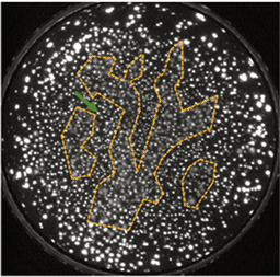
Three-dimensional field ion microscopy is a powerful technique to analyze material at a truly atomic scale. Most previous studies have been made on pure, crystalline materials such as tungsten or iron. In this article, we study more complex materials, and we present the first images of an amorphous sample, showing the capability to visualize the compositional fluctuations compatible with theoretical medium order in a metallic glass (FeBSi), which is extremely challenging to observe directly using other microscopy techniques. The intensity of the spots of the atoms at the moment of field evaporation in a field ion micrograph can be used as a proxy for identifying the elemental identity of the imaged atoms. By exploiting the elemental identification and positioning information from field ion images, we show the capability of this technique to provide imaging of recrystallized phases in the annealed sample with a superior spatial resolution compared with atom probe tomography.

Type
Field Ion Microscopy
Copyright
Copyright © The Author(s), 2021. Published by Cambridge University Press on behalf of the Microscopy Society of America

Access options

Get access to the full version of this content by using one of the access options below. (Log in options will check for institutional or personal access. Content may require purchase if you do not have access.)

References

Bernal, JD (1960). Geometry of the structure of monatomic liquids. Nature 185, 6870. doi:10.1038/185068a0CrossRefGoogle Scholar
Cazottes, S, Vurpillot, F, Fnidiki, A, Lemarchand, D, Baricco, M & Danoix, F (2012). Nanometer scale tomographic investigation of fine scale precipitates in a CuFeNi granular system by three-dimensional field Ion microscopy. Microsc Microanal 18, 11291134. doi:10.1017/S1431927612000517CrossRefGoogle Scholar
Cerezo, A, Hetherington, MG, Hyde, JM, Miller, MK, Smith, GDW & Underkoffler, JS (1992). Visualisation of three-dimensional microstructures. Surf Sci 266, 471480. doi:10.1016/0039-6028(92)91063-HCrossRefGoogle Scholar
Costa, GD, Wang, H, Duguay, S, Bostel, A, Blavette, D & Deconihout, B (2012). Advance in multi-hit detection and quantization in atom probe tomography. Rev Sci Instrum 83, 123709. doi:10.1063/1.4770120CrossRefGoogle ScholarPubMed
Da Costa, G, Vurpillot, F, Bostel, A, Bouet, M & Deconihout, B (2005). Design of a delay-line position-sensitive detector with improved performance. Rev Sci Instrum 76, 013304. doi:10.1063/1.1829975CrossRefGoogle Scholar
Dagan, M, Gault, B, Smith, GDW, Bagot, PAJ & Moody, MP (2017). Automated atom-by-atom three-dimensional (3D) reconstruction of field ion microscopy data. Microsc Microanal 23, 255268. doi:10.1017/S1431927617000277CrossRefGoogle ScholarPubMed
Dagan, M, Hanna, LR, Xu, A, Roberts, SG, Smith, GDW, Gault, B, Edmondson, PD, Bagot, PAJ & Moody, MP (2015). Imaging of radiation damage using complementary field ion microscopy and atom probe tomography. Ultramicroscopy 159, 387394. doi:10.1016/j.ultramic.2015.02.017CrossRefGoogle ScholarPubMed
Danoix, F, Epicier, T, Vurpillot, F & Blavette, D (2012). Atomic-scale imaging and analysis of single layer GP zones in a model steel. Journal of Materials Science 47(3), 15671571. doi: 10.1007/s10853-011-6008-4.CrossRefGoogle Scholar
Dong, B, Zhou, S, Qin, J, Li, Y, Chen, H & Wang, Y (2018). The hidden disintegration of cluster heterogeneity in Fe-based glass-forming alloy melt. Prog Nat Sci Mater Int 28, 696703. doi:10.1016/j.pnsc.2018.09.005CrossRefGoogle Scholar
Gault, B (Ed.) (2012). Atom Probe Microscopy. Springer Series in Materials Science. New York: Springer.CrossRefGoogle Scholar
Hono, K (1999). Atom probe microanalysis and nanoscale microstructures in metallic materials. Acta Mater 47, 31273145. doi:10.1016/S1359-6454(99)00175-5CrossRefGoogle Scholar
Joy, DC, Newbury, DE & Davidson, DL (1982). Electron channeling patterns in the scanning electron microscope. J Appl Phys 53, R81R122. doi:10.1063/1.331668CrossRefGoogle Scholar
Katnagallu, S, Nematollahi, A, Dagan, M, Moody, M, Grabowski, B, Gault, B, Raabe, D & Neugebauer, J (2017). High fidelity reconstruction of experimental field ion microscopy data by atomic relaxation simulations. Microsc Microanal 23, 642643. doi:10.1017/S1431927617003877CrossRefGoogle Scholar
Katnagallu, S, Stephenson, LT, Mouton, I, Freysoldt, C, Subramanyam, APA, Jenke, J, Ladines, AN, Neumeier, S, Hammerschmidt, T, Drautz, R, Neugebauer, J, Vurpillot, F, Raabe, D & Gault, B (2019). Imaging individual solute atoms at crystalline imperfections in metals. New J Phys 21, 123020. doi:10.1088/1367-2630/ab5cc4CrossRefGoogle Scholar
Kaufman, L & Bernstein, H (1970). Computer Calculation of Phase Diagrams with Special Reference to Refractory Metals, Refractory Materials, V. 4. New York: Academic Press.Google Scholar
Klaes, B, Lardé, R, Delaroche, F, Hatzoglou, C, Parviainen, S, Houard, J, Da Costa, G, Normand, A, Brault, M, Radiguet, B & Vurpillot, F (2021). Development of wide field of view three-dimensional field Ion microscopy and high-fidelity reconstruction algorithms to the study of defects in nuclear materials. Microsc Microanal. doi:10.1017/ S1431927621000131CrossRefGoogle Scholar
Klaes, B, Lardé, R, Delaroche, F, Parviainen, S, Rolland, N, Katnagallu, S, Gault, B & Vurpillot, F (2020). A model to predict image formation in the three-dimensional field ion microscope. Comput Phys Commun, 107317. doi:10.1016/j.cpc.2020.107317Google Scholar
Lefebvre-Ulrikson, W (Ed.) (2016). Atom Probe Tomography: Put Theory into Practice. London: Academic Press.Google Scholar
Mat'ko, I, Illeková, E, Švec, P & Duhaj, P (1997). Crystallization characteristics in the FeSiB glassy ribbon system. Mater Sci Eng A 225, 145152. doi:10.1016/S0921-5093(96)10567-0CrossRefGoogle Scholar
Miller, MK (1996). Atom Probe Field Ion Microscopy, Monographs on the Physics and Chemistry of Materials. Oxford, New York: Clarendon Press; Oxford University Press.Google Scholar
Miller, MK (2014). Atom-Probe Tomography: The Local Electrode Atom Probe. New York: Springer.CrossRefGoogle Scholar
Moody, MP, Stephenson, LT, Ceguerra, AV & Ringer, SP (2008). Quantitative binomial distribution analyses of nanoscale like-solute atom clustering and segregation in atom probe tomography data. Microsc Res Tech 71, 542550. doi:10.1002/jemt.20582CrossRefGoogle ScholarPubMed
Morgado, FF, Katnagallu, S, Freysoldt, C, Klaes, B, Vurpillot, F, Neugebauer, J, Raabe, D, Neumeier, S, Gault, B & Stephenson, LT (2021). Revealing atomic-scale vacancy-solute interaction in nickel. arXiv:2103.01639.Google Scholar
Müller, EW (1951). Das Feldionenmikroskop. Z Phys 131, 136142. doi:10.1007/BF01329651CrossRefGoogle Scholar
Muller, EW (1965). Field ion microscopy. Science 149, 591601. doi:10.1126/science.149.3684.591CrossRefGoogle ScholarPubMed
Prosa, TJ & Larson, DJ (2017). Modern focused-ion-beam-based site-specific specimen preparation for atom probe tomography. Microsc Microanal 23, 194209. doi:10.1017/S1431927616012642CrossRefGoogle ScholarPubMed
Rendulic, KD (1971). Measurements on field adsorption of neon and helium and the field ionization of a helium-neon mixture. Surf Sci 28, 285298. doi:10.1016/0039-6028(71)90100-2CrossRefGoogle Scholar
Rendulic, KD (1973). On field adsorption, ionization rates and gas supply. Surf Sci 34, 581587. doi:10.1016/0039-6028(73)90027-7CrossRefGoogle Scholar
Schröder, H, Samwer, K & Köster, U (1985). Micromechanism for metallic-glass formation by solid-state reactions. Phys Rev Lett 54, 197200. doi:10.1103/PhysRevLett.54.197CrossRefGoogle ScholarPubMed
Seidman, DN, Current, MI, Pramanik, D & Wei, C-Y (1981). Direct observations of the primary state of radiation damage of ion-irradiated tungsten and platinum. Nucl Instrum Methods 182–183, 477481. doi:10.1016/0029-554X(81)90718-7CrossRefGoogle Scholar
Sheng, HW, Luo, WK, Alamgir, FM, Bai, JM & Ma, E (2006). Atomic packing and short-to-medium-range order in metallic glasses. Nature 439, 419425. doi:10.1038/nature04421CrossRefGoogle ScholarPubMed
ur Rehman, H, Durst, K, Neumeier, S, Sato, A, Reed, R & Göken, M (2017). On the temperature dependent strengthening of nickel by transition metal solutes. Acta Mater 137, 5463. doi:10.1016/j.actamat.2017.05.038CrossRefGoogle Scholar
Vurpillot, F, Bostel, A & Blavette, D (2001). A new approach to the interpretation of atom probe field-ion microscopy images. Ultramicroscopy 89, 137144. doi:10.1016/S0304-3991(01)00097-3CrossRefGoogle Scholar
Vurpillot, F, Danoix, F, Gilbert, M, Koelling, S, Dagan, M & Seidman, DN (2017). True atomic-scale imaging in three dimensions: A review of the rebirth of field-ion microscopy. Microsc Microanal 23, 210220. doi:10.1017/S1431927617000198CrossRefGoogle ScholarPubMed
Wille, C, Al-Kassab, T, Heinrich, A & Kirchheim, R (2006). Nanostructured materials studied by means of the computed field ion image tomography (CFIIT). Presented at the 2006 19th International Vacuum Nanoelectronics Conference, IEEE, Lijiang Waterfall Hotel, Guilin, China, pp. 17–18. doi:10.1109/IVNC.2006.335312CrossRefGoogle Scholar