Hostname: page-component-84b7d79bbc-5lx2p Total loading time: 0 Render date: 2024-07-30T00:35:01.917Z Has data issue: false hasContentIssue false

Atom Probe Tomography Investigations of Ag Nanoparticles Embedded in Pulse-Electrodeposited Ni Films

Published online by Cambridge University Press:  29 June 2021

Hosun Jun
Affiliation:
Department of Materials Science and Engineering, Korea Advanced Institute of Science and Technology (KAIST), 291 Daehak-ro, Yuseong-gu, Daejeon34141, Republic of Korea
Kyuseon Jang
Affiliation:
Department of Materials Science and Engineering, Korea Advanced Institute of Science and Technology (KAIST), 291 Daehak-ro, Yuseong-gu, Daejeon34141, Republic of Korea
Chanwon Jung
Affiliation:
Department of Materials Science and Engineering, Korea Advanced Institute of Science and Technology (KAIST), 291 Daehak-ro, Yuseong-gu, Daejeon34141, Republic of Korea
Pyuck-Pa Choi*
Affiliation:
Department of Materials Science and Engineering, Korea Advanced Institute of Science and Technology (KAIST), 291 Daehak-ro, Yuseong-gu, Daejeon34141, Republic of Korea
*
*Corresponding author: Pyuck-Pa Choi, E-mail: p.choi@kaist.ac.kr
Get access

Abstract

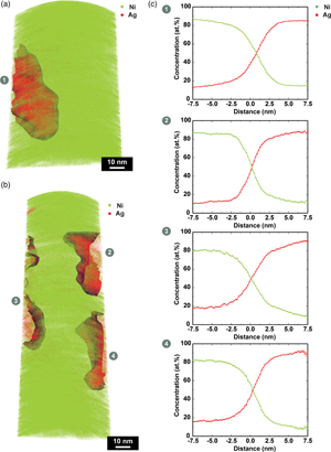
Atomic mapping of nanomaterials, in particular nanoparticles, using atom probe tomography (APT) is of great interest, as their properties strongly depend on shape, size, and composition. However, APT analyses of nanoparticles are extremely challenging, and there is an urgent need for developing robust and universally applicable sample preparation methods. Herein, we explored a method based on pulse electrodeposition to embed Ag nanoparticles in a Ni matrix and prepare APT specimens from the resulting composite film. By systematically varying the duty cycle during pulse electrodeposition, the dispersion and number density of the nanoparticles within the matrix was significantly enhanced as compared to DC electrodeposition. Several Ag nanoparticles were analyzed with APT from such samples. Shape distortions and biased compositions were observed for the Ag nanoparticles after applying a standard data reconstruction protocol. Numerical simulations of the field evaporation process showed that such artifacts were caused by a difference in the evaporation field of Ni and Ag and a local magnification effect. We expect such detrimental effects to be mitigated by a careful selection of the matrix material, matching the evaporation field of the nanoparticles. Furthermore, we anticipate that the method presented herein can be extended to a wider range of nanomaterials.

Type
Materials Science Applications
Copyright
Copyright © The Author(s), 2021. Published by Cambridge University Press on behalf of the Microscopy Society of America

Access options

Get access to the full version of this content by using one of the access options below. (Log in options will check for institutional or personal access. Content may require purchase if you do not have access.)

References

Beinke, D, Bürger, F, Solodenko, H, Acharya, R, Klauk, H & Schmitz, G (2020). Extracting the shape of nanometric field emitters. Nanoscale 12, 28202832.CrossRefGoogle ScholarPubMed
Beinke, D, Oberdorfer, C & Schmitz, G (2016). Towards an accurate volume reconstruction in atom probe tomography. Ultramicroscopy 165, 3441. doi:10.1016/j.ultramic.2016.03.008CrossRefGoogle ScholarPubMed
Clogston, JD & Patri, AK (2011). Zeta potential measurement. In Characterization of Nanoparticles Intended for Drug Delivery, McNeil, SE (Ed.), pp. 6370. Totowa, NJ: Humana Press. doi:10.1007/978-1-60327-198-1_6CrossRefGoogle Scholar
Dhara, S, Marceau, RKW, Wood, K, Dorin, T, Timokhina, IB & Hodgson, PD (2018). Atom probe tomography data analysis procedure for precipitate and cluster identification in a Ti-Mo steel. Data Brief 18, 968982. doi:10.1016/j.dib.2018.03.094CrossRefGoogle Scholar
Dmitrieva, O, Ponge, D, Inden, G, Millán, J, Choi, P, Sietsma, J & Raabe, D (2011). Chemical gradients across phase boundaries between martensite and austenite in steel studied by atom probe tomography and simulation. Acta Mater 59, 364374.CrossRefGoogle Scholar
Felfer, P, Benndorf, P, Masters, A, Maschmeyer, T & Cairney, JM (2014). Revealing the distribution of the atoms within individual bimetallic catalyst nanoparticles. Angew Chem Int Ed 53, 1119011193.CrossRefGoogle ScholarPubMed
Folcke, E, Lardé, R, Le Breton, JM, Gruber, M, Vurpillot, F, Shield, JE, Rui, X & Patterson, MM (2012). Laser-assisted atom probe tomography investigation of magnetic FePt nanoclusters: First experiments. J Alloys Compd 517, 4044. doi:10.1016/j.jallcom.2011.11.134CrossRefGoogle Scholar
Gault, B (2016). A brief overview of atom probe tomography research. Appl Microsc 46, 117126.CrossRefGoogle Scholar
Gault, B, Moody, MP, Cairney, JM & Ringer, SP (2012). Atom Probe Microscopy. New York: Springer.CrossRefGoogle Scholar
Gnaser, H (2014). Atom probe tomography of nanostructures. Surf Interface Anal 46, 383388.CrossRefGoogle Scholar
Hellman, OC, Vandenbroucke, JA, Rüsing, J, Isheim, D & Seidman, DN (2000). Analysis of three-dimensional atom-probe data by the proximity histogram. Microsc Microanal 6, 437444.CrossRefGoogle ScholarPubMed
Herbig, M, Raabe, D, Li, YJ, Choi, P, Zaefferer, S & Goto, S (2013). Atomic-scale quantification of grain boundary segregation in nanocrystalline material. Phys Rev Lett 112, 15.Google Scholar
Jung, C, Kang, K, Marshal, A, Pradeep, KG, Seol, JB, Lee, HM & Choi, PP (2019). Effects of phase composition and elemental partitioning on soft magnetic properties of AlFeCoCrMn high entropy alloys. Acta Mater 171, 3139. doi:10.1016/j.actamat.2019.04.007CrossRefGoogle Scholar
Katayama, K, Nomura, H, Ogata, H & Eitoku, T (2009). Diffusion coefficients for nanoparticles under flow and stop-flow conditions. Phys Chem Chem Phys 11, 1049410499.CrossRefGoogle ScholarPubMed
Kim, SH, Kang, PW, Park, OO, Seol, JB, Ahn, JP, Lee, JY & Choi, PP (2018). A new method for mapping the three-dimensional atomic distribution within nanoparticles by atom probe tomography (APT). Ultramicroscopy 190, 3038. doi:10.1016/j.ultramic.2018.04.005CrossRefGoogle Scholar
Kim, SH, Lee, JY, Ahn, JP & Choi, PP (2019). Fabrication of atom probe tomography specimens from nanoparticles using a fusible Bi-In-Sn alloy as an embedding Medium. Microsc Microanal 25, 438446.CrossRefGoogle ScholarPubMed
Kim, SH, Lim, J, Sahu, R, Kasian, O, Stephenson, LT, Scheu, C & Gault, B (2020). Direct imaging of dopant and impurity distributions in 2D MoS2. Adv Mater 32(8), 1907235Google ScholarPubMed
Larson, DJ, Prosa, TJ, Ulfig, RM, Geiser, BP & Kelly, TF (2013). Local Electrode Atom Probe Tomography. New York: Springer Science.CrossRefGoogle Scholar
Li, T, Bagot, PAJ, Christian, E, Theobald, BRC, Sharman, JDB, Ozkaya, D, Moody, MP, Tsang, SCE & Smith, GDW (2014). Atomic imaging of carbon-supported Pt, Pt/Co, and Ir@Pt nanocatalysts by atom-probe tomography. ACS Catal 4, 695702.CrossRefGoogle Scholar
Lim, J, Kim, SH, Aymerich Armengol, R, Kasian, O, Choi, PP, Stephenson, LT, Gault, B & Scheu, C (2020). Atomic-Scale mapping of impurities in partially reduced hollow TiO2 nanowires. Angew Chem Int Ed 59, 56515655.CrossRefGoogle ScholarPubMed
Melkonyan, D, Fleischmann, C, Arnoldi, L, Demeulemeester, J, Kumar, A, Bogdanowicz, J, Vurpillot, F & Vandervorst, W (2017). Atom probe tomography analysis of SiGe fins embedded in SiO2: Facts and artefacts. Ultramicroscopy 179, 100107.CrossRefGoogle ScholarPubMed
Millán, J, Sandlöbes, S, Al-Zubi, A, Hickel, T, Choi, P, Neugebauer, J, Ponge, D & Raabe, D (2014). Designing Heusler nanoprecipitates by elastic misfit stabilization in Fe-Mn maraging steels. Acta Mater 76, 94105.CrossRefGoogle Scholar
Miller, MK & Forbes, RG (2009). Atom probe tomography. Mater Charact 60, 461469. doi:10.1016/j.matchar.2009.02.007CrossRefGoogle Scholar
Miller, MK & Hetherington, MG (1991). Local magnification effects in the atom probe. Surf Sci 246, 442449.CrossRefGoogle Scholar
Miller, MK & Kenik, EA (2004). Atom probe tomography: A technique for nanoscale characterization. Microsc Microanal 10, 336341.CrossRefGoogle ScholarPubMed
Müller, EW (1956). Field desorption. Phys Rev 102, 618624.CrossRefGoogle Scholar
Oberdorfer, C, Eich, SM & Schmitz, G (2013). A full-scale simulation approach for atom probe tomography. Ultramicroscopy 128, 5567. doi:10.1016/j.ultramic.2013.01.005CrossRefGoogle ScholarPubMed
Pinheiro, JP, Domingos, R, Lopez, R, Brayner, R, Fiévet, F & Wilkinson, K (2007). Determination of diffusion coefficients of nanoparticles and humic substances using scanning stripping chronopotentiometry (SSCP). Colloids Surf, A 295, 200208.CrossRefGoogle Scholar
Povstugar, I, Zenk, CH, Li, R, Choi, PP, Neumeier, S, Dolotko, O, Hoelzel, M, Göken, M & Raabe, D (2016). Elemental partitioning, lattice misfit and creep behaviour of Cr containing gammaprime strengthened Co base superalloys. Mater Sci Technol 32, 220225.CrossRefGoogle Scholar
Raabe, D, Herbig, M, Sandlöbes, S, Li, Y, Tytko, D, Kuzmina, M, Ponge, D & Choi, PP (2014). Grain boundary segregation engineering in metallic alloys: A pathway to the design of interfaces. Curr Opin Solid State Mater Sci 18, 253261.CrossRefGoogle Scholar
Salopek, B, Krasic, D & Filipovic, S (1992). Measurement and application of zeta-potential. Rudarsko-geolosko-naftni Zbornik 4, 147151.Google Scholar
Schlesinger, M & Paunovic, M (2011). Modern Electroplating, 5th Ed, New Jersey: Wiley.Google Scholar
Singleton, M & Nash, P (1987). The Ag-Ni (silver-nickel) system. J Phase Equilib 8, 119121.CrossRefGoogle Scholar
Tedsree, K, Li, T, Jones, S, Chan, CWA, Yu, KMK, Bagot, PAJ, Marquis, EA, Smith, GDW & Tsang, SCE (2011). Hydrogen production from formic acid decomposition at room temperature using a Ag-Pd core-shell nanocatalyst. Nat Nanotechnol 6, 302307. doi:10.1038/nnano.2011.42CrossRefGoogle ScholarPubMed
Thompson, K, Lawrence, D, Larson, DJ, Olson, JD, Kelly, TF & Gorman, B (2007). In situ site-specific specimen preparation for atom probe tomography. Ultramicroscopy 107, 131139.CrossRefGoogle ScholarPubMed
Tsong, TT (1990). Atom-Probe Field Ion Microscopy: Field Ion Emission, and Surfaces and Interfaces at Atomic Resolution. Cambridge: Cambridge University Press.CrossRefGoogle Scholar
Vurpillot, F, Bostel, A & Blavette, D (2000). Trajectory overlaps and local magnification in three-dimensional atom probe. Appl Phys Lett 76, 31273129.CrossRefGoogle Scholar
Walker, R & Holt, NS (1984). Determination of the nernst diffusion layer thickness in the hydroson agitation tank. Surface Technol 22, 165174.CrossRefGoogle Scholar
Xiang, Y, Chitry, V, Liddicoat, P, Felfer, P, Cairney, J, Ringer, S & Kruse, N (2013). Long-chain terminal alcohols through catalytic CO hydrogenation. J Am Chem Soc 135, 71147117.CrossRefGoogle ScholarPubMed
Yang, Q, Joyce, DE, Saranu, S, Hughes, GM, Varambhia, A, Moody, MP & Bagot, PAJ (2015). A combined approach for deposition and characterization of atomically engineered catalyst nanoparticles. Catal Struct React 1, 125131. doi:10.1179/2055075815Y.0000000006.Google Scholar
Yao, MJ, Dey, P, Seol, JB, Choi, P, Herbig, M, Marceau, RKW, Hickel, T, Neugebauer, J & Raabe, D (2016). Combined atom probe tomography and density functional theory investigation of the Al off-stoichiometry of κ-carbides in an austenitic Fe-Mn-Al-C low density steel. Acta Mater 106, 229238. doi:10.1016/j.actamat.2016.01.007CrossRefGoogle Scholar
Yu, KMK, Tong, W, West, A, Cheung, K, Li, T, Smith, G, Guo, Y & Tsang, SCE (2012). Non-syngas direct steam reforming of methanol to hydrogen and carbon dioxide at low temperature. Nat Commun 3(1), 17.CrossRefGoogle ScholarPubMed
Supplementary material: File

Jun et al. supplementary material

Figures S1-S4

Download Jun et al. supplementary material(File)
File 3 MB