


Crossref Citations
This article has been cited by the following publications. This list is generated based on data provided by Crossref.
陆, 叶
2022.
Advances of the Side Effects of Prostaglandin Eye Drops.
Hans Journal of Ophthalmology,
Vol. 11,
Issue. 01,
p.
55.
Chiang, Jeremy Chung Bo
Tran, Vincent
and
Wolffsohn, James S.
2023.
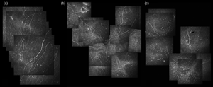
The impact of dry eye disease on corneal nerve parameters: A systematic review and meta‐analysis.
Ophthalmic and Physiological Optics,
Vol. 43,
Issue. 5,
p.
1079.
Oddone, Francesco
Tanga, Lucia
Giammaria, Sara
Sabbatini, Lorenzo
Strianese, Alfonso
Ferrazza, Manuela
and
Rossetti, Luca
2024.
24-Hour Evaluation of the Effectiveness and Tolerability of Preservative-Free Tafluprost-Timolol Fixed Combination in Open-Angle Glaucoma or Ocular Hypertensive Patients Previously Treated with Preserved Latanoprost.
Clinical Ophthalmology,
Vol. Volume 18,
Issue. ,
p.
1751.