


No CrossRef data available.
Article contents
Performance Evaluation of Focal Plane Array (FPA)-FTIR and Synchrotron Radiation (SR)-FTIR Microspectroscopy to Classify Rice Components
Published online by Cambridge University Press: 05 September 2022
Abstract
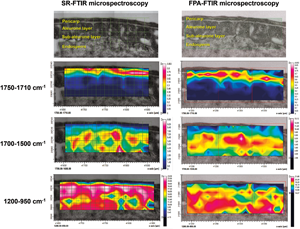
The development of biochemical analysis techniques to study heterogeneous biological samples is increasing. These techniques include synchrotron radiation Fourier transform infrared (SR-FTIR) microspectroscopy. This method has been applied to analyze biological tissue with multivariate statistical analysis to classify the components revealed by the spectral data. This study aims to compare the efficiencies of SR-FTIR microspectroscopy and focal plane array (FPA)-FTIR microspectroscopy when classifying rice tissue components. Spectral data were acquired for mapping the same sample areas from both techniques. Principal component analysis and cluster imaging were used to investigate the biochemical variations of the tissue types. The classification was based on the functional groups of pectin, protein, and polysaccharide. Four layers from SR-FTIR microspectroscopy including pericarp, aleurone layer, sub-aleurone layer, and endosperm were classified using cluster imaging, while FPA-FTIR microspectroscopy could classify only three layers of pericarp, aleurone layer, and endosperm. Moreover, SR-FTIR microspectroscopy increased the image contrast of the biochemical distribution in rice tissue more efficiently than FPA-FTIR microspectroscopy. We have demonstrated the capability of the high-resolution synchrotron technique and its ability to clarify small structures in rice tissue. The use of this technique might increase in future studies of tissue characterization.
Keywords
- Type
- Biological Applications
- Information
- Copyright
- Copyright © The Author(s), 2022. Published by Cambridge University Press on behalf of the Microscopy Society of America
References
Achten, E, Schütz, D, Fischer, M, Fauhl-Hassek, C, Riedl, J & Horn, B (2019). Classification of grain maize (Zea mays L.) from different geographical origins with FTIR spectroscopy a suitable analytical tool for feed authentication? Food Anal Methods 12(10), 2172–2184. doi:10.1007/s12161-019-01558-9CrossRefGoogle Scholar
Amir, RAI, Anjum, F, Khan, M, Khan, MR, Pasha, I & Nadeem, M (2013). Application of Fourier transform infrared (FTIR) spectroscopy for the identification of wheat varieties. J Food Sci Technol 50, 1018–1023. doi:10.1007/s13197-011-0424-yCrossRefGoogle ScholarPubMed
Andriotis, VME, Rejzek, M, Barclay, E, Rugen, MD, Field, RA & Smith, AM (2016). Cell wall degradation is required for normal starch mobilisation in barley endosperm. Sci Rep 6(1), 1–15. doi:10.1038/srep33215CrossRefGoogle ScholarPubMed
Bambery, KR, Schültke, E, Wood, BR, Rigley MacDonald, ST, Ataelmannan, K, Griebel, RW, Juurlink, BHJ & McNaughton, D (2006). A Fourier transform infrared microspectroscopic imaging investigation into an animal model exhibiting glioblastoma multiforme. Biochim Biophys Acta 1758(7), 900–907. doi:10.1016/j.bbamem.2006.05.004CrossRefGoogle ScholarPubMed
Barron, C, Parker, ML, Mills, ENC, Rouau, X & Wilson, RH (2005). FTIR imaging of wheat endosperm cell walls in situ reveals compositional and architectural heterogeneity related to grain hardness. Planta 220(5), 667–677. doi:10.1007/s00425-004-1383-6CrossRefGoogle ScholarPubMed
Barron, C & Rouau, X (2008). FTIR and Raman signatures of wheat grain peripheral tissues. Cereal Chem 85, 619–625. doi:10.1094/CCHEM-85-5-0619CrossRefGoogle Scholar
Bayari, S, Utku, H, Ikemoto, Y, Celasun, B & Kömürcü, M (2007). Synchrotron FT-IR microspectroscopic analysis of necrotic bone. Spectroscopy 21, 227–234. doi:10.1155/2007/732013CrossRefGoogle Scholar
Bhargava, R (2012). Infrared spectroscopic imaging: The next generation. Appl Spectrosc 66(10), 1091–1120. doi:10.1366/12-06801CrossRefGoogle ScholarPubMed
Câmara, J, Alves, A & Marques, JC (2006). Multivariate analysis for the classification and differentiation of Madeira wines according to main grape varieties. Talanta 68, 1512–1521. doi:10.1016/j.talanta.2005.08.012CrossRefGoogle ScholarPubMed
Chan, KL & Kazarian, SG (2006). Detection of trace materials with Fourier transform infrared spectroscopy using a multi-channel detector. Analyst 131(1), 126–131. doi:10.1039/b511243eCrossRefGoogle ScholarPubMed
Cheeseman, S, Truong, VK, Vongsvivut, J, Tobin, MJ, Crawford, R & Ivanova, EP (2019). Applications of synchrotron-source ir spectroscopy for the investigation of insect wings. In Synchrotron Radiation – Useful and Interesting Applications, Joseph, D (Ed.). IntechOpen. doi:10.5772/intechopen.84591Google Scholar
Chen, X, Chen, M, Lin, G, Yang, Y, Yu, X, Wu, Y & Xiong, F (2019). Structural development and physicochemical properties of starch in caryopsis of super rice with different types of panicle. BMC Plant Biol 19(1), 482–482. doi:10.1186/s12870-019-2101-7CrossRefGoogle ScholarPubMed
Cinque, G, Frogley, MD, Wehbe, K, Nguyen, TNQ, Fitzpatrick, A & Kelley, CS (2017). Synchrotron-based infrared spectral imaging at the MIRIAM beamline of diamond light source. Synchrotron Radiat News 30(4), 11–16. doi:10.1080/08940886.2017.1338416CrossRefGoogle Scholar
Deegan, A, Cinque, G, Wehbe, K & Konduru, S (2015). Tracking calcification in tissue-engineered bone using synchrotron micro-FTIR and SEM. Anal Bioanal Chem 407(4), 1097–1105. doi:10.1007/s00216-014-8316-4CrossRefGoogle ScholarPubMed
Deng, Z, Gong, C & Wang, T (2013). Use of proteomics to understand seed development in rice. Proteomics 13, 1784–1800. doi:10.1002/pmic.201200389CrossRefGoogle ScholarPubMed
Dokken, KM & Davis, LC (2011). Infrared monitoring of dinitrotoluenes in sunflower and maize roots. J Environ Qual 40(3), 719–730. doi:10.2134/jeq2010.0143CrossRefGoogle ScholarPubMed
Dowell, FE, Maghirang, EB, Xie, F, Lookhart, GL, Pierce, RO, Seabourn, BW, Wilson, JD & Chung, OK (2006). Predicting wheat quality characteristics and functionality using near-infrared spectroscopy. Cereal Chem 83(5), 529–536. doi:10.1094/CC-83-0529CrossRefGoogle Scholar
Dumas, P, Sockalingum, GD & Sulé-Suso, J (2007). Adding synchrotron radiation to infrared microspectroscopy: What's new in biomedical applications? Trends Biotechnol 25(1), 40–44. doi:10.1016/j.tibtech.2006.11.002CrossRefGoogle ScholarPubMed
Faoláin, EÓ, Hunter, MB, Byrne, JM, Kelehan, P, McNamara, M, Byrne, HJ & Lyng, FM (2005). A study examining the effects of tissue processing on human tissue sections using vibrational spectroscopy. Vib Spectrosc 38(1), 121–127. doi:10.1016/j.vibspec.2005.02.013CrossRefGoogle Scholar
Geisler-Lee, J & Gallie, DR (2005). Aleurone cell identity is suppressed following connation in maize kernels. Plant Physiol 139(1), 204–212. doi:10.1104/pp.105.064295CrossRefGoogle ScholarPubMed
Guendel, A, Rolletschek, H, Wagner, S, Muszynska, A & Borisjuk, L (2018). Micro imaging displays the sucrose landscape within and along its allocation pathways. Plant Physiol 178(4), 1448–1460. doi:10.1104/pp.18.00947CrossRefGoogle ScholarPubMed
Heraud, P, Caine, S, Sanson, G, Gleadow, R, Wood, BR & McNaughton, D (2007). Focal plane array infrared imaging: A new way to analyse leaf tissue. New Phytol 173(1), 216–225. doi:10.1111/j.1469-8137.2006.01881.xCrossRefGoogle ScholarPubMed
Huffman, SW, Bhargava, R & Levin I, W (2002). Generalized implementation of rapid-scan Fourier transform infrared spectroscopic imaging. Appl Spectrosc 56(8), 965–969. doi:10.1366/000370202760249684CrossRefGoogle Scholar
Hughes, C, Gaunt, L, Brown, M, Clarke, NW & Gardner, P (2014). Assessment of paraffin removal from prostate FFPE sections using transmission mode FTIR-FPA imaging. Anal Methods 6(4), 1028–1035. doi:10.1039/C3AY41308JCrossRefGoogle Scholar
Igisu, M, Komiya, T, Awramik, SM, Ikemoto, Y, Geng, Y, Uehara, H & Takai, K (2019). Fourier transform infrared microspectroscopic characterization of Neoproterozoic organic microfossils from the Fifteenmile Group in Yukon, Canada. Island Arc 28(5), e12310. doi:10.1111/iar.12310CrossRefGoogle Scholar
Ishimaru, T, Ida, M, Hirose, S, Shimamura, S, Masumura, T, Nishizawa, NK, Nakazono, M & Kondo, M (2015). Laser microdissection-based gene expression analysis in the aleurone layer and starchy endosperm of developing rice caryopses in the early storage phase. Rice 8(1), 57. doi:10.1186/s12284-015-0057-2CrossRefGoogle ScholarPubMed
Ishimaru, T, Nakazono, M, Masumura, T, Abiko, M, San-oh, Y, Nishizawa, NK & Kondo, M (2007). A method for obtaining high integrity RNA from developing aleurone cells and starchy endosperm in rice (Oryza sativa L.) by laser microdissection. Plant Sci 173(3), 321–326. doi:10.1016/j.plantsci.2007.06.004CrossRefGoogle Scholar
Kimber, JA & Kazarian, SG (2017). Spectroscopic imaging of biomaterials and biological systems with FTIR microscopy or with quantum cascade lasers. Anal Bioanal Chem 409(25), 5813–5820. doi:10.1007/s00216-017-0574-5CrossRefGoogle ScholarPubMed
Lahlali, R, Kumar, S, Wang, L, Forseille, L, Sylvain, N, Korbas, M, Muir, D, Swerhone, G, Lawrence, JR, Fobert, PR, Peng, G & Karunakaran, C (2016). Cell wall biomolecular composition plays a potential role in the host type II resistance to fusarium head blight in wheat. Front Microbiol 7(910), 1–12. doi:10.3389/fmicb.2016.00910CrossRefGoogle Scholar
Lasch, P, Haensch, W, Naumann, D & Diem, M (2004). Imaging of colorectal adenocarcinoma using FT-IR microspectroscopy and cluster analysis. Biochim Biophys Acta Mol Basis Dis 1688(2), 176–186. https://doi.org/10.1016/j.bbadis.2003.12.006.CrossRefGoogle ScholarPubMed
Lasch, P (2012). Spectral Pre-processing for Biomedical Vibrational Spectroscopy and Microspectroscopic Imaging. Chemometr Intell Lab Syst 117, 100–114. https://doi.org/10.1016/j.chemolab.2012.03.011.CrossRefGoogle Scholar
Lasch, P, Stämmler, M, Zhang, M, Baranska, M, Bosch, A & Majzner, K (2018). FT-IR hyperspectral imaging and artificial neural network analysis for identification of pathogenic bacteria. Anal Chem 90(15), 8896–8904. doi:10.1021/acs.analchem.8b01024CrossRefGoogle ScholarPubMed
Le Naour, F, Bralet, MP, Debois, D, Sandt, C, Guettier, C, Dumas, P, Brunelle, A & Laprévote, O (2009). Chemical imaging on liver steatosis using synchrotron infrared and ToF-SIMS microspectroscopies. PLoS One 4(10), e7408–e7408. doi:10.1371/journal.pone.0007408CrossRefGoogle ScholarPubMed
Liberda, D, Kosowska, K, Koziol, P & Wrobel, TP (2021). Spatial sampling effect on data structure and random forest classification of tissue types in high definition and standard definition FT-IR imaging. Chemometr Intell Lab Syst 217, 104407. doi:10.1016/j.chemolab.2021.104407CrossRefGoogle Scholar
Lloyd, G, Nallala, J & Stone, N (2016). Investigating the effect of pixel size of high spatial resolution FTIR imaging for detection of colorectal cancer. Proc SPIE 9703. doi:10.1117/12.2210844.Google Scholar
Lupoi, JS, Smith-Moritz, A, Singh, S, McQualter, R, Scheller, HV, Simmons, BA & Henry, RJ (2015). Localization of polyhydroxybutyrate in sugarcane using Fourier-transform infrared microspectroscopy and multivariate imaging. Biotechnol Biofuels 8, 98–98. doi:10.1186/s13068-015-0279-yCrossRefGoogle ScholarPubMed
Manley, M, Van Zyl, L & Osborne, BG (2002). Using Fourier transform near infrared spectroscopy in determining kernel hardness, protein and moisture content of whole wheat flour. J Near Infrared Spec 10(1), 71–76. doi:10.1255/jnirs.323CrossRefGoogle Scholar
Miller, LM & Dumas, P (2006). Chemical imaging of biological tissue with synchrotron infrared light. Biochim Biophys Acta 1758(7), 846–857. doi:10.1016/j.bbamem.2006.04.010CrossRefGoogle ScholarPubMed
Miller, LM & Smith, RJ (2005). Synchrotrons versus Globars, point-detectors versus focal plane arrays: Selecting the best source and detector for specific infrared microspectroscopy and imaging applications. Vib Spectrosc 38(1), 237–240. doi:10.1016/j.vibspec.2005.03.010CrossRefGoogle Scholar
Moss, DA, Keese, M & Pepperkok, R (2005). IR microspectroscopy of live cells. Vib Spectrosc 38(1), 185–191. doi:10.1016/j.vibspec.2005.04.004CrossRefGoogle Scholar
Ogawa, Y, Orts, WJ, Glenn, GM & Wood, DF (2003). A simple method for studying whole sections of rice grain. Biotech Histochem 78(5), 237–242. doi:10.1080/10520290310001630467CrossRefGoogle ScholarPubMed
Park, HY, Lee, KW & Choi, HD (2017). Rice bran constituents: Immunomodulatory and therapeutic activities. Food Funct 8(3), 935–943. doi:10.1039/c6fo01763kCrossRefGoogle ScholarPubMed
Petibois, C, Piccinini, M, Guidi, MC & Marcelli, A (2010). Facing the challenge of biosample imaging by FTIR with a synchrotron radiation source. J Synchrotron Radiat 17(1), 1–11. doi:10.1107/s0909049509046056CrossRefGoogle ScholarPubMed
Phansak, P, Siriwong, S, Sangpueak, R, Kanawapee, N, Thumanu, K & Buensanteai, N (2021). Screening rice blast-resistant cultivars via synchrotron Fourier transform infrared (SR-FTIR) microspectroscopy. Emir J Food Agric 33(9), 726–741. doi:10.9755/ejfa.2021.v33.i9.2758Google Scholar
Rajalahti, T & Kvalheim, OM (2011). Multivariate data analysis in pharmaceutics: A tutorial review. Int J Pharm 417(1), 280–290. doi:10.1016/j.ijpharm.2011.02.019CrossRefGoogle ScholarPubMed
Robert, P, Marquis, M, Barron, C, Guillon, F & Saulnier, L (2005). FT-IR investigation of cell wall polysaccharides from cereal grains. Arabinoxylan infrared assignment. J Agric Food Chem 53(18), 7014–7018. doi:10.1021/jf051145yCrossRefGoogle ScholarPubMed
Sadhana, B (2014). FTIR - technique for harvested seeds of chickpea and cowpea plants grown under Rhizobium and AM fungi inoculated condition. Int J Adv Res Biol Sci 1(9), 214–222.Google Scholar
Severcan, F, Toyran, N, Kaptan, N & Turan, B (2000). Fourier transform infrared study of the effect of diabetes on rat liver and heart tissues in the CH region. Talanta 53(1), 55–59. doi:10.1016/s0039-9140(00)00379-9CrossRefGoogle ScholarPubMed
Sharma, S & Singh, R (2020). Detection and discrimination of seminal fluid using attenuated total reflectance Fourier transform infrared (ATR FT-IR) spectroscopy combined with chemometrics. Int J Legal Med 134(2), 411–432. doi:10.1007/s00414-019-02222-xCrossRefGoogle ScholarPubMed
Singh, VK, Devi, A, Pathania, S, Kumar, V, Tripathi, DK, Sharma, S, Chauhan, DK, Singh, VK & Zorba, V (2017). Spectroscopic investigation of wheat grains (Triticum aestivum) infected by wheat seed gall nematodes (Anguina tritici). Biocatal Agric Biotechnol 9, 58–66. doi:10.1016/j.bcab.2016.11.005CrossRefGoogle Scholar
Smith, TI (2002). The source issue in infrared microspectroscopy. Nucl Instrum Methods 483(1), 565–570. doi:10.1016/S0168-9002(02)00383-2CrossRefGoogle Scholar
Snively, CM & Koenig, JL (1999). Characterizing the performance of a fast FT-IR imaging spectrometer. Appl Spectrosc 53(2), 170–177. doi:10.1366/0003702991946497CrossRefGoogle Scholar
Szymanska-Chargot, M & Zdunek, A (2013). Use of FT-IR spectra and PCA to the bulk characterization of cell wall residues of fruits and vegetables along a fraction process. Food Biophys 8(1), 29–42. doi:10.1007/s11483-012-9279-7CrossRefGoogle Scholar
Theophilou, G, Morais, CLM, Halliwell, DE, Lima, KMG, Drury, J, Martin-Hirsch, PL, Stringfellow, F, Hapangama, DK & Martin, FL (2018). Synchrotron- and focal plane array-based Fourier-transform infrared spectroscopy differentiates the basalis and functionalis epithelial endometrial regions and identifies putative stem cell regions of human endometrial glands. Anal Bioanal Chem 410(18), 4541–4554. doi:10.1007/s00216-018-1111-xCrossRefGoogle ScholarPubMed
Thumanu, K, Wongchalee, D, Sompong, M, Phansak, P, Le Thanh, T, Namanusart, W, Vechklang, K, Kaewnum, S & Buensanteai, N (2017). Synchrotron-based FTIR microspectroscopy of chili resistance induced by Bacillus subtilis strain D604 against anthracnose disease. J Plant Interact 12(1), 255–263. doi:10.1080/17429145.2017.1325523CrossRefGoogle Scholar
Vijayan, P, Willick, IR, Lahlali, R, Karunakaran, C & Tanino, KK (2015). Synchrotron radiation sheds fresh light on plant research: The use of powerful techniques to probe structure and composition of plants. Plant Cell Physiol 56(7), 1252–1263. doi:10.1093/pcp/pcv080CrossRefGoogle Scholar
Vongsvivut, J, Pérez-Guaita, D, Wood, BR, Heraud, P, Khambatta, K, Hartnell, D, Hackett, MJ & Tobin, MJ (2019). Synchrotron macro ATR-FTIR microspectroscopy for high-resolution chemical mapping of single cells. Analyst 144(10), 3226–3238. doi:10.1039/C8AN01543KCrossRefGoogle ScholarPubMed
Wehbe, K, Forfar, I, Eimer, S & Cinque, G (2015). Discrimination between two different grades of human glioma based on blood vessel infrared spectral imaging. Anal Bioanal Chem 407(24), 7295–7305. doi:10.1007/s00216-015-8891-zCrossRefGoogle ScholarPubMed
Wei, Z, Jiao, D & Xu, J (2015). Using Fourier transform infrared spectroscopy to study effects of magnetic field treatment on wheat (Triticum aestivum L.) seedlings. J Spectrosc 2015, 570190. doi:10.1155/2015/570190CrossRefGoogle Scholar
Wiercigroch, E, Szafraniec, E, Czamara, K, Pacia, MZ, Majzner, K, Kochan, K, Baranska, M & Malek, K (2017). Raman and infrared spectroscopy of carbohydrates: A review. Acta A Mol Biomol Spectrosc 185, 317–335. doi:10.1016/j.saa.2017.05.045CrossRefGoogle ScholarPubMed
Wrobel, TP, Koziol, P, Raczkowska, MK, Liberda, D, Paluszkiewicz, C & Kwiatek, WM (2019). Noise-free simulation of an FT-IR imaging hyperspectral dataset of pancreatic biopsy core bound by experiment. Sci Data 6(1), 239. doi:10.1038/s41597-019-0260-xCrossRefGoogle Scholar
Wu, X, Liu, J, Li, D & Liu, CM (2016). Rice caryopsis development II: Dynamic changes in the endosperm. J Integr Plant Biol 58(9), 786–798. doi:10.1111/jipb.12488CrossRefGoogle ScholarPubMed
Yu, P (2004). Application of advanced synchrotron radiation-based Fourier transform infrared (SR-FTIR) microspectroscopy to animal nutrition and feed science: A novel approach. Br J Nutr 92(6), 869–885. doi:10.1079/BJN20041298CrossRefGoogle ScholarPubMed
Yuliyanda, I, Masithoh, RE, Khuriyati, N & Saputro, AD (2019). Classification of crop flours based on protein contents using near infra-red spectroscopy and principle component analysis. IOP Conf Ser Earth Environ Sci 355, 012002. doi:10.1088/1755-1315/355/1/012002CrossRefGoogle Scholar
Zohdi, V, Whelan, D, Wood, B, Pearson, J, Bambery, K & Black, M (2015). Importance of tissue preparation methods in FTIR micro-spectroscopical analysis of biological tissues: “traps for new users”. PLoS One 10, e0116491. doi:10.1371/journal.pone.0116491CrossRefGoogle Scholar