Hostname: page-component-7bb8b95d7b-l4ctd Total loading time: 0 Render date: 2024-09-11T21:12:02.122Z Has data issue: false hasContentIssue false

Fluorescence Polarization Microscopy Enables Spatial Mapping of the 3D Orientation of Piconewton Integrin Traction Forces

Published online by Cambridge University Press:  05 August 2019

Joshua M. Brockman
Affiliation:
Wallace H. Coulter Department of Biomedical Engineering, Georgia Institute of Technology and Emory University, Atlanta, GeorgiaUSA
Aaron T. Blanchard
Affiliation:
Wallace H. Coulter Department of Biomedical Engineering, Georgia Institute of Technology and Emory University, Atlanta, GeorgiaUSA
Khalid Salaita
Affiliation:
Wallace H. Coulter Department of Biomedical Engineering, Georgia Institute of Technology and Emory University, Atlanta, GeorgiaUSA Department of Chemistry, Emory University, Atlanta, GAUSA
Alexa L. Mattheyses*
Affiliation:
Department of Cell, Developmental, and Integrative Biology, University of Alabama at Birmingham, Birmingham, ALUSA
*
*Corresponding author: mattheyses@uab.edu

Abstract

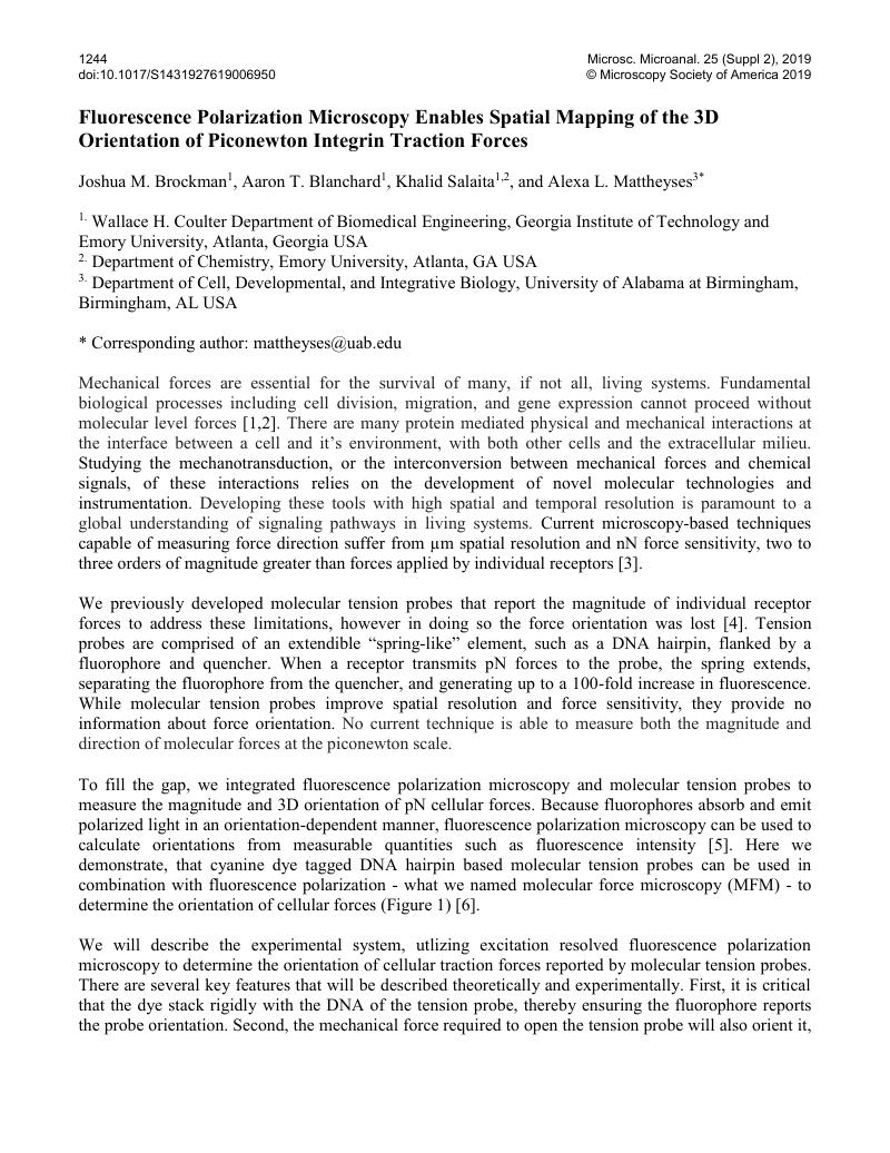
Image of the first page of this content. For PDF version, please use the ‘Save PDF’ preceeding this image.'
Type
Light and Fluorescence Microscopy for Imaging Cell Surface and Cell Structure
Copyright
Copyright © Microscopy Society of America 2019 

References

[1]Vogel, V and Sheetz, M, Nat. Rev. Mol. Cell Biol. 7, (2006) 265.Google Scholar
[2]DuFort, C, Paszek, M, and Weaver, V, Nat Rev Mol Cell Biol 12, (2011) 308.Google Scholar
[3]Polacheck, W and Chen, C, Nat Methods 13, (2016) 415.Google Scholar
[4]Stabley, D et al. , Nat Methods 9, (2012) 64.Google Scholar
[5]Bradley, S et al. , Biophys J 101, (2011) 985.Google Scholar
[6]Brockman, J et al. , Nat Methods 15 (2018) 115.Google Scholar
[7]The authors acknowledge funding from NIGMS R01 GM124472 (K.S.), NSF 1350829 (K.S.), NSF CAREER 1553344 (A.L.M.) and NSF IDBR 1353939 (K.S. and A.L.M.).Google Scholar