Hostname: page-component-848d4c4894-5nwft Total loading time: 0 Render date: 2024-06-09T22:55:06.062Z Has data issue: false hasContentIssue false

In Situ Observation of the Early Stages of Rapid Solid–Liquid Reaction in Closed Liquid Cell TEM Using Graphene Encapsulation

Published online by Cambridge University Press:  10 December 2021

Hyun Woo Cha
Affiliation:
School of Advanced Materials Science & Engineering, Sungkyunkwan University, Suwon, Gyeonggi-do 16419, Korea
Byeong-Seon An
Affiliation:
School of Advanced Materials Science & Engineering, Sungkyunkwan University, Suwon, Gyeonggi-do 16419, Korea
Cheol-Woong Yang*
Affiliation:
School of Advanced Materials Science & Engineering, Sungkyunkwan University, Suwon, Gyeonggi-do 16419, Korea
*
*Corresponding author: Cheol-Woong Yang, E-mail: cwyang@skku.edu
Get access

Abstract

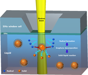
In situ liquid cell transmission electron microscopy (TEM) is a very useful tool for investigating dynamic solid–liquid reactions. However, there are challenges to observe the early stages of spontaneous solid–liquid reactions using a closed-type liquid cell system, the most popular and simple liquid cell system. We propose a graphene encapsulation method to overcome this limitation of closed-type liquid cell TEM. The solid and liquid are separated using graphene to suspend the reaction until the graphene layer is destroyed. Graphene can be decomposed by the high-energy electron beam used in TEM, allowing the reaction to proceed. Fast dissolution of graphene-capped copper nanoparticles in an FeCl3 solution was demonstrated via in situ liquid cell TEM at 300 kV using a cell with closed-type SiNx windows.

Type
Materials Science Applications
Copyright
Copyright © The Author(s), 2021. Published by Cambridge University Press on behalf of the Microscopy Society of America

Access options

Get access to the full version of this content by using one of the access options below. (Log in options will check for institutional or personal access. Content may require purchase if you do not have access.)

References

Ambrožič, B, Prašnikar, A, Hodnik, N, Kostevšek, N, Likozar, B, Rožman, & Šturm, S (2019). Controlling the radical-induced redox chemistry inside a liquid-cell TEM. Chem Sci 10(38), 87358743.CrossRefGoogle ScholarPubMed
Bharda, AV & Jung, HS (2019). Liquid electron microscopy: Then, now and future. Applied Microscopy 49(1), 1–6.CrossRefGoogle Scholar
Bryce, C & Berk, D (1995). Kinetics of the dissolution of copper in iron (III) chloride solutions. Ind Eng Chem Res 34(4), 14121418.CrossRefGoogle Scholar
Cançado, LG, Jorio, A, Ferreira, EM, Stavale, F, Achete, CA, Capaz, RB & Ferrari, AC (2011). Quantifying defects in graphene via Raman spectroscopy at different excitation energies. Nano Lett 11(8), 31903196.CrossRefGoogle ScholarPubMed
Chen, FC, Chen, JY, Lin, YH, Kuo, MY, Hsu, YJ & Wu, WW (2019). In situ TEM observation of Au–Cu2O core–shell growth in liquids. Nanoscale 11(21), 1048610492.CrossRefGoogle ScholarPubMed
Cho, H, Jones, MR, Nguyen, SC, Hauwiller, MR, Zettl, A & Alivisatos, AP (2017). The use of graphene and its derivatives for liquid-phase transmission electron microscopy of radiation-sensitive specimens. Nano Lett 17, 414420.CrossRefGoogle ScholarPubMed
Denis, PA & Iribarne, F (2013). Comparative study of defect reactivity in graphene. J Phys Chem C 117(37), 1904819055.CrossRefGoogle Scholar
Drescher, D, Traub, H, Büchner, T, Jakubowski, N & Kneipp, J (2017). Properties of in situ generated gold nanoparticles in the cellular context. Nanoscale 9(32), 1164711656.CrossRefGoogle ScholarPubMed
Dresselhaus, MS, Jorio, A, Souza Filho, AG & Saito, R (2010). Defect characterization in graphene and carbon nanotubes using Raman spectroscopy. Philos Trans R Soc A 368(1932), 53555377.CrossRefGoogle ScholarPubMed
Egerton, RF (2012). Mechanisms of radiation damage in beam-sensitive specimens, for TEM accelerating voltages between 10 and 300 kV. Microsc Res Tech 75(11), 15501556.CrossRefGoogle ScholarPubMed
Gupta, T, Schneider, NM, Park, JH, Steingart, D & Ross, FM (2018). Spatially dependent dose rate in liquid cell transmission electron microscopy. Nanoscale 10(16), 77027710.CrossRefGoogle ScholarPubMed
Hodnik, N, Dehm, G & Mayrhofer, KJ (2016). Importance and challenges of electrochemical in situ liquid cell electron microscopy for energy conversion research. Acc Chem Res 49(9), 20152022.CrossRefGoogle ScholarPubMed
Huang, JY, Zhong, L, Wang, CM, Sullivan, JP, Xu, W, Zhang, LQ & Fan, H (2010). In situ observation of the electrochemical lithiation of a single SnO2 nanowire electrode. Science 330(6010), 15151520.CrossRefGoogle Scholar
Jeong, M, Yuk, JM & Lee, JY (2015). Observation of surface atoms during platinum nanocrystal growth by monomer attachment. Chem Mater 27(9), 32003202.CrossRefGoogle Scholar
Jiang, Y, Zhu, G, Lin, F, Zhang, H, Jin, C, Yuan, J, Yang, D & Zhang, Z (2014). In situ study of oxidative etching of palladium nanocrystals by liquid cell electron microscopy. Nano Lett 14(7), 37613765.CrossRefGoogle ScholarPubMed
Kang, J, Shin, D, Bae, S & Hong, BH (2012). Graphene transfer: Key for applications. Nanoscale 4(18), 55275537.CrossRefGoogle ScholarPubMed
Klein, KL, Anderson, IM & De Jonge, N (2011). Transmission electron microscopy with a liquid flow cell. J Microsc 242(2), 117123.CrossRefGoogle ScholarPubMed
Kostov, MK, Santiso, EE, George, AM, Gubbins, KE & Nardelli, MB (2005). Dissociation of water on defective carbon substrates. Phys Rev Lett 95(13), 136105.CrossRefGoogle ScholarPubMed
Lee, JW, Kim, SW, Cho, YL, Jeong, HY, Song, YI & Suh, SJ (2016). Dispersion stabilities of multi-layer graphene-coated copper prepared by electrical wire-explosion method. J Nanosci Nanotechnol 16(11), 1128611291.CrossRefGoogle Scholar
Lee, JW, Kim, TY, Cho, YL, Na, YI, Kim, YS, Song, YI & Suh, SJ (2015). Effect of liquid media on the formation of multi-layer graphene-synthesized metal particles. J Nanosci Nanotechnol 15(11), 90149019.CrossRefGoogle ScholarPubMed
Leonhardt, A, Hampel, S, Mueller, C, Moench, I, Koseva, R, Ritschel, M & Buechner, B (2006). Synthesis, properties, and applications of ferromagnetic-filled carbon nanotubes. Chem Vap Deposition 12(6), 380387.CrossRefGoogle Scholar
Li, J, Nie, Z, Zheng, YY, Dong, S & Loh, ZH (2013). Elementary electron and ion dynamics in ionized liquid water. J Phys Chem Lett 4(21), 36983703.10.1021/jz401987fCrossRefGoogle Scholar
Liao, HG & Zheng, H (2016). Liquid cell transmission electron microscopy. Annu Rev Phys Chem 67, 719747.CrossRefGoogle ScholarPubMed
Marton, L (1935). La microscopie electronique des objets biologiques. Bull Acad Roy Med Belg 21, 600617.Google Scholar
Nicholls, D, Lee, J, Amari, H, Stevens, AJ, Mehdi, BL & Browning, ND (2020). Minimising damage in high resolution scanning transmission electron microscope images of nanoscale structures and processes. Nanoscale 12(41), 2124821254.CrossRefGoogle ScholarPubMed
Parent, LR, Bakalis, E, Proetto, M, Li, Y, Park, C, Zerbetto, F & Gianneschi, NC (2018). Tackling the challenges of dynamic experiments using liquid-cell transmission electron microscopy. Acc Chem Res 51(1), 311.CrossRefGoogle ScholarPubMed
Park, JH, Schneider, NM, Grogan, JM, Reuter, MC, Bau, HH, Kodambaka, S & Ross, FM (2015). Control of electron beam-induced Au nanocrystal growth kinetics through solution chemistry. Nano Lett 15, 53145320.CrossRefGoogle ScholarPubMed
Powers, AS, Liao, H, Raja, S, Bronstein, N, Alivisatos, A & Zheng, H (2017). Tracking nanoparticle diffusion and interaction during self-assembly in a liquid cell. Nano Lett 17(1), 1520.CrossRefGoogle Scholar
Schneider, NM, Norton, MM, Mendel, BJ, Grogan, JM, Ross, FM & Bau, HH (2014). Electron–water interactions and implications for liquid cell electron microscopy. J Phys Chem C 118(38), 2237322382. (2014)CrossRefGoogle Scholar
Stoppiello, CT, Biskupek, J, Li, ZY, Rance, GA, Botos, A, Fogarty, RM, Bourne, RA, Yuan, J, Lovelock, KRJ, Thompson, P, Fay, MW, Kaiser, U, Chamberlain, TW & Khlobystov, AN (2017). A one-pot-one-reactant synthesis of platinum compounds at the nanoscale. Nanoscale 9(38), 1438514394.CrossRefGoogle ScholarPubMed
Sun, M, Li, X, Tang, Z, Wei, X & Chen, Q (2018). Constant-rate dissolution of InAs nanowires in radiolytic water observed by in situ liquid cell TEM. Nanoscale 10(42), 1973319741.CrossRefGoogle ScholarPubMed
Sung, J, Choi, BK, Kim, B, Kim, J, Lee, D, Kim, S, Kang, K, Hyeon, T & Park, J (2019). Redox-sensitive facet dependency in etching of ceria nanocrystals directly observed by liquid cell TEM. J Am Chem Soc 141(46), 1839518399.CrossRefGoogle ScholarPubMed
Tao, S, Liu, HT, Yan, LM, Yue, BH & Li, AJ (2017). Hydrogenation of graphene nanoflakes and C–H bond dissociation of hydrogenated graphene nanoflakes: A density functional theory study. Adv Manuf 5(3), 289298.CrossRefGoogle Scholar
Wang, N, Wang, L, Tan, Q & Pan, YX (2013). Effects of hydroxyl group on H2 dissociation on graphene: A density functional theory study. J Energy Chem 22(3), 493497.CrossRefGoogle Scholar
Wang, Y, Peng, X, Abelson, A, Xiao, P, Qian, C, Yu, L, Ophus, C, Ercius, P, Wang, LW, Law, M & Zheng, H (2019). Dynamic deformability of individual PbSe nanocrystals during superlattice phase transitions. Sci Adv 5(6), eaaw5623.CrossRefGoogle ScholarPubMed
Zeng, Z, Liang, WI, Liao, HG, Xin, HL, Chu, YH & Zheng, H (2014). Visualization of electrode-electrolyte interfaces in LiPF6/EC/DEC electrolyte for lithium ion batteries via in-situ TEM. Nano Lett 14, 17451750.CrossRefGoogle ScholarPubMed
Zhu, C, Liang, S, Song, E, Zhou, Y, Wang, W, Shan, F, Shi, Y, Hao, C, Yin, K, Zhang, T, Liu, J, Zheng, H & Sun, L (2018). In-situ liquid cell transmission electron microscopy investigation on oriented attachment of gold nanoparticles. Nature Comm 9, 421.CrossRefGoogle ScholarPubMed
Supplementary material: File

Cha et al. supplementary material

Cha et al. supplementary material 1

Download Cha et al. supplementary material(File)
File 319 KB

Cha et al. supplementary material

Cha et al. supplementary material 2

Download Cha et al. supplementary material(Video)
Video 6 MB

Cha et al. supplementary material

Cha et al. supplementary material 3

Download Cha et al. supplementary material(Video)
Video 11.6 MB