


No CrossRef data available.
Article contents
Patterns of Organization of Cerebellum and Spinal Cord of the Red-Tail Shark (Epalzeorhynchos bicolor): Histological, Morphometrical, and Immunohistochemical Studies
Part of:
Micrographia Collection
Published online by Cambridge University Press: 14 October 2020
Abstract
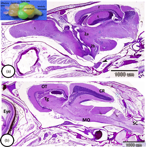
Teleosts exhibit enormous heterogeneity in brain morphology, especially in the patterns of the organization of cerebellum. The cerebellum of a red-tail shark that we analyzed was well-developed and included three main divisions: the valvula cerebelli, the corpus cerebelli, and the vestibulolateral lobe. Characteristically, the cerebellar cortex contained three well-distinct layers: an outer molecular, intermediate ganglionic, and inner granular layer. The ganglionic layer possessed irregularly arranged Purkinje cells and eurydendroid cells that extended their processes into the molecular layer. Both Purkinje cells and eurydendroid cells showed immunoreactivity for iNOS2. Moreover, astrocytes in the cerebellum showed the expression of glial fibrillary acidic protein. The most striking observation in the cerebellum of shark was the lack of deep cerebellar nuclei and a well-identified white matter. On the other hand, the gray substance in the spinal cord displays a characteristic pattern in its organization, in which the dorsal horns lie quite close together, giving the gray substance the shape of an inverted Y and possessing large neurons. Notably, the white matter possessed myelinated nerve fibers. The current study provides the first report on the organization of layers and neurons in the cerebellum and spinal cord of red-tail shark. This research will contribute to the neuroanatomy and evolutionary studies of the brain of Cyprinidae.
- Type
- Micrographia
- Information
- Copyright
- Copyright © The Author(s), 2020. Published by Cambridge University Press on behalf of the Microscopy Society of America
References
Altman, J & Bayer, SA (1997). Development of the Cerebellar System: In Relation to its Evolution, Structure, and Functions. Boca Raton: CRC Press.Google Scholar
Bae, YK, Kani, S, Shimizu, T, Tanabe, K, Nojima, H, Kimura, Y, Higashijima, S & Hibi, M (2009). Anatomy of zebrafish cerebellum and screen for mutations affecting its development. Dev Biol 330, 406–426.CrossRefGoogle ScholarPubMed
Bancroft, JD & Gamble, M (2002). Theory and Practice of Histological and Histochemical Techniques, 3rd ed. London: Churchill Livingstone.Google Scholar
Cerdá-Revertera, JM, Muriacha, B, Zanuya, S & Mưñoz-Cueto, JA (2008). A cytoarchitectonic study of the brain of a perciform species, the seabass (Dicentrarchus labrax): The midbrain and hindbrain. Acta Histochem 110, 433–450.CrossRefGoogle Scholar
Eastman, JT & Lannoo, MJ (2001). Anatomy and histology of the brain and sense organs of the antarctic eel cod Muraenolepis microps (Gadiformes; Muraenolepididae). J Morphol 250(1), 34–50.CrossRefGoogle ScholarPubMed
Eastman, JT & Lannoo, MJ (2003). Anatomy and histology of the brain and sense organs of the antarctic plunderfish Dolloidraco longedorsalis (Perciformes: Notothenioidei: Artedidraconidae), with comments on the brain morphology of other artedidraconids and closely related harpagiferids. J Morphol 255(3), 358.CrossRefGoogle ScholarPubMed
Eastman, JT & Lannoo, MJ (2008). Brain and sense organ anatomy and histology of the Falkland Islands mullet, Eleginops maclovinus (Eleginopidae), the sister group of the Antarctic notothenioid fishes (Perciformes: Notothenioidei). J Morphol 269(1), 84.CrossRefGoogle ScholarPubMed
Folgueira, M, Anadon, R & Yanez, J (2006). Afferent and efferent connections of the cerebellum of a salmonid, the rainbow trout (Oncorhynchus mykiss): A tract-tracing study. J Comp Neurol 497, 542–565.CrossRefGoogle ScholarPubMed
Hans, S, Kaslin, J, Freudenreich, D & Brand, M (2009). Temporally-controlled site-specific recombination in zebrafish. PLoS One 4, e4640.CrossRefGoogle ScholarPubMed
Ikenaga, T, Yoshida, M & Uematsu, K (2005). Morphology and immunohistochemistry of efferent neurons of the goldfish corpus cerebelli. J Comp Neurol 487, 300–311.CrossRefGoogle ScholarPubMed
Ikenaga, T, Yoshida, M & Uematsu, K (2006). Cerebellar efferent neurons in teleost fish. Cerebellum 5, 268–274.CrossRefGoogle ScholarPubMed
Ito, H, Ishikawa, Y, Yoshimoto, M & Yamamoto, N (2007). Diversity of brain morphology in teleost: Brain and ecological niche. Brain Behav Evol 69, 76–86.CrossRefGoogle ScholarPubMed
Kaslin, J, Kroehne, V, Benato, F, Argenton, F & Brand, M (2013). Development and specification of cerebellar stem and progenitor cells in zebrafish: From embryo to adult. Neural Dev 8, 9.CrossRefGoogle ScholarPubMed
Kriegstein, AR & Götz, M (2003). Radial glia diversity: A matter of cell fate. Glia 43(1), 37–43.CrossRefGoogle ScholarPubMed
Matsui, H, Namikawa, K, Babaryka, A & Koster, RW (2014). Functional regionalization of the teleost cerebellum analyzed in vivo. Proc Natl Acad Sci USA 111, 11846–11851.CrossRefGoogle ScholarPubMed
Meek, J (1992). Comparative aspects of cerebellar organization. From mormyrids to mammals. Eur J Morphol 30, 37–51.Google ScholarPubMed
Meek, J, Yang, JY, Han, VZ & Bell, CC (2008). Morphological analysis of the mormyrid cerebellum using immunohistochemistry, with emphasis on the unusual neuronal organization of the valvula. J Comp Neurol 510, 396–421.CrossRefGoogle ScholarPubMed
Mokhtar, DM (2020). Fish Histology From Cells to Organs. 2nd ed. Canada: Apple Academic Press.Google Scholar
Murakami, T & Morita, Y (1987). Morphology and distribution of the projection neurons in the cerebellum in a teleost, Sebastiscus marmoratus. J Comp Neurol 256, 607–623.CrossRefGoogle Scholar
Prasada, PD, Jadhao, AG & Sharma, SC (1987). Descending projection neurons to the spinal cord of goldfish, Carassius auratus. J Comp Neurol 265, 96–108.Google Scholar
Uttenthal, LO, Alonso, D, Fernandez, AP, Campbell, RO, Moro, MA, Leza, JC, Lizasoain, I, Esteban, FJ, Barroso, JB & Valderrama, R (1998). Neuronal and inducible nitric oxide synthase and nitrotyrosine immunoreactivities in the cerebral cortex of the aging rat. Microsc Res Tech 43, 75–88.3.0.CO;2-0>CrossRefGoogle ScholarPubMed
Wullimann, MF (1997). The central nervous system. In Physiology of Fishes, Evans, DH, Claiborne, JB & Currie, S (Eds.), pp. 245–282. Boca Raton: CRC Press.Google Scholar
Wullimann, MF & Northcutt, RG (1988). Connections of the corpus cerebelli in the green sunfish and the common goldfish: A comparison of perciform and cypriniform teleosts. Brain Behav Evol 32, 293–316.CrossRefGoogle ScholarPubMed
Yang, JX & Winterbottom, R (1998). Phylogeny and zoogeography of the cyprinid genus Epalzeorhynchos Bleeker (Cyprinidae: Ostariophysi). J Copeia 1, 48–63.CrossRefGoogle Scholar