


Crossref Citations
This article has been cited by the following publications. This list is generated based on data provided by Crossref.
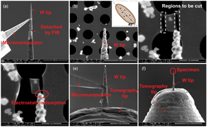
Hettler, Simon
Furqan, Mohammad
and
Arenal, Raul
2024.
Support‐Based Transfer and Contacting of Individual Nanomaterials for In Situ Nanoscale Investigations.
Small Methods,