Hostname: page-component-848d4c4894-x5gtn Total loading time: 0 Render date: 2024-05-19T12:42:25.932Z Has data issue: false hasContentIssue false

Special Stains Used to Identify Fungal Infections

Published online by Cambridge University Press:  30 July 2021

Abstract

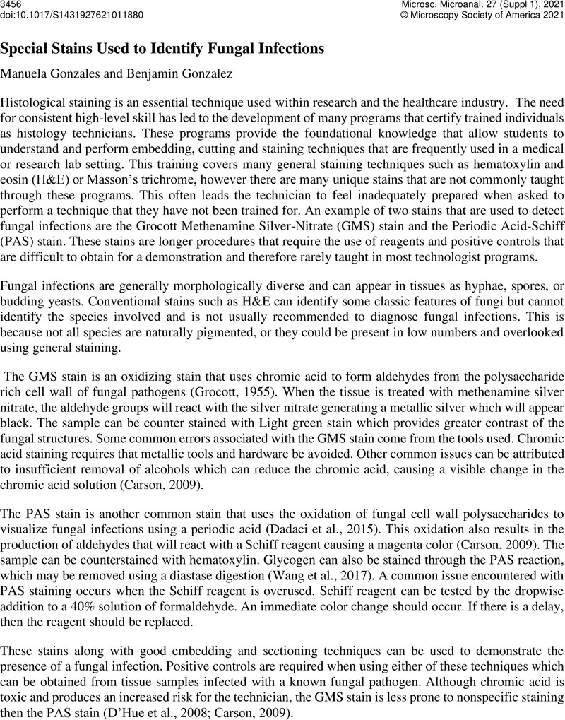
Image of the first page of this content. For PDF version, please use the ‘Save PDF’ preceeding this image.'
Type
Technologists' Forum Workshop Technique Tips: Special Stains and Serial Sectioning
Copyright
Copyright © The Author(s), 2021. Published by Cambridge University Press on behalf of the Microscopy Society of America

References

Grocott, R.G. 1955. A Stain for Fungi in Tissue Sections and Smears. American Journal of Clinical Pathology. 25:975979. doi:10.1093/ajcp/25.8_ts.0975.CrossRefGoogle ScholarPubMed
Carson, F.L., and Cappellano, C.H.. 2009. Histotechnology. ASCP Press, Chicago.Google Scholar
Dadaci, Z., Kılınç, F., Ozer, T.T., Sahin, G.O., Acir, N.O., and Borazan, M.. 2015. Periodic acid–Schiff staining demonstrates fungi in chronic anterior blepharitis. Eye. 29:15221527. doi:10.1038/eye.2015.144.CrossRefGoogle ScholarPubMed
DHue, Z., Perkins, S.M., and Billings, S.D.. 2008. GMS is superior to PAS for diagnosis of onychomycosis. Journal of Cutaneous Pathology. 35:745747. doi:10.1111/j.1600-0560.2007.00890.x.CrossRefGoogle Scholar