Hostname: page-component-78c5997874-g7gxr Total loading time: 0 Render date: 2024-11-18T00:59:15.820Z Has data issue: false hasContentIssue false

X-ray Spectrometry in the Era of Aberration-Corrected Electron Optical Beam Lines

Published online by Cambridge University Press:  10 May 2022

Nestor J. Zaluzec*
Affiliation:
Photon Sciences Directorate, Argonne National Laboratory, Lemont, IL, USA
Get access

Abstract

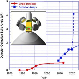
Aberration correction in the analytical transmission electron microscope is most closely associated with improvements in high-resolution imaging. In this paper, the combination of that technology with new system designs, which optimize both electron optics and x-ray detection, is shown to provide more than a tenfold increase in performance over the last 25 years.

Type
Original Article
Copyright
Copyright © The Author(s), 2022. Published by Cambridge University Press on behalf of the Microscopy Society of America

Access options

Get access to the full version of this content by using one of the access options below. (Log in options will check for institutional or personal access. Content may require purchase if you do not have access.)

References

Bowman, HR, Hyde, EK, Thompson, SG & Jared, C (1966). Application of high-resolution semiconductor detectors in X-ray emission spectrography. Science 151, 562568.CrossRefGoogle ScholarPubMed
Castaing, R (1952). Application des sondes électroniques à une méthode d'analyse ponctuelle chimique et cristallographique: publication ONERA (Office national d’études et de recherches aéronautiques/Institute for Aeronautical Research) No. 55. PhD Thesis. University of Paris.Google Scholar
Chen, W, Kraner, H, Li, Z, Rehak, P, Gatti, E, Longni, A, Sampietro, M, Holl, P, Kemmer, J, Faschingbauer, U, Schmitt, B, Woner, A & Wurm, JP (1992). Large area cylindrical silicon drift detector. IEEE Trans Nucl Sci 39, 619.CrossRefGoogle Scholar
Crewe, AV & Kopf, D (1980). A sextupole system for the correction of spherical aberration. Optik 55, 110.Google Scholar
Egerton, RF (1980). The design of an aberration corrected electron spectrometer for the TEM. Optic 57, 229242.Google Scholar
Egerton, RF (1995). Electron Energy-Loss Spectroscopy in the Electron Microscope. New York: Springer-Verlag. ISBN: 978-0-306-42158-7.CrossRefGoogle Scholar
Fiori, CE, Swyt, CR & Willis, JR (1982). The Theoretical to Characteristic Continuum Ratio in Energy Dispersive Analysis in the Analytical Electron Microscope in Microbeam Analysis-1982, Heinrich KFJ (Ed.), pp. 57–71. San Francisco Press.Google Scholar
Fitzgerald, R, Keil, K & Heinrich, KFJ (1968). Solid-state energy-dispersion spectrometer for electron-microprobe X-ray analysis. Science 159(3814), 528530.CrossRefGoogle ScholarPubMed
Garbrecht, M & Zaluzec, N (2020). Sensitivity and figure of merit measurements of EELS vs XEDS in the analytical electron microscope. Microsc Microanal 26(S2), 15181521. doi:10.1017/S1431927620018383Google Scholar
Haider, M, Braunshausen, G & Schwan, E (1995). Correction of the spherical aberration of a 200 kV TEM by means of a hexapole corrector. Optik 99, 167179.Google Scholar
Hawkes, PW (2009). Aberration correction past and present. Philos Trans R Soc A 367, 36373664.CrossRefGoogle ScholarPubMed
Howe, JY, Ramprasad, T, Hanawa, A, Inada, H, Jimenez, J, Hoyle, D, Voelkl, E & Zega, T (2017). Collection efficiency of the twin EDS detectors for quantitative X-ray analysis on a new probe-corrected TEM/STEM. Microsc Microanal 23(Suppl 1), 520521.CrossRefGoogle Scholar
Iwanczyk, JS, Patt, BE, Tull, CR & Barkan, S (2001). High-throughput, large area silicon X-ray detectors for high-resolution spectroscopy applications. Microsc Microanal 7(S2), 1052.CrossRefGoogle Scholar
Jimbo, Y, Ohnishi, I, Hashiguchi, H, Iwasawa, Y, Morishita, S, Miyatake, K, Mukai, M & Sawada, H (2020). Development of ultrahigh resolution objective lens enabling high analytical sensitivity. Microsc Microanal 26(Suppl 2), 31263128. doi:10.1017/S1431927620023892CrossRefGoogle Scholar
Kabius, B, Hartel, P, Haider, M, Müller, H, Uhlemann, S, Loebau, U, Zach, J & Rose, H (2009). First application of CC-corrected imaging for high-resolution and energy-filtered TEM. J Electron Microsc 58, 147.CrossRefGoogle ScholarPubMed
Kahl, F, Gerheim, V, Linck, M, Müller, H, Schillinger, R & Uhlemann, S (2019). Chapter three—test and characterization of a new post-column imaging energy filter. Adv Imag Electron Phys 212, 3570. doi:10.1016/bs.aiep.2019.08.005CrossRefGoogle Scholar
Kisielowski, C, Freitag, B, Bischoff, M, van Lin, H, Lazar, S, Knippels, G, Tiemeijer, P, van der Stam, M, von Harrach, S, Stekelenburg, M, Haider, M, Uhlemann, S, Müller, H, Hartel, P, Kabius, B, Miller, DJ, Petrov, I, Olson, EA, Donchev, T, Kenik, EA, Lupini, AR, Bentley, J, Pennycook, SJ, Anderson, IM, Minor, AM, Schmid, AK, Duden, T, Radmilovic, V, Ramasse, QM, Watanabe, M, Erni, R, Stach, EA, Denes, P & Dahmen, U (2008). Aberration-corrected electron microscope with 0.5-Å information limit. Microsc Microanal 14, 469477. doi:10.1017/S1431927608080902CrossRefGoogle ScholarPubMed
Kociak, M & Zagonel, LF (2017). Cathodoluminescence in the scanning transmission electron microscope. Ultramicroscopy 176, 112131. doi:10.1016/j.ultramic.2017.03.014CrossRefGoogle ScholarPubMed
Krivanek, O, Dellby, N, Spence, AJ, Camps, RA & Brown, LM (1997). In Aberration Correction in the STEM. Proc. EMAG-1997, Cambridge, UK, Rodenburg, JM (Ed.), pp. 3539. Bristol, UK: Institute of Physics.Google Scholar
Lorimer, GW, Razik, NA & Cliff, G (1973). The use of the analytical electron microscope EMMA-4 to study the solute distribution in thin foils: Some applications to metals and minerals. J Microsc 99, 153164.CrossRefGoogle Scholar
Miller, D, Dahmen, U & Stach, E (2011). New opportunities for in situ science based on the TEAM platform. Microsc Microanal 17(S2), 450451. doi:10.1017/S1431927611003126CrossRefGoogle Scholar
Niculae, A, Lechner, P, Soltau, H, Lutz, G, Struder, L, Fiorini, C & Longoni, A (2006). Optimized readout methods of silicon drift detectors for high-resolution X-ray spectroscopy. Nucl Instrum Methods Phys Res Sec A 568(1), 336342. doi:10.1016/j.nima.2006.06.025CrossRefGoogle Scholar
Rose, H (1990). Outline of a spherically corrected semi-aplanatic medium-voltage transmission electron microscope. Optik 85, 1924.Google Scholar
Rose, H (1994). Correction of aberrations, a promising means for improving the spatial and energy resolution of energy-filtering electron microscopes. Ultramicroscopy 56, 11.CrossRefGoogle Scholar
Scherzer, O (1947). Sphrische und chromatische Korrektur von Elektronen-Linsen. Optik 2, 114132.Google Scholar
Shuman, H & Somlyo, AP (1982). Energy-filtered transmission electron microscopy of ferritin. Proc Natl Acad Sci USA 79, 106.CrossRefGoogle ScholarPubMed
Sommerfeld, A (1931). Uber die beugung und bremsung der electronen. Ann D Phys 11(3), 256330.Google Scholar
Tordoff, B, Beam, S, Schweitzer, M, Hill, E, Kugler, V & Png, K (2012). Introducing twin X-ray detectors and fast backscattered electron imaging through a new field emission SEM from Carl Zeiss. Proceedings of EMC-2012, Manchester, September, PS2.2.Google Scholar
Von Harrach, HS, Dona, P, Freitag, B, Soltau, H, Niculae, A & Rohde, M (2009). An integrated silicon drift detector system for FEI Schottky field emission transmission electron microscopes. Microsc Microanal 15(S2), 208209.CrossRefGoogle Scholar
Watanabe, M & Wade, CA (2013). Practical measurement of X-ray detection performance of a large solid-angle silicon drift detector in an aberration-corrected STEM. Microsc Microanal 19(S2), 1264.CrossRefGoogle Scholar
Wen, JG, Miller, DJ, Cook, RE, & Zaluzec, NJ (2014). Amplitude contrast imaging: High resolution electron microscopy using a spherical and chromatic aberration corrected TEM. Microsc Microanal 20(S3), 942-943. doi:10.1017/S1431927614006436CrossRefGoogle Scholar
Wen, JG, Miller, DJ, Zaluzec, NJ, Hiller, JM, Cook, RE, Shah, AB & Zuo, JM (2012). Atomic resolution energy-filtered HREM at high-loss region using Cs- and Cc-corrected TEM. Microsc Microanal 18(Suppl 2), 384385. doi:10.1017/S1431927612003777CrossRefGoogle Scholar
Yankovich, A, Zeng, L, Olsson, E & Zaluzec, NJ (2021). On the dependence of the sensitivity of EELS vs XEDS in the AEM with thickness and beam energy. Proceedings of Microscience Microscopy Congress 2021. Paper #257. doi:10.22443/rms.mmc2021.257.CrossRefGoogle Scholar
Zaluzec, NJ (1991). Progress on the ANL Advanced AEM Project. Proceedings of Microbeam Analysis Society, pp. 137142. San Francisco Press.Google Scholar
Zaluzec, NJ (2004). XEDS systems for the next generation analytical electron microscope. Microsc Microanal 10(S2), 122. doi:10.1017/S1431927604883247CrossRefGoogle Scholar
Zaluzec, NJ (2015). The influence of Cs/Cc correction in analytical imaging and spectroscopy in scanning and transmission electron microscopy. Ultramicroscopy 151, 240249. doi:10.1016/j.ultramic.2014.09.012CrossRefGoogle ScholarPubMed
Zaluzec, NJ (2016). Theoretical and experimental X-ray peak/background ratios and implications for energy-dispersive spectrometry in the next-generation analytical electron microscope. Microsc Microanal 22, 230236. doi:10.1017/S1431927615015755CrossRefGoogle ScholarPubMed
Zaluzec, NJ (2019). Improving the sensitivity of X-ray microanalysis in the analytical electron microscope. Ultramicroscopy 203, 163169.CrossRefGoogle ScholarPubMed
Zaluzec, NJ (2021 a). First light on the argonne PicoProbe and the X-ray perimeter array detector (XPAD). Microsc Microanal 27(S1), 20702073. doi:10.1017/S1431927621007492CrossRefGoogle Scholar
Zaluzec, NJ (2021 b). Quantitative assessment and measurement of X-ray detector performance and solid angle in the analytical electron microscope. Microsc Microanal 113. doi:10.1017/S143192762101360XGoogle Scholar
Zaluzec, NJ, Iwanczyk, JS, Patt, BE, Barkan, S & Feng, L (2003). Performance of a high-count rate silicon drift X-ray detector on the ANL 300 kV advanced analytical electron microscope. Microsc Microanal 9(S2), 892893. doi:10.1017/S1431927603444462CrossRefGoogle Scholar