Introduction
Although the last decade in electron microscopy has seen tremendous gains in image resolution, new challenges in the field have come to the forefront. First, new ultra-sensitive instruments bring about unprecedented environmental specifications and facility needs for their optimal use. Second, in the quest for higher spatial resolutions, the importance of developing and sharing crucial expertise—from sample preparation to scientific vision—has perhaps been deemphasized. Finally, for imaging to accelerate discoveries related to large scientific and societal problems, in situ capabilities that replicate real-world process conditions are often required to deliver necessary information. This decade, these are among the hurdles leaders in the field are striving to overcome.
The Department of Energy's (DOE) Environmental Molecular Sciences Laboratory (EMSL) has a newly developed suite of microscopy capabilities that are being applied to various fields across the EMSL user community. Broad examples of these fields include catalysis, energy storage, and biology.
EMSL's “Quiet Wing,” depicted in Figure 1, is a new addition to the user facility to be completed in September 2011. Two years prior, in September 2009, EMSL director Allison Campbell testified before an Energy and Environment subcommittee at the U.S. House of Representatives, offering the scientific rationale for an expanded footprint: “EMSL will build a new space that will house ultra-high-resolution instruments for providing physical and chemical information at unprecedented spatial or energy resolution. Called the Quiet Wing, it will house new microscopy capabilities that require extremely low electromagnetic field and vibrational interference as well as high temperature-stability [Reference Campbell1].”

Figure 1: The EMSL Quiet Wing will initially house five microscopy instruments, and planning for further additions is ongoing.
The electron optical advancements of the last decades have enabled crystal structure analysis on an atomic level. However, improved electron optics alone are not sufficient for this progress. The whole system—instrument and environment—must be stable enough to allow sub-Ångstrom resolution within realistic exposure times (1–10 seconds). The results of these increases in resolution were more stringent specifications for the stability of the environment, including low acoustic noise and low floor vibrations.
To meet these requirements each instrument cell in the Quiet Wing is built with a separate three-foot-thick foundation isolated from the foundations of the other rooms. The facility also features noise-dampening materials, a laminar air-flow dispersion system to provide uniform temperatures and to minimize air movement at the microscope columns, and specialized shielding to prevent electromagnetic interference (EMI). In addition, the wing's location was chosen for its very low EMI and vibration background, and the structure itself was designed to not create disruptive EMI. This environment allows scientific users to make optimal use of several new instruments, many of which were acquired during a $60-million investment in EMSL through the American Recovery and Reinvestment Act (ARRA).
Capabilities
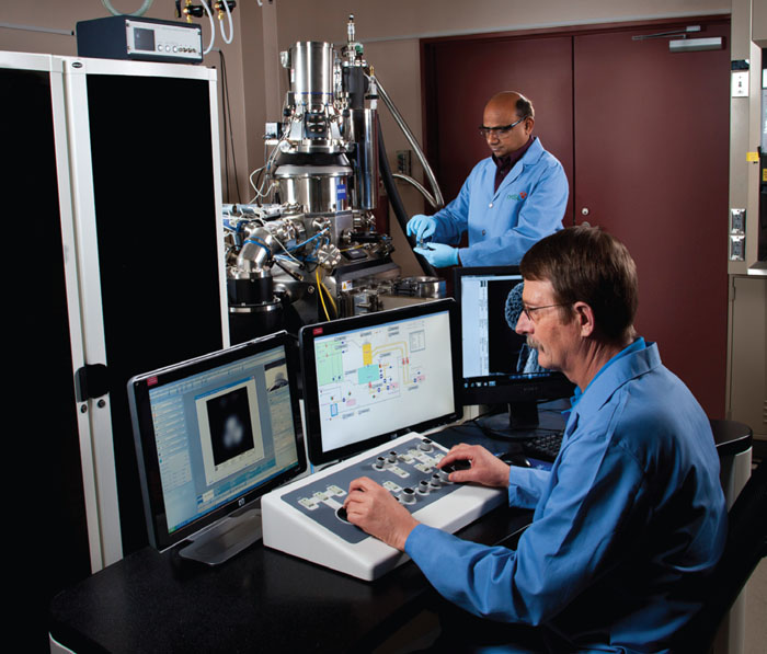
The Quiet Wing will house only a handful of the eighteen instruments within EMSL's microscopy capability group. Here we introduce the five instruments that will initially occupy the wing, which themselves form a unique collection for the research community. The tools and techniques listed—and others in EMSL—are meant to be complementary. In a number of research areas, integrating them, as well as incorporating theoretical modeling and simulation, greatly enhances experimental results. For this reason, one can consider them different components or aspects of one large imaging capability.
Scanning/Transmission Electron Microscope (S/TEM)
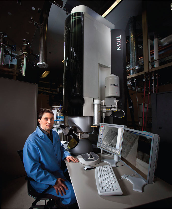
Figure 2 shows the FEI Titan S/TEM, a high-demand instrument in EMSL that will be moved into the Quiet Wing upon its completion. This aberration-corrected instrument offers spatial resolution of 0.1 nm (STEM mode) and energy resolution of 0.3 eV. It is referred to as a “multi-purpose tool” for research involving energy materials, catalysis, interfacial phenomena, and nanostructured materials because of its superior structural imaging and chemical analysis on an atomic level. The instrument is equipped with a monochromator that improves energy resolution for electron energy-loss spectroscopy (EELS) to a level where not only chemical elements can be analyzed but also their binding state. The electron probe corrector in combination with the monochromator allows elemental and chemical analysis with high spatial resolution on an atomic level. Energy-dispersive X-ray spectroscopy (EDXS) is also available for detection and quantification of all elements heavier than boron. The three attachments mentioned above convert this instrument into a powerful tool for analytical electron microscopy. The scope of available experiments can also be extended to in situ studies of dynamic processes by using sample holders that can control temperature, electrical current, and gaseous environment.

Figure 2: EMSL's aberration-corrected FEI Titan scanning/transmission electron microscope (S/TEM) provides high-resolution imaging with sub-angstrom resolution and spectroscopic capabilities.
Environmental Transmission Electron Microscope (ETEM)
The Quiet Wing will host an FEI ETEM, which is dedicated to in situ experiments for a wide range of research applications, including interfacial phenomena, chemical science and engineering, nanoscience technology, materials science, environmental science, and biogeoscience. For this purpose, it is equipped with an integrated environmental cell, which allows precise control of pressure and gas composition while simultaneously allowing control of electrical current, magnetic field, and temperature. Pressures up to 20 mbar and temperatures up to 1000°C are available for dynamic studies. In situ experiments require high time resolution, which makes TEM imaging the preferred operational mode. Therefore, an image corrector is integrated into the instrument to eliminate image artifacts such as contrast delocalization, which scramble structural information. These features provide in situ capabilities with atomic-resolution imaging to study dynamic processes. Through these features and the ETEM's advantages in large-field imaging for structural interface analysis, the instrument will accelerate research in catalysis, growth of nanowires, evolution of nanoparticle morphology, and organization of molecules.
Helium Ion Microscope (HIM)
Figure 3 shows the first-ever helium-ion microscope to become available at a national scientific user facility. We believe this tool, manufactured by Carl Zeiss NTS, opens the door to vital new experimentation in catalyst nanostructures; biological, geochemical, and biogeochemical processes; and surface/interface studies. The HIM provides ultra-high-resolution images (with spatial resolution down to 0.35 nm) on a wide range of materials, including insulators, through high contrast and superior depth-of-field. Specifically, this instrument minimizes damage to biological samples and offers a Rutherford backscattering spectrometry (RBS) detector to identify atomic elements and determine material composition.

Figure 3: The Quiet Wing will house the first helium ion microscope (Carl Zeiss) available at a national scientific user facility.
Ultra-High Vacuum, Low-Temperature, Scanning Probe Microscope (UHV LT SPM)
Manufactured by Omicron NanoTechnology, this tool provides a unique combination of capabilities to examine the molecular details of chemical reactions—particularly those important for heterogeneous catalysis and photocatalysis. With atomic-level spatial resolution and vertical resolution better than .01 nm, the UHV LT SPM allows in situ imaging of spatially resolved reaction kinetics and dynamics at specific catalytic reaction sites. In general, catalytic reactions are activated and have to be carried out at elevated temperatures. Using LT SPM in combination with collimated beams of reagents with well-defined hyperthermal kinetic energies allows probing reactions at low temperatures. The instrument also enables detailed measurements of nucleation and growth processes important for materials synthesis, offers true cryo (5 K) scanning tunneling and non-contact atomic force microscopes, and can perform single-molecule vibrational spectroscopy. It combines in situ operation with traditional, ensemble-averaging surface analytical tools, providing a truly unique, state-of-the art capability.
Ultra-High Vacuum, Variable-Temperature Scanning Probe Microscope (UHV VT SPM)
This existing EMSL instrument will be moved into the Quiet Wing to optimize its performance. Manufactured by Omicron NanoTechnology, it is primarily used for studies of model catalytic systems and associated surface thermal and photochemistry under ultrahigh vacuum conditions. It includes a pair of interlocked ultrahigh vacuum chambers: a scanning probe chamber and a sample preparation/characterization chamber with ensemble-averaged surface analysis capabilities. Both the scanning tunneling microscope (STM) and non-contact atomic force microscope provide atomic resolution in a full temperature range of 50–500 K.
Science Made Possible
The singular motivation for deploying these instruments within the Quiet Wing is to enhance observation of scientific processes, with a specific focus on pushing the boundaries of in situ imaging for science areas of national and global importance. As scientists at EMSL, we are eager to collaborate with users to attain better images and spectra that lead to new discoveries. Although the Quiet Wing has yet to open, several of the tools described above have already yielded promising results for catalysis, energy storage, and health-related biology.
Catalysis
A major challenge in TEM is to enable a deeper understanding of catalytic reactions and interfacial/surface properties. Addressing this need calls for 3-D structural and chemical analyses of nanoparticles and nanostructures and, ideally, for the position and type of each atom involved. The difficulty in obtaining this information using standard tomographic techniques is that a large number of images (~150) is required for 3-D reconstruction. The total exposure time can be several hours, which results in an electron dose that causes severe radiation damage in many systems of scientific interest, such as nanoparticles with catalytic properties. Much of today's catalysis-focused TEM is indeed hampered by low time-resolution, strong radiation damage, and poor stability. However, positive steps are taking place through atomic-level characterizations of transition metal catalytic clusters on oxide substrates in order to determine the structure-property relationships of small clusters. For example, Figure 4 shows iridium nanoscale clusters supported by MgAl2O4 in an image collected with EMSL's aberration-corrected S/TEM. With high-angle annular dark field (HAADF) imaging, contrast is highly sensitive to the atomic number (Z1.7) [Reference Hartel, Rose and Dinges2]. A very high signal-to-noise ratio in Figure 4 enables correlation between the electron count rate at the position of atomic columns and the number of atoms in these columns, which subsequently enables approximation of the full three-dimensional shape of the catalytic nanoclusters. Although this approach requires an acquisition of only a single image, it can lead to several possible structural models. Thus, ab initio calculations using supercomputing resources should be further used to identify the most likely model and achieve higher accuracy for atomic positions. Additional integration of the results with those from other tools can provide unprecedented analysis of how the catalyst interacts with its support. For example, high-resolution TEM phase contrast reveals the epitaxial relationship between the Ir clusters and the MgAl2O4 substrate, allowing quantitative analysis of the clusters. The in situ challenge is to reveal how the shapes of catalytic nanoparticles change in real time, in realistic, gaseous environments. In addition to its microscopy efforts, EMSL is making strides in this area within the ultrahigh-field nuclear magnetic resonance capability.

Figure 4: Iridium clusters supported by MgAl2O4.
Energy Storage
Rechargeable lithium-ion batteries are ubiquitous across today's technologies, from laptops to cars, and they play a key role in the overarching goal of improving energy storage applications. In exploring why these batteries succeed or fail under operating conditions, in situ TEM has played a vital role. In a recent collaboration with the Center for Integrated Nanotechnologies, a concept was developed at EMSL to create a working lithium-ion nanobattery that used a single nanowire as an electrode and an ionic liquid-based electrolyte, designed specifically for placement inside a high-vacuum TEM to allow observation of the electrode's structural evolution during charging of the nanobattery [Reference Huang, Zhong, Wang, Sullivan, Xu, Zhang, Mao, Hudak, Liu, Subramanian, Fan, Qi, Kushima and Li3]. One outcome of the effort was a video [Reference Huang, Zhong, Wang, Sullivan, Xu, Zhang, Mao, Hudak, Liu, Subramanian, Fan, Qi, Kushima and Li4] that captured the microstructural evolution of a tin oxide (SnO2) nanowire anode, which measured 100 nm in diameter and 10 μm in length. With the progression of the lithium injection into the SnO2, the nanowire exhibited swelling, elongation, and contortion in a spiral fashion, as illustrated in Figure 5. The lithiation reaction front is characterized by continuous generation and annihilation of dislocations, which apparently is coupled with the lithium diffusion process. These unprecedented in situ observations provide vital information for a deeper understanding of how and why rechargeable batteries wear out over time. Thus, the creation and real-time observation of this “world's smallest battery” [5] has uncovered mechanistic insights to stimulate new thinking to increase battery performance and longevity—results that were only possible by pushing the boundaries of in situ TEM. Future plans by scientists involved in this research will rely on access to the EMSL Quiet Wing. Specifically, further emphasis will be placed on in situ atomic-level structural and chemical observations of batteries during their operation, for the purpose of discovering new materials for better batteries.

Figure 5: An artist's rendering of in situ observations of a single SnO2 nanowire battery during charging with lithium ions (copper color). The diameter of the nanowire was ~200 nm.
Biology: Nanomaterials and Human Health
As nanomaterials are more widely used in commercial applications, increased human exposure to these materials is expected. However, the impact of such exposure on health remains unclear, especially with respect to underlying mechanisms. A recent investigation [Reference Arey, Shutthanandan, Xie, Tolic, Williams and Orr6] used EMSL's HIM to collect images that characterize the interactions of engineered nanoparticles at the surface of alveolar epithelial cells from mouse lungs. The use of HIM for biological samples is fairly new, but it presents key advantages, including sub-nanometer resolution, efficient charge control, small beam damage, and high depth of field. The growth of the cells at the air-liquid interface and their exposure to aerosolized nanomaterials in ambient air closely mimicked in vivo exposure conditions, and the use of the Rutherford backscattered ion imaging mode—and an added Rutherford backscattering spectrometer—provided the contrast necessary for optimal specimen analysis. Zinc oxide (ZnO) engineered nanoparticles were chosen because they are used extensively in a wide variety of commercial applications. The HIM enabled us to identify the spatial and temporal distribution of the particles and their dissolved ions at the cell membrane with unprecedented resolution and to bring new insights to the origin of their toxicity. This constitutes a very useful complement to SEM imaging for biological samples (Figure 6), establishing HIM as an excellent emerging tool for the life sciences. In addition to tracing processes at the interface of living cells with nanomaterials and other health-related studies, the HIM will enhance various biological disciplines and applications, including the study of microbial interactions with minerals, a high-impact area for environmental contaminant cleanup.

Figure 6: Twenty-four-hour exposure images show the contrast difference between the secondary (A) and the Rutherford backscatter image (B). In the RBI Image, ZnO particles are shown in the filopodia and also in the cellular membrane. In both images, the field of view is 10 microns.
Conclusion and Future Prospects
The Quiet Wing provides an environment in which ultra-sensitive instrumentation can exhibit its optimum performance unlimited by external disturbances. There are many experimental investigations that simply are not possible for our users without this facility. Examples include chemical (EELS) mapping on an atomic scale for catalytic, spintronics applications, and direct structure imaging in TEM. STM applications benefit from orbital-mediated and inelastic electron tunneling spectroscopy for electronic and vibrational structure characterization of adsorbed molecules.
It should be stated that environmental limitations are not the only constraints on imaging. The extent to which users can closely collaborate, share expertise, and integrate various techniques will also largely determine the science impact. Beyond a new building, we are committed to helping accelerate critical science through a focus on both people and capabilities. EMSL continues to build and maintain a staff of experienced scientists for deep collaboration with users and is currently engaging scientific leaders around the world to not only enhance in situ capabilities but to set new “grand challenge” targets for unprecedented time resolution—microscopy's next revolution.
Acknowledgments
EMSL is a national scientific user facility sponsored by the Department of Energy's Office of Biological and Environmental Research and located at Pacific Northwest National Laboratory. More information on the EMSL Quiet Wing is available at http://www.emsl.pnl.gov/capabilities/facilities/quiet_wing.jsp.


