


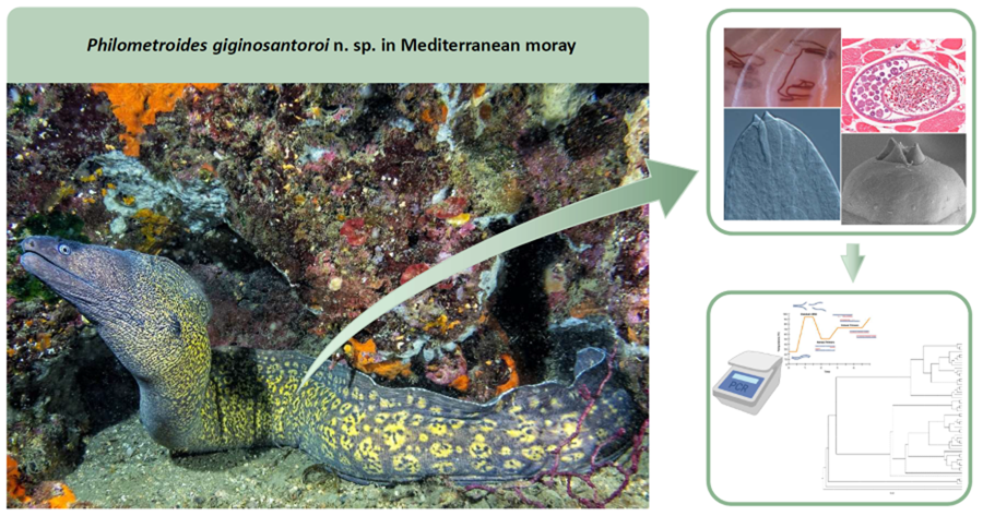
Introduction
Philometridae Baylis & Daubney, 1926 is a family of dracunculoid nematodes mostly comprising gonad, body cavity and tissue-infecting species restricted to teleost fishes. Members of the family are characterized by a simple mouth surrounded by a variable number of cephalic papillae arranged in 2 circles; the buccal capsule is generally reduced or absent, and have an oesophagus, often bulbously inflated at its anterior end, provided with a large oesophageal gland (Moravec, Reference Moravec2004, Reference Moravec2006, Reference Moravec2024). Their life cycles involve copepods as intermediate hosts and fishes as paratenic and definitive hosts (Moravec, Reference Moravec2004). Male philometrids are considerably smaller and have a shorter life span than females (Moravec, Reference Moravec2004, Reference Moravec2006, Reference Moravec2024). A recent experimental study revealed that, in their definitive host, females of Philometroides seriolae (Ishii, 1931) Yamaguti, 1935 can take more than a year to be able to produce and disseminate larvae (Ogawa et al., Reference Ogawa, Sata, Sugihara, Miyazaki, Ueno, Kuramochi and Shirakash2023).
With a total of 217 valid species, the genus-level classification within Philometridae remains among the most problematic and unsatisfactory in the phylum Nematoda (Moravec and de Buron, Reference Moravec and de Buron2013; Moravec, Reference Moravec2024). Their taxonomic diagnoses mostly rely on female morphology, as males are unknown for most species and even genera, or on molecular tools (Moravec, Reference Moravec2004, Reference Moravec2006, Reference Moravec2024).
The Mediterranean moray Muraena helena Linnaeus, 1758 (Anguilliformes: Muraenidae) is a nocturnal and territorial carnivorous species distributed throughout the whole Mediterranean Sea and the eastern Atlantic and mainly feeding on fish, crustaceans and cephalopods (Matić-Skoko et al., Reference Matić-Skoko, Tutman, Bojanić Varezić, Skaramuca, Đikić, Lisičić and Skaramuca2014; Sallami et al., Reference Sallami, Ben Salem, Reynaud and Capapé2014). Its meat is used and appreciated for human consumption since the Roman Empire, when it was farmed and raised in specific fishponds especially in the Gulf of Naples (Italy, Tyrrhenian Sea) (Pesando and Stefanile, Reference Pesando and Stefanile2016). Notwithstanding that, data on its helminth fauna are scarce and mostly limited to descriptions or reports of few specialist gastrointestinal trematodes (Bartoli et al., Reference Bartoli, Overstreet and Gibson2003, Reference Bartoli, Gibson and Bray2005; Bartoli and Gibson, Reference Bartoli and Gibson2007).
Within a long-term project aiming to study the parasite fauna of the marine fishes of the Gulf of Naples through interdisciplinary approaches, unusual philometrid specimens were found infecting the skeletal muscles of the Mediterranean moray. These resulted to be a morphologically different and previously unknown species of the genus Philometroides Yamaguti, 1935, herein described based on an integrative taxonomic approach.
Materials and methods
Collection data and parasitological study
A total of 32 Mediterranean moray specimens were obtained between July 2021 and March 2023 from professional fishermen operating in 3 different localities of the Gulf of Naples (Nisida Island: 40.7991N, 14.1535E; Pozzuoli: 40.8161N, 14.144E; Procida Island: 40.7494N, 14.0167E) with nets, pots or longlines at depths ranging between 5 and 30 m. Fish were immediately refrigerated and studied within 24 h after death. They were measured (total length – TL) to the nearest 0.1 cm and sex was determined by gonadal examination at necropsy. Then, eyes, skin, gills, mouth cavity, digestive tract (stomach and intestine), liver, heart, gonads, visceral cavity, mesenteries and skeletal muscles of each specimen were examined for parasites under a dissecting microscope (Zeiss Axio Zoom V16, Zeiss, Switzerland) using the methods described in Santoro et al. (Reference Santoro, Palomba, Aco Alburqueque and Mattiucci2022, Reference Santoro, Bellisario, Fernández-Álvarez, Crocetta and Palomba2023). When philometrid nematodes were observed in muscles, they were extracted with tweezers, washed in physiological saline solution and preserved in 70% ethanol, 2.5% glutaraldehyde or frozen for subsequent morphological and molecular analyses.
For the light microscopy, nematodes were cleared with Amman's lactophenol, and measurements were obtained using a compound microscope (Axio Imager M1, Zeiss) and a dissecting microscope equipped with the ZEN 3.1 imaging system (Zeiss). Measurements are reported in micrometres unless otherwise indicated and are expressed as mean value ± standard deviation, followed by ranges and the number of measurements for each character in parenthesis. Drawings were made with the aid of a XP PEN Deco 02 drawing tablet and the software Adobe Illustrator and Adobe Photoshop. For the scanning electronic microscopy (SEM), anterior and posterior extremities and intermediate portions of 2 individual nematodes fixed in 2.5% glutaraldehyde were transferred to 40% ethanol (10 min), rinsed in 0.1 M cacodylate buffer, postfixed in 1% OsO4 for 2 h and dehydrated in ethanol series, critical point dried and sputter-coated with platinum. Observations were made using a JEOL JSM 6700F scanning electron microscope operating at 5.0 kV (JEOL, Italy). For the histopathological study, selected samples of skeletal muscles of infected fish were fixed in 10% neutral phosphate-buffered formalin and processed by routine methods into paraffin blocks which were cut into 3 μm-thick sections and stained with haematoxylin and eosin.
Molecular and phylogenetic analyses
Genomic DNA was extracted from 3 frozen specimens using the Quick-gDNA Miniprep Kit (Zymo Research, USA). The 18S rRNA sequences were amplified and sequenced using the combination of 3 sets of primers [WormA (5′-GCGAATGGCTCATTAAATCAG-3′) and 1270R (5′-CCGTCAATTCCTTTAAGTTT-3′); 1100F (5′-CAGAGATTCGAAGACGATC-3′) and WormB (5′-CTTGTTACGACTTTTACTTCC-3′)] of which 1 was internal pair [930F (5′-GCATGGAATAATGGAATAGG-3′) and 1262R (5′-AACGGCCATGCACCACCACCC-3′)] (Littlewood and Olson, Reference Littlewood, Olson, Littlewood and Bray2001).
Polymerase chain reactions (PCRs) were performed in 25 μL volume containing 1.5 μL of each primer 10 mm, 3 μL of MgCl2 25 mm (Promega, USA), 5 μL of 5× buffer (Promega), 0.6 μL of dNTPs 10 mm (Promega), 0.2 μL of Go-Taq Polymerase (5U μL−1) (Promega) and 2 μL of total DNA. PCR cycling parameters for primer sets WormA and 1270R and 1100F and WormB were as follows: 94°C for 2 min, followed by 40 cycles of 94°C for 30 s, 50°C for 30 s, 72°C for 2 min and a final 72°C extension for 7 min. PCR cycling parameters for 930F and 1262R were as follows: 94°C for 5 min, followed by 40 cycles of 94°C for 40 s, 50°C for 40 s, 72°C for 2 min and a final 72°C extension for 10 min.
Successful PCR products were purified using Agencourt AMPure XP (Beckman Coulter, USA), following the standard manufacturer-recommended protocol. Clean PCR products were Sanger sequenced from both strands using an Automated Capillary Electrophoresis Sequencer 3730 DNA Analyzer (Applied Biosystems, USA) and the BigDye® Terminator v. 3.1 Cycle Sequencing Kit (Life Technologies, USA). All 3 sets of primers were used for sequencing to maximize sequences quality. The obtained contiguous sequences were assembled and edited using MEGAX v. 11 (Kumar et al., Reference Kumar, Stecher, Li, Knyaz and Tamura2018). Sequence identity was verified using the Nucleotide Basic Local Alignment Search Tool (BLASTn) (Morgulis et al., Reference Morgulis, Coulouris, Raytselis, Madden, Agarwala and Schaffer2008).
Sequences representatives of Philometridae were downloaded from GenBank for phylogenetic analysis according to Barton et al. (Reference Barton, Moravec, Zhu and Shamsi2022), who provided an updated phylogeny of philometrid nematodes based on the 18S rRNA nuclear gene (Table 1). Sequences of analogous length to the ones generated in this study were selected (>1600 bp). Two dracunculoids (family Dracunculoidae Stiles, 1907), namely Dracunculus medinensis (Linnaeus, 1758) (MK163617) and Philonema sp. (U81574), were selected as outgroups (Sokolov et al., Reference Sokolov, Kalmykov and Malysheva2020; Barton et al., Reference Barton, Moravec, Zhu and Shamsi2022). The sequences were aligned using the MAFFT algorithm (Katoh et al., Reference Katoh, Rozewicki and Yamada2019) implemented in T-Coffee (Notredame et al., Reference Notredame, Higgins and Heringa2000), then submitted to the transitive consistency score to verify the reliability of aligned positions and optimize the phylogenetic topology (Chang et al., Reference Chang, Di Tommaso, Lefort, Gascuel and Notredame2015). In total, 55 sequences were analysed. Maximum likelihood (ML) and Bayesian inference (BI) phylogenetic trees were calculated using iQtree v. 1.6.12 (Nguyen et al., Reference Nguyen, Schmidt, Von Haeseler and Minh2015) and MrBayes v. 3.2.7 (Ronquist and Huelsenbeck, Reference Ronquist and Huelsenbeck2003), respectively. The best fitted evolutionary models were HKY + I + G as suggested by JmodelTest v. 2.1.10 (Darriba et al., Reference Darriba, Taboada, Doallo and Posada2012). For the ML analyses, 5000 ultrafast bootstrap approximations were performed to test the phylogenetic reliability. Posterior probability distributions for the Bayesian analysis were generated using Markov Chain Monte Carlo (MCMC) method. MCMC searches were run for 10.000.000 generations on 2 simultaneous runs of 4 chains and sampled every 1.000 generations; the first 25% of samples from the MCMC algorithm were discarded as burn in. The quality of the Bayesian analysis (parameter densities, effective sample size and burn-in) and the chain convergence were examined in Tracer (Rambaut et al., Reference Rambaut, Drummond, Xie, Baele and Suchard2018). Trees were visualized using Figtree v. 1.4.4 (Rambaut, Reference Rambaut2012). The definition of the major clades in all phylogenies of this work follows Barton et al. (Reference Barton, Moravec, Zhu and Shamsi2022).
Table 1. Information about sequences obtained from GenBank used in the phylogenetic analysis

FW, freshwater; NS, not stated; SW, saltwater; T, terrestrial.
a Outgroup.
b Listed as Philometra lateolabracis in Table 1 of Wijová et al. (Reference Wijová, Moravec, Horák and Lukeš2006).
Results
General data
A total of 14 philometrid nematodes (all subgravid females) were found in the skeletal muscles (Fig. 1A and B) of 3 out of the 32 (9.4%) Mediterranean morays examined, accounting for 2 females (TL: 70.5 and 54 cm) from Nisida and 1 male (TL: 71 cm) from Pozzuoli. Histological analysis revealed that the parasites elicited no host inflammatory response in the skeletal muscles of its host (Fig. 1C and D). Cross-sections of the parasites showed the presence of red blood cells of the fish host in their intestine, confirming that these philometrids are hematophagous (Fig. 1C and D). Eggs at different development stages were observed in all histological sections inside uteri (Fig. 1C and D).

Figure 1. Philometroides giginosantoroi n. sp. in the skeletal muscles of the Mediterranean moray. (A) Alive specimens; (B) a 70% ethanol preserved specimen; (C) cross-sections of specimens from the skeletal muscles; (D) magnification of a cross-section of P. giginosantoroi n. sp. showing uterus with eggs and intestine filled by host erythrocytes.
Description
Family Philometridae Baylis and Daubney, 1926
Subfamily Philometrinae Baylis and Daubney, 1926
Genus Philometroides Yamaguti, 1935
Philometroides giginosantoroi n. sp. López-Verdejo, Occhibove & Santoro, 2024 (Figs 1–4) in López-Verdejo, Occhibove, degli Uberti, Crocetta & Santoro, 2024

Figure 2. Line drawings of Philometroides giginosantoroi n. sp. Anterior (A and C) and posterior (B and D) ends; lateral (E) and apical (F) view of cephalic end. (E, F) (a) Submedian cephalic papilla of external circle; (b) submedian cephalic papilla of internal circle; (c) lateral cephalic papilla of internal circle.

Figure 3. Light micrographs of Philometroides giginosantoroi n. sp. (A) Anterior end; (B) posterior end showing cuticular bosses; (C) median portion of the body showing uterus and intestine; (D) median portion of the body showing cuticular bosses; (E) ventriculus of a dissected specimen; (F) oesophageal gland of a dissected specimen showing the cell nucleus.

Figure 4. Scanning electron micrographs of Philometroides giginosantoroi n. sp. (A) Lateral view of anterior end showing papillae; (B) magnification of lateral view of anterior end showing papilla and oesophageal teeth; (C) apical view of anterior end showing papillae; (D) apical view of oesophageal teeth; (E) posterior end; (F) cuticular bosses in a young individual. (A–C) (a) Submedian cephalic papilla of external circle; (b) submedian cephalic papilla of internal circle; (c) lateral cephalic papilla of internal circle.
ZooBank LSID: urn:lsid:zoobank.org:act:5190867B-2131-4170-9CED-24017092E460.
Type-host: Mediterranean moray M. helena Linnaeus, 1758 (Anguilliformes: Muraenidae).
Type-locality: Off Pozzuoli (40.8161N, 14.144E; Gulf of Naples, Italy, Tyrrhenian Sea, central-western Mediterranean Sea) (collected on July 2021).
Other localities: Off Nisida (40.7991N, 14.1535E; Gulf of Naples) (collected on March 2023).
Site in host: Skeletal muscles.
Prevalence and intensity: Overall prevalence 9.4%; 1–12 (mean 4.6) nematodes per infected fish.
Type-material: Holotype (MHNG-INVE-0159470), 3 paratypes and 2 SEM preparations (MHNG-INVE-0159471) in the Parasite Collection of the Natural History Museum of Geneva in Geneva (Switzerland).
Etymology: The species is named in memory of Luigi (Gigino) Santoro, father of the last author recently passed away.
Description (based on 7 complete and 4 incomplete – with no posterior extremity – ovigerous specimens, and selected portions of 2 incomplete specimens studied by SEM). Body of live specimens reddish, filiform, with rounded anterior and posterior extremities (Fig. 1A and B). Small oval-shaped cuticular bosses 8.2 ± 2.1 (4.5–12.3; n = 15) in diameter (Fig. 3B and D), irregularly distributed on ventral surface (observed only in larger specimens) from approximately nerve ring area to posterior extremity, conversely cuticular depressions observed on smaller individuals (Fig. 4F). Body length 62.7 ± 16.9 (42.3–90.4; n = 7) mm long, maximum width at middle 208 ± 33.4 (175–250; n = 7). Maximum width/body length ratio 1:296 (240–362; n = 7). Width of cephalic extremity 116 ± 12.5 (103–141; n = 9). Oral aperture circular with 3 sclerotized triangular oesophageal teeth 20 ± 3.5 (15–28; n = 21) wide at base, and 14 ± 2.1 (10–18; n = 20) height, protruding out of mouth (Figs 2C, 2E, 3A, 4A, 4B, 4C and 4D). Cephalic papillae small, arranged in 2 circles: external circle formed by 4 pairs of submedian papillae; internal circle consisting of 6 papillae (4 submedian and 2 lateral) surrounding oral aperture (Figs 2E, 2F, 4A and 4C). Oesophagus muscular, including anterior inflation 1487 ± 200.1 long (1318–1850), representing 2.4% (2.2–3%) of body length (n = 7), anterior oesophageal inflation 79 ± 11.2 (67–95) long, 87 ± 8.8 wide (76–104) (Figs 2C and 3A). Ventriculus opening into intestine through valve, 50 ± 5.9 (40–60; n = 9) long, 58 ± 11.4 (44–77; n = 9) wide (2A and 2E). Oesophageal gland large, 1341 ± 191.6 (1132–1686; n = 9) long, starting short anteriorly nerve ring and extending posteriorly to oesophageal end (Fig. 2A), with a large, cell nucleus 1022 ± 185.4 (890–1303; n = 7) from anterior extremity (Fig. 3F). Nerve ring 272 ± 33.4 (234–326; n = 9), from anterior extremity. Length of intestinal ligament 323 ± 80.8 (197–422). Vulva and anus absent. Uterus filled with numerous eggs (Figs 2A, 2B, 2D and 3C). Posterior end rounded, 112 ± 21.8 wide (72–142; n = 7), without caudal projections (Figs 2B, 2D, 3B and 4E).
Male: not known.
Molecular and phylogenetic analyses
Three identical 18S sequences (1771 bp) were obtained for P. giginosantoroi n. sp. BLASTn queries against the NCBI database showed, among others, a 93.4% identity with Philometra gracilis Moravec & Barton, Reference Moravec and Barton2016 (|MZ274362; 1818 bp), and 93.2% identity with Philometra lati Moravec, Charo-Karisa & Jirků, 2009 (|JF803945; 1739 bp). A representative sequence of P. giginosantoroi n. sp. was deposited in GenBank under the accession number PP746031.
Phylogenetic analyses incorporated 55 sequences ascribed to 53 nominal species (Fig. 5) (alignment length 2079 bp). Topologies of ML and BI trees were identical. Philometridae formed a fully supported monophyletic assemblage. In agreement with previous studies, both ML and BI (Fig. 5) grouped the sequences into 4 fully supported clades (Barton et al., Reference Barton, Moravec, Zhu and Shamsi2022; Ailán-Choke et al., Reference Ailán-Choke, Paschoal, Couto and Pereira2023). Clade A included parasites located either in host body cavity or in subcutaneous tissues of freshwater fishes from South America (Neotropical Region). Clade B represented philometrids of freshwater Cypriniformes from Europe (Palearctic Region), mostly parasitizing host body cavity. Clade C resulted sister to clade B and comprised parasites of a variety of marine fish from different ecoregions; these were mostly found in host ovaries, but also in the body cavity. Clade D, which included the studied Philometroides specimens, represented parasites with a diverse array of host taxa, habitat/ecoregion and site of infection of gravid females (Fig. 5). Within clade D, the 4 groups identified in previous phylogenies were also confirmed by our analyses. They accounted for: group (1) parasites of subcutaneous tissues of marine fish; group (2) parasite species belonging to different genera and characterized by a wide range of features; group (3) philometrids with a variety of host orders, site of infections and geographic origins (all Philometra spp. except Caranginema americanum Moravec, Montoya-Mendoza & Salgado-Maldonado, 2008); group (4) including Philometra and Philometroides species. Noteworthy, Afrophilometra hydrocyoni (Fahmy, Mandour & El-Nafar, 1976) Moravec, Charo-Karisa & Jirků, 2009, Dentiphilometroides marinus Moravec & de Buron, 2009 and P. lati Moravec, Charo-Karisa & Jirků, 2009 were not assigned to any group within clade D in the previous studies.

Figure 5. Phylogenetic tree of the family of Philometridae based on partial 18S rRNA sequence alignment of 2079 bp in length. Tree was calculated through maximum likelihood and Bayesian algorithm and shown as Bayesian tree. Ultrafast bootstrap support (maximum likelihood tree) over 90% and posterior probabilities (Bayesian tree) over 0.90 are shown on the nodes (e.g. 90/0.90).
Philometroides giginosantoroi n. sp. grouped with tissue-infecting philometrids from group 1 of clade D (D1) (Fig. 5). In particular, it seemed closely related to P. gracilis Moravec & Barton, Reference Moravec and Barton2016 and Dentiphilometra lutjani González-Solís, Moravec & Tuz-Paredes, 2007, which were collected from the head tissues and ovary of their marine fish hosts (both Perciformes), respectively.
Remarks
The genus Philometroides includes so far 35 tissue-dwelling species. Out of those, 20 taxa are parasites of freshwater fishes, 12 of marine fishes and 3 of brackish-water fishes (Moravec and de Buron, Reference Moravec and de Buron2013; Montes et al., Reference Montes, Plaul and Martorelli2016; Cavalcante et al., Reference Cavalcante, Moravec and Santos2018; Moravec, Reference Moravec2024). Philometroides giginosantoroi n. sp. differs from the congeneric species by the presence of sclerotized oesophageal teeth, absent in all species except Philometroides branchiarum Moravec & Barton, Reference Moravec and Barton2016, a parasite of gill arches of the John's snapper Lutjanus johnii (Perciformes: Lutjanidae), described from Australia on the basis of 2 larvigerous females (Moravec and Barton, Reference Moravec and Barton2016). However, the females of P. branchiarum have a smaller body (6–9 vs 42.3–90.4 mm), a different shape (conical vs triangular shape) and height (3 vs 20.1) of the oesophageal teeth, a smaller maximum width/length ratio of the body (1:28–33 vs 1:240–362), a different morphology and distribution of the cuticular ornamentations, a different distribution of cephalic papillae, a different position of the oesophageal gland (starting just posteriorly to the nerve-ring vs anteriorly to the nerve ring), a longer oesophagus length ratio with respect to the body length (13–17 vs 2.2–3%), a posterior extremity bearing 2 large lateral caudal projections (absent in the newly described species). In addition, the hosts of P. branchiarum and of P. giginosantoroi n. sp. live in different ecoregions (Indo-Pacific vs Mediterranean) and belong to different orders and families (Perciformes: Lutjanidae vs Anguilliformes: Muraenidae), while the parasite species have different tissue tropisms (arch gills vs skeletal muscles).
Obvious morphological differences also exist between the new species and Philometroides oveni Parukhin, Reference Parukhin1975, the only congeneric species reported in the Mediterranean Sea (from off Lampedusa). This species infects the oculo-orbit and the eye of Serranus hepatus (Perciformes: Serranidae) (Parukhin, Reference Parukhin1975; Moravec and de Buron, Reference Moravec and de Buron2013; Moravec, Reference Moravec2024).
Philometroides oveni differs from the new species by having smaller body (28 vs 62.7 mm), absence vs presence of oesophageal teeth, different number and pattern distribution of cephalic papillae (P. oveni has 4 pairs of papillae arranged in a single circle) (Parukhin, Reference Parukhin1975), maximum width/body length ratio (1:36 vs 1:296), oesophagus/body length ratio (0.03 vs 2.4%), different host orders and families and different tissue tropism.
Discussion
According to Moravec (Reference Moravec2024), the family Philometridae includes 3 subfamilies with 16 genera and 217 valid species: Alineminae (1 genus with 1 species), Neophilometroidinae (1 genus with 2 species) and Philometrinae (14 genera with 214 species). Within Philometrinae, the genera Philometroides and Philometra are quite similar, with the main features used to distinguish the genera being the presence/absence of cuticular bosses and oesophageal teeth (Moravec, Reference Moravec2006; Anderson et al., Reference Anderson, Chabaud and Willmott2009). The general morphology of P. giginosantoroi n. sp. well corresponded to the diagnosis of Philometroides, as it showed cuticular bosses and presence of well-developed oesophageal teeth. Therefore, P. giginosantoroi n. sp. raises to 218 species of the family and to 36 species of the genus.
Members of Philometridae, moreover, exhibit a high degree of host specificity and tissue tropism (Rasheed, Reference Rasheed1963; Ivashkin et al., Reference Ivashkin, Sobolev and Khromova1971; Moravec, Reference Moravec2004, Reference Moravec2006, Reference Moravec2024; Moravec and Justine, Reference Moravec and Justine2009; Moravec and de Buron, Reference Moravec and de Buron2013; Moravec et al., Reference Moravec, Nagasawa, Nitta and Tawa2019). The discovery of P. giginosantoroi n. sp. is also remarkable because it represents the second species of Philometroides found in the Mediterranean and in general in European waters and the third species of Philometridae infecting the family Muraenidae worldwide (Moravec, Reference Moravec2024). These are Philometra gymnothoracis Moravec & de Buron, Reference Moravec and de Buron2009, described using 2 gravid females collected from the body cavity of the spotted moray Gymnothorax moringa from off the Atlantic coast of South Carolina, USA (Moravec and de Buron, Reference Moravec and de Buron2009), and Philometra kidakoi Moravec, Nagasawa, Nitta & Tawa, 2019, described using a subgravid and an incomplete gravid female from the ovary of the Kidako moray Gymnothorax kidako from Western North Pacific Ocean, Japan (Moravec et al., Reference Moravec, Nagasawa, Nitta and Tawa2019). Worth a mention, some fragments of a female of an unidentified species of Philometroides were also reported under the skin of a honeycomb moray Muraena melanotis off Senegal, although no other information or figures were provided by Campana-Rouget (Reference Campana-Rouget1956).
Notwithstanding the morphological assignment to the genus Philometroides, recent phylogenetic studies suggested that at least some of the morphological characters used to distinguish the genera may be not reliable. This is apparently confirmed also by our phylogenetic analyses, consistent with previous studies and highlighting the presence of 4 main clades (named from A to D), characterized by a combination of features related to the site of infection within the host and the host habitat (freshwater or marine) (Negreiros et al., Reference Negreiros, Tavares-Dias, Elisei, Tavares and Pereira2019; Barton et al., Reference Barton, Moravec, Zhu and Shamsi2022; Ailán-Choke et al., Reference Ailán-Choke, Paschoal, Couto and Pereira2023). Within clade D, Barton et al. (Reference Barton, Moravec, Zhu and Shamsi2022) and Ailán-Choke et al. (Reference Ailán-Choke, Paschoal, Couto and Pereira2023) identified 4 sub-clades (named from D1 to D4). The present new species consistently grouped with tissue-infecting philometrids from clade D1, comprising species of 2 additional genera (Dentiphilometra and Philometra) parasitizing the subcutaneous tissues or muscles of marine fishes. Within this group, D. lutjani infects musculature of Lutjanus griseus (Lutjanidae), Philometra ocularis infects the ocular cavity of Serranidae, Philometra brevispicula was found from the mouth and buccal epithelium of L. griseus, P. gracilis infects the tissues behind the gills of L. johnii, Philometra morii infects the subcutaneous tissue of buccal cavity and sinuses of Epinephelus morio (Serranidae) and Philometra sp. infects the subcutaneous tissue of buccal cavity of Mycteroperca microlepis (Serranidae). The genus Dentiphilometra is mainly characterized by the presence of a sclerotized oral ring armed on its inner surface by numerous small peribuccal teeth in female, while differences between Philometroides and Philometra were already listed above (Moravec, Reference Moravec2024). Unfortunately, due to the current scarcity of data regarding the host association, life cycle and relevant taxonomic characters of most of philometrids, their phylogenetic relationships is still mostly unresolved (Negreiros et al., Reference Negreiros, Tavares-Dias, Elisei, Tavares and Pereira2019; Barton et al., Reference Barton, Moravec, Zhu and Shamsi2022; Montes et al., Reference Montes, Albarracin, Barneche, Croci, Balcazar, Cardarella and Martorelli2022; Ailán-Choke et al., Reference Ailán-Choke, Paschoal, Couto and Pereira2023; Moravec, Reference Moravec2024). For instance, not considering the sequence of the present species, the current phylogenetic analysis only included 18S sequences of 6 species of Philometroides and other 41 species of philometrids because of the exclusion of shorter sequences, while we included only sequences of comparable length (~1700 bp). Thus, further data on molecular and morphological features are of pivotal relevance to understand the true relationships within the family and shed light on correct genera assignments.
Data availability statement
The authors confirm that the data supporting the findings of this study are available within the article.
Acknowledgements
We thank Dr František Moravec for providing important bibliography and Fabio Russo for the picture of the Mediterranean moray used for the graphical abstract.
Author contributions
A. L.-V. performed the molecular analysis and the line drawings; F. O. performed the phylogenetic analyses; B. D. U. performed the histological analysis; F. C. provided the fish; M. S. conceived the study, conducted the research, performed the species description, supervised the molecular and phylogenetic data and wrote the manuscript; all authors reviewed and approved the final manuscript.
Financial support
This work was partially funded by the project ADViSE (PG/2018/0494374) (sampling and by-catch analyses) and the National Biodiversity Future Centre (NBFC) Program, Italian Ministry of University and Research, PNRR, Missione 4 Componente 2 Investimento 1.4 (Project: CN00000033).
Competing interests
None.
Ethical standards
Not applicable.


 Open access