Hostname: page-component-7479d7b7d-t6hkb Total loading time: 0 Render date: 2024-07-15T12:56:56.588Z Has data issue: false hasContentIssue false

The contribution of environmental enteropathy to the global problem of micronutrient deficiency

Published online by Cambridge University Press:  05 March 2021

Paul Kelly*
Affiliation:
Blizard Institute, Barts & The London School of Medicine, Queen Mary University of London, London, UK Tropical Gastroenterology and Nutrition Group, University of Zambia School of Medicine, Lusaka, Zambia
*
Corresponding author: Paul Kelly, email m.p.kelly@qmul.ac.uk
Rights & Permissions [Opens in a new window]

Abstract

Sometimes referred to as hidden hunger, micronutrient deficiencies persist on a global scale. For some micronutrients this appears to be due to inadequate intake, for others intake may not match increased requirements. However, for most micronutrient deficiencies there is uncertainty as to the dominant driver, and the question about the contribution of malabsorption is open. Environmental enteropathy (EE), formerly referred to as tropical enteropathy and also referred to as environmental enteric dysfunction, is an asymptomatic disorder of small intestinal structure and function which is very highly prevalent in many disadvantaged populations. Recent studies of the pathology and microbiology of this disorder suggest that it is driven by very high pathogen burdens in children and adults living in insanitary environments and is characterised by major derangements of the epithelial cells of the intestinal mucosa. Transcriptomic data suggest that it may lead to impaired digestion and absorption of macronutrients. Given the very high prevalence of EE, marginal malabsorption could have large impacts at population scales. However, the relative contributions of inadequate soil and crop micronutrient contents, inadequate intake, malabsorption and increased requirements are unknown. Malabsorption may compromise attempts to improve micronutrient status, but with the exception of zinc there is currently little evidence to confirm that malabsorption contributes to micronutrient deficiency. Much further research is required to understand the role of malabsorption in hidden hunger, especially in very disadvantaged populations where these deficiencies are most prevalent.

Type
Conference on ‘Micronutrient malnutrition across the life course, sarcopenia and frailty’
Copyright
Copyright © The Author(s), 2021. Published by Cambridge University Press on behalf of The Nutrition Society.

The global burden of micronutrient malnutrition

Micronutrient deficiencies affect about one-third of the world's population(Reference Muthayya, Rah and Sugimoto1,Reference Bailey, West and Black2 ). In many parts of Africa, the burden of disability-adjusted life-years attributable to deficiencies of iron, zinc and vitamin A exceeds 5000 per 100 000 population(Reference Muthayya, Rah and Sugimoto1). Micronutrient deficiencies may persist despite economic growth, as in South Asia(Reference Harding, Aguayo and Webb3). Deficiencies in sub-Saharan Africa are widespread(Reference Kihara, Bolo and Kinyua4). The purpose of this review is to set out the reasons why micronutrient deficiencies may have become so widespread, and to explore the contribution of environmental enteropathy (EE) to these deficiencies.

Why might an individual develop a micronutrient deficiency?

Geography and geochemistry

For wealthier people, food may be purchased from outside their region of residence but for poor people the majority of their diet comprises foods grown very locally. There are two important micronutrients, iodine and selenium, for which good evidence exists that human nutritional status depends to a large degree on where you live and the soil on which your crops grow. Iodine deficiency, which causes thyroid disease(Reference Zimmermann and Boelaert5), is well known to be common where soils and foods have low iodine content, such as in mountainous and landlocked areas(Reference Kelly and Snedden6). Selenium seems to be similar, with clear evidence of a relationship between soil selenium and human selenium status in Malawi(Reference Hurst, Siyame and Young7). However, in Kenya the relationship between soil selenium and crop selenium content was more complex and was also dependent on pH, organic matter and the presence of other inorganic chemicals(Reference Ngigi, Du Laing and Masinde8).

Food processing and anti-nutrients

In addition to deficiencies in soils and therefore crops, anti-nutrients and toxic compounds may also be present(Reference Frazzoli and Mantovani9). Examples include thyroid disrupting pollutants which exacerbate functional iodine deficiency, and fumonisins and arsenic which antagonise physiological effects of folate(Reference Frazzoli and Mantovani9). Fumonisins are fungal toxins which contaminate crops during harvesting and storage. The best-known anti-nutrient is phytate, which reduces bioavailability of iron and zinc(Reference Gibson, Raboy and King10). Anti-thiamin compounds are another example(Reference Titcomb and Tanumihardjo11). A striking example of the impact of food processing on micronutrient content of foods is the relationship between the type of maize milling and losses of zinc(Reference Chomba, Westcott and Westcott12). Processing of rice also impacts nutrient composition(Reference Atungulu and Pan13). Detailed discussion is beyond the scope of this review, but there is also evidence that appropriate food processing can enhance micronutrient availability(Reference Platel and Srinivasan14).

Clinical gastrointestinal disease

Individuals with clinical gastrointestinal disease, especially oesophageal disease, can lose weight very rapidly and develop severe wasting in a few weeks and such patients will have micronutrient deficiencies commensurate with their weight loss. Patients with anorexia nervosa also have micronutrient deficiencies. More subtly, patients who have had gastrointestinal surgery are at heightened risk of deficiencies. Gastrectomy is followed by iron and vitamin B12 deficiency(Reference Jun, Yoo and Lee15); ileal resection can be followed by vitamin B12 deficiency; jejunoileal bypass has been reported as leading to deficiencies of vitamins A, D, K, zinc, selenium and copper(Reference Joshi, McLarney and Abramoff16,Reference Haria, Sibonga and Taylor17) and multiple deficiencies have been reported both before and after bariatric surgery(Reference Bal, Finelli and Shope18). As the number of people undergoing surgery for obesity increases, this will become a significant clinical burden, but none of these disorders make a significant contribution to the scale of the problem outlined earlier.

Dietary diversity and poverty

Both food insecurity and limited dietary diversity, largely related to poverty, make a much greater contribution to the global burden of micronutrient deficiency than the clinical disorders described earlier. Even in populations with a double burden of malnutrition, including poor ones, micronutrient deficiencies can be prevalent(Reference Wells, Sawaya and Wibaek19). A lack of dietary diversity has been identified as a driver of micronutrient deficiency in Sri Lanka(Reference Weerasekara, Withanachchi and Ginigaddara20), but dietary diversity is a remediable problem, as recently demonstrated in Malawi(Reference Kansanga, Kangmennaang and Bezner Kerr21). The problem of dietary diversity in poor populations is a large subject, beyond the scope of this paper, but major initiatives are being implemented to attempt to improve micronutrient status around the world through drives to increase dietary diversity(Reference Thompson and Amoroso22). Poverty also predisposes to EE, to which we now turn.

Environmental enteropathy: what is it?

History

Studies of diarrhoea and malabsorption in the tropics in the early 1960s used apparently asymptomatic, healthy and well-nourished adults and children from the same population as control subjects. Unexpectedly, they identified a high prevalence of abnormal intestinal permeability (as measured by urinary sugar recovery) and/or histological abnormalities (villous blunting; crypt hypertrophy; villous fusion and mucosal inflammation) in these controls(Reference Louis-Auguste and Kelly23). Studies in American soldiers and Peace Corps volunteers stationed in Thailand and in Peace Corps volunteers in Bangladesh showed that the condition was acquired, and that these abnormalities were similar to those observed in the indigenous population(Reference Louis-Auguste and Kelly23). Furthermore, histological examination of fetal and neonatal intestine also showed that these abnormalities were not present during development, and only became apparent post-natally(Reference Baker24,Reference Walker-Smith25) .

These changes were reversible, as demonstrated by prospective assessment of small bowel histology and absorption. Peace Corps volunteers who had lived in India or Pakistan returned to histological and absorptive normality, usually within 2 years after returning to the United States(Reference Lindenbaum, Gerson and Kent26). Furthermore, adult students from endemic areas moving to the United States to study also normalised intestinal structure and function(Reference Gerson, Kent and Saha27), and in the UK it was noted that there was a relationship between villus morphology and the time from last visit to the tropics(Reference Wood, Gearty and Cooper28). Based on the initial studies which were exclusively conducted in the tropics, the condition was labelled tropical enteropathy(Reference Cook29).

The adoption of environmental enteropathy

However, an extensive global study of intestinal function using lactulose permeation and sugar absorption demonstrated that the observed abnormalities were not observed in some affluent, tropical populations (such as Singapore and Qatar)(Reference Menzies, Zuckerman and Nukajam30), and the condition is therefore more correctly termed EE. Brunser et al., working in Santiago which is not tropical, noted the same enteropathy in children living in insanitary slums(Reference Brunser, Araya and Espinoza31). Numerous subsequent studies have confirmed that EE is highly prevalent throughout the developing world irrespective of climate, and it is seasonal(Reference Kelly, Menzies and Crane32). The increased intestinal permeability to small sugars as measured by sugar absorption assays is correlated with significant and pathological abnormalities of intestinal barrier function, for example as measured by plasma lipopolysaccharide levels(Reference Lunn, Northrop-Clewes and Downes33,Reference Campbell, Elia and Lunn34) . More recently, the term ‘environmental enteric dysfunction’ has been used to emphasise the functional consequences of the enteropathy for child health, as it appears to be a major contributor to stunting(Reference Keusch, Denno and Black35Reference Prendergast and Humphrey38). There is some evidence that EE may also contribute to the well-established impairment of responses to oral vaccines seen in many tropical and disadvantaged populations(Reference Marie, Ali and Chandwe39,Reference Church, Parker and Kosek40) .

Aetiology of environmental enteropathy

There is increasing evidence that EE is characterised by polymicrobial infection with a wide range of enteropathogens. The eight-country Mal-ED study revealed that asymptomatic infection is a major contributor to EE and to adverse growth outcomes in children(Reference Kosek41). This study recognised the impact of enteroaggregative Escherichia coli (Reference Rogawski, Guerrant and Havt42), and identified that Campylobacter infections make a major contribution to EE(Reference Amour, Gratz and Mduma43). Major impacts of Shigella, Giardia, norovirus, enteropathogenic E. coli, Cryptosporidium, and Enterocytozoon bieneusi were revealed by the quantitative measurement of pathogen burden(Reference Rogawski, Liu and Platts-Mills44). These effects probably interact with nutritional deficiencies, including those of micronutrients, in complex ways.

Given the apparent key role of enteropathogens, which are largely transmitted faeco-orally, in the genesis of EE, the failure of three large recent water and sanitation trials to solve the problem of stunting(Reference Null, Stewart and Pickering45Reference Humphrey, Mbuya and Ntozini47) is surprising and disappointing. There is still debate as to why this is, but there are good grounds for thinking that water and sanitation interventions may need to be more transformative to achieve the desired effect(Reference Cumming, Arnold and Ban48). It would certainly be inappropriate to conclude that sanitation does not matter as there is little doubt that the sanitation transformations of the 19th century were a major contributor to massively improved public health in Europe.

The polymicrobial infections which characterise EE may reflect a general disturbance of the microbiota and the normal host–commensal relationship. Recent analysis of the duodenal microbiota in stunted children in Bangladesh identified fourteen taxa which were closely related to growth failure, and which could confer a malnutrition phenotype when adoptively transferred into gnotobiotic mice(Reference Chen, Kung and Das49). This study adds to a growing body of evidence that the microbiota can drive metabolic and other changes in the gut leading to malnutrition, playing a key role in driving, or at least perpetuating, malnutrition(Reference Stine50).

Mucosal abnormalities in environmental enteropathy

Mucosal biopsies from adults and children in disadvantaged populations show consistent villus blunting, leading to reduced absorptive surface area, and crypt hypertrophy(Reference Kelly, Menzies and Crane32,Reference Campbell, Murch and Elia51,Reference Kelly, Besa and Zyambo52) . There is also a marked lymphocytic inflammatory infiltrate in the lamina propria and the epithelium (Fig. 1). This predominantly affects the proximal small intestine; the author has never observed villus blunting in the ileum during hundreds of ileoscopies in Zambia. As the majority of micronutrients are absorbed in the proximal small intestine this may help explain how EE would affect micronutrient status. Recent, and ongoing, comparative histological analysis of intestinal biopsies from Bangladesh, Pakistan, Zambia and the United States have revealed that EE has other key features in addition to reduced surface area and inflammation. These include markedly reduced numbers of goblet cells and Paneth cells(Reference Liu, VanBuskirk and Ali53), both of which make a major contribution to barrier function by maintenance of a protective mucus layer on the epithelial surface which has antimicrobial properties. Defining the cellular and transcriptional changes in EE is currently the focus of intense research of which much will be published in the next 2–3 years. It is clear already that EE is characterised by increased gene expression of mucosal barrier function genes (mucins and NADPH oxidases(Reference Chama, Amadi and Chandwe54)) which are also transcriptional features of inflammatory bowel disease(Reference Haberman, Tickle and Dexheimer55). There is also emerging evidence of changes in solute transporters which mediate the intestinal absorption of micronutrients(Reference Amadi, Zyambo and Chandwe56). Another prominent feature of EE is the increased permeability referred to earlier, which over the past 40 years has been most extensively studied using lactulose permeation(Reference Menzies, Zuckerman and Nukajam30,Reference Lee, McCormick and Seidman57) . More recent studies have used fluorescein as a marker of the leakiness of the mucosa, either by showing leakage of fluorescein from the systemic circulation into the gut lumen during endoscopy(Reference Kelly, Besa and Zyambo52,Reference Amadi, Besa and Zyambo58) , or by showing leakage of oral fluorescein into the systemic circulation(Reference Dorshow, Hall-Moore and Shaikh59,Reference Maurice, Lett and Skinner60) . In an ingenious approach, transcriptomic analysis of shed enterocytes in stool samples identified mucin and inflammatory genes associated with EE in an entirely non-invasive way(Reference Ordiz, Wold and Kaimila61).

Fig. 1. Mucosal abnormalities in environmental enteropathy (EE). (a) Finger-like villi imaged endoscopically in the terminal ileum of a Zambian adult, which contrasts with marked villus fusion and reduced height in an adult with EE (b). (c) Histological image of duodenal biopsy showing enteropathy with inflammatory infiltrate but with preserved villus height, which contrasts with severe inflammation and loss of villus height (d). (e) Confocal laser endomicroscopy image of duodenal mucosa showing good barrier function, intact capillaries (white arrow) and no fluorescein in the intestinal lumen following systemic injection of fluorescein. (f and g) Focal leakage of fluorescein (black arrow), and a microerosion is seen where cellular integrity is lost (hollow arrow, g). (h) Gross leakage of fluorescein seen filling the gut lumen (*).

Maldigestion in environmental enteropathy

Impaired digestion could follow from achlorhydria (reduced or absent gastric acid secretion), exocrine pancreatic insufficiency or enterocyte damage leading to impaired activity of enzymes of the brush border. Achlorhydria would be expected to diminish the efficiency of protein digestion, and it is very common in Zambian adults(Reference Kelly, Shawa and Mwanamakondo62), but there have been few publications on this aspect of malnutrition. It has been proposed that impairment of the gastric acid barrier could lead to small intestinal bacterial overgrowth and contribute to malnutrition(Reference Sarker, Ahmed and Brüssow63). Pancreatic insufficiency would lead to malabsorption of lipids and lipid-soluble vitamins. Reduced expression of brush border digestive enzyme genes was apparent in Zambian adults and children (P Kelly, unpublished results) but the functional implications of these early findings for micronutrient status are not yet apparent.

Malabsorption in environmental enteropathy

The clearest evidence that EE is associated with malabsorption comes from stable isotope studies of zinc (using 68Zn and 70Zn). In Malawian children, fractional absorption of zinc was reduced and endogenous losses increased(Reference Manary, Abrams and Griffin64). For most micronutrients, direct evidence that EE causes or exacerbates the deficiency is lacking, and the only evidence of an association is their widespread prevalence (Table 1). Clearly, interpretation of geographical overlap data is problematic as inadequate intake is very common(Reference Harika, Faber and Samuel65). Stable isotopes remain the most promising way to evaluate directly the contribution of malabsorption to deficiency(Reference Davidsson and Kaskell66).

Table 1. Evidence of micronutrient deficiency in environmental enteropathy (EE)

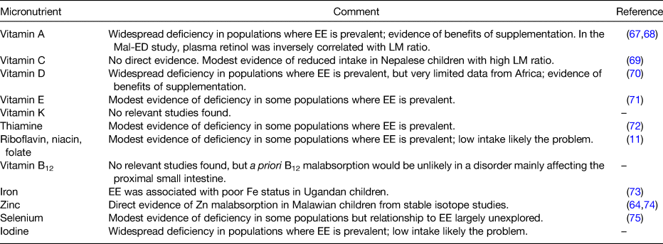
LM, lactulose:rhamnose. Summary of evidence of micronutrient malabsorption in EE. Only for zinc the evidence is direct; for vitamin A the evidence is correlational only.

Treatment of environmental enteropathy

In a trial of multiple micronutrient supplementation over 2 years in Zambian adults, plasma concentrations of folate increased over 2 years, plasma zinc showed a marginal increase, but vitamin A did not increase at all(Reference Kelly, Katubulushi and Todd76). Although not incompatible with the play of chance, or inadequate compliance, these data might suggest that malabsorption due to EE may blunt the efficacy of supplementation programmes. They do not support the idea that EE can be reversed by micronutrient supplementation. Trials of interventions to improve EE have included antibiotic therapy with rifaximin(Reference Trehan, Shulman and Ou77), albendazole(Reference Wang, Shulman and Crocker78), probiotic therapy(Reference Galpin, Manary and Fleming79), zinc(Reference Lauer, McDonald and Kisenge80) or multiple micronutrients(Reference Lauer, McDonald and Kisenge80). However, glutamine supplementation has been reported to have some efficacy in improving intestinal barrier function(Reference Lima, Anstead and Zhang81). In Zambian adults, a combination of glutamine, tryptophan and leucine led to a modest but significant increase in villus height(Reference Louis-Auguste, Besa and Zyambo82). Given this somewhat patchy and disappointing record, novel approaches are urgently required to treat the underlying causes of EE.

Solutions

If it is a valid generalisation that micronutrient malabsorption contributes to hidden hunger, there are two possible approaches to ameliorating the problem: either increase intake, or treat the enteropathy. Neither of these solutions would be easy to implement at scale. Treatment of EE would be highly desirable, as it may improve stunting and help with vaccine responses in addition to improving micronutrient status. However, those treatments which have been tried so far have been disappointing, as noted earlier. Combating the influence of the complex polymicrobial infections now known to be very common in EE will require novel approaches, either to restoring the normal balance of microbiota or to improving mucosal barrier function.

It is axiomatic that flux through a membrane transporter or enzyme system can be overcome by providing more substrate, at least up to a point of saturation, so it is likely to be possible to overcome degrees of malabsorption by increasing the concentration of micronutrients in the gut lumen. Shouldn't we just increase the reference nutrient intake of micronutrients in vulnerable populations? To match such increases in the reference nutrient intake, however, would almost certainly require biofortification(83) and/or supplementation strategies. Biofortification can be very effective, as demonstrated for iodine and thiamine(Reference Keats, Neufeld and Garrett84). Supplementation with folate, as an example, has dramatically reduced neural tube defects in many countries. More controversially, there are suggestions that increased meat consumption, especially by young children, may be an efficient way to increase micronutrient intakes(Reference Wyness85).

Conclusions

EE is a very widespread alteration in gut structure and function which predisposes disadvantaged populations to the consequences of inflammation, leakiness and reduced surface area. These include micronutrient deficiencies, through maldigestion, malabsorption, wastage of plasma constituents into the gut and possibly through inflammation-induced anorexia. The direct connections between enteropathy and malabsorption are just beginning to be explored; currently there is direct evidence only for zinc. These may require revision of fundamental assumptions about micronutrient requirements in disadvantaged populations, and in turn this is likely to necessitate major investments in micronutrient biofortification and/or supplementation. Ideally, a solution should be found to improve gut function in disadvantaged populations with the objective of improving micronutrient status globally.

Financial Support

None.

Conflict of Interest

None.

Authorship

The author takes full responsibility for the entire paper and all content.

References

Muthayya, S, Rah, JH, Sugimoto, JD, et al. (2013) The global hidden hunger indices and maps: an advocacy tool for action. PLoS ONE 8, e67860.CrossRefGoogle Scholar
Bailey, RL, West, KP Jr & Black, RE (2015) The epidemiology of global micronutrient deficiencies. Ann Nutr Metab 66(Suppl 2), 2233.CrossRefGoogle ScholarPubMed
Harding, KL, Aguayo, VM & Webb, P (2018) Hidden hunger in South Asia: a review of recent trends and persistent challenges. Public Health Nutr 21, 785795.CrossRefGoogle ScholarPubMed
Kihara, J, Bolo, P, Kinyua, M et al. (2020) Micronutrient deficiencies in African soils and the human nutritional nexus: opportunities with staple crops. Environ Geochem Health 42, 30153033.CrossRefGoogle ScholarPubMed
Zimmermann, MB & Boelaert, K (2015) Iodine deficiency and thyroid disorders. Lancet Diabetes Endocrinol 3, 286295.CrossRefGoogle ScholarPubMed
Kelly, FC & Snedden, WW (editors) (1962) Le Goitre Endemique (pp. 188190). WHO Edition, Geneva: Ed. Clements.Google Scholar
Hurst, R, Siyame, EW, Young, SD et al. (2013) Soil-type influences human selenium status and underlies widespread selenium deficiency risks in Malawi. Sci Rep 3, 1425.CrossRefGoogle ScholarPubMed
Ngigi, PB, Du Laing, G, Masinde, PW et al. (2020) Selenium deficiency risk in central Kenya highlands: an assessment from the soil to the body. Environ Geochem Health 42, 22332250.CrossRefGoogle ScholarPubMed
Frazzoli, C & Mantovani, A (2020) Toxicological risk factors in the burden of malnutrition: the case of nutrition (and risk) transition in sub-Saharan Africa. Food Chem Toxicol 146, 111789.CrossRefGoogle ScholarPubMed
Gibson, RS, Raboy, V & King, JC (2018) Implications of phytate in plant-based foods for iron and zinc bioavailability, setting dietary requirements, and formulating programs and policies. Nutr Rev 76, 793804.CrossRefGoogle ScholarPubMed
Titcomb, TJ & Tanumihardjo, SA (2019) Global concerns with B vitamin statuses: biofortification, fortification, hidden hunger, interactions, and toxicity. Compr Rev Food Sci Food Saf 18, 19681984.CrossRefGoogle ScholarPubMed
Chomba, E, Westcott, CM, Westcott, JE et al. (2015) Zinc absorption from biofortified maize meets the requirements of young rural Zambian children. J Nutr 145, 514519.CrossRefGoogle ScholarPubMed
Atungulu, GG & Pan, Z (2014) Rice industrial processing worldwide and impact on macro- and micronutrient content, stability, and retention. Ann N Y Acad Sci 1324, 1528.CrossRefGoogle ScholarPubMed
Platel, K & Srinivasan, K (2016) Bioavailability of micronutrients from plant foods: an update. Crit Rev Food Sci Nutr 56, 16081619.CrossRefGoogle ScholarPubMed
Jun, J-H, Yoo, JE, Lee, JA et al. (2016) Anemia after gastrectomy in long-term survivors of gastric cancer: a retrospective cohort study. Int J Surg 28, 162168.CrossRefGoogle ScholarPubMed
Joshi, S, McLarney, M & Abramoff, B (2019) Copper deficiency related myelopathy 40 years following a jejunoileal bypass. Spinal Cord Ser Cases 5, 104.CrossRefGoogle ScholarPubMed
Haria, DM, Sibonga, JD & Taylor, HC (2005) Hypocalcemia, hypovitaminosis D osteopathy, osteopenia, and secondary hyperparathyroidism 32 years after jejunoileal bypass. Endocr Pract 11, 335340.CrossRefGoogle ScholarPubMed
Bal, BS, Finelli, FC, Shope, TR et al. (2012) Nutritional deficiencies after bariatric surgery. Nat Rev Endocrinol 8, 544556.CrossRefGoogle ScholarPubMed
Wells, JC, Sawaya, AL, Wibaek, R et al. (2020) The double burden of malnutrition: aetiological pathways and consequences for health. Lancet 395, 7588.CrossRefGoogle ScholarPubMed
Weerasekara, PC, Withanachchi, CR, Ginigaddara, GAS et al. (2020) Understanding dietary diversity, dietary practices and changes in food patterns in marginalised societies in Sri Lanka. Foods 9, 1659.CrossRefGoogle ScholarPubMed
Kansanga, MM, Kangmennaang, J, Bezner Kerr, R et al. (2020) Agroecology and household production diversity and dietary diversity: evidence from a five-year agroecological intervention in rural Malawi. Soc Sci Med [Epublication ahead of print version].CrossRefGoogle ScholarPubMed
Thompson, B & Amoroso, L (editors) (2011) Combating Micronutrient Deficiencies: Food-Based Approaches. Rome: FAO. Available online at: http://www.fao.org/3/a-am027e.pdf.CrossRefGoogle Scholar
Louis-Auguste, J & Kelly, P (2017) Tropical enteropathies. Curr Gastroenterol Rep 19, 29, 10.1007/s11894-017-0570-0.CrossRefGoogle ScholarPubMed
Baker, SJ (1976) Subclinical intestinal malabsorption in developing countries. Bull World Health Org 54, 485494.Google ScholarPubMed
Walker-Smith, JA (1967) Dissecting microscope appearance of small bowel mucosa in childhood. Arch Dis Child 42, 626630.CrossRefGoogle Scholar
Lindenbaum, J, Gerson, CD & Kent, TH (1971) Recovery of small-intestinal structure and function after residence in the tropics. I. Studies in peace corps volunteers. Ann Intern Med 74, 218222.CrossRefGoogle ScholarPubMed
Gerson, CD, Kent, TH, Saha, JR et al. (1971) Recovery of small-intestinal structure and function after residence in the tropics. II. Studies in Indians and Pakistanis living in New York city. Ann Intern Med 75, 4148.CrossRefGoogle ScholarPubMed
Wood, GM, Gearty, JC & Cooper, BT (1991) Small bowel morphology in British Indian and afro-Caribbean subjects: evidence of tropical enteropathy. Gut 32, 256259.CrossRefGoogle ScholarPubMed
Cook, GC (1980) Tropical Gastroenterology. Oxford: Oxford University Press.Google Scholar
Menzies, I, Zuckerman, MJ, Nukajam, WS et al. (1999) Geography of intestinal permeability and absorption. Gut 44, 483489.CrossRefGoogle ScholarPubMed
Brunser, O, Araya, M, Espinoza, J et al. (1987) Chronic environmental enteropathy in a temperate climate. Hum Nutr Clin Nutr 41, 251261.Google Scholar
Kelly, P, Menzies, I, Crane, R et al. (2004) Responses of small intestinal architecture and function over time to environmental factors in a tropical population. Am J Trop Med Hyg 70, 412419.CrossRefGoogle Scholar
Lunn, PG, Northrop-Clewes, CA & Downes, RM (1991) Intestinal permeability, mucosal injury, and growth faltering in Gambian infants. Lancet 338, 907910.CrossRefGoogle ScholarPubMed
Campbell, DI, Elia, M & Lunn, PG (2003) Growth faltering in rural Gambian infants is associated with impaired small intestinal barrier function, leading to endotoxemia and systemic inflammation. J Nutr 133, 13321338.CrossRefGoogle ScholarPubMed
Keusch, GT, Denno, DM, Black, RE et al. (2014) Environmental enteric dysfunction: pathogenesis, diagnosis, and clinical consequences. Clin Infect Dis 59(Suppl 4), S207S212.CrossRefGoogle ScholarPubMed
Guerrant, RL, DeBoer, MD, Moore, SR et al. (2013) The impoverished gut – a triple burden of diarrhoea, stunting and chronic disease. Nat Rev Gastroenterol Hepatol 10, 220229.CrossRefGoogle Scholar
Prendergast, A & Kelly, P (2012) Enteropathies in the developing world: neglected effects on global health. Am J Trop Med Hyg 86, 756763.CrossRefGoogle ScholarPubMed
Prendergast, AJ & Humphrey, JH (2014) The stunting syndrome in developing countries. Paediatr Int Child Health 34, 250265.CrossRefGoogle ScholarPubMed
Marie, C, Ali, A, Chandwe, K et al. (2018) Pathophysiology of environmental enteric dysfunction and its impact on oral vaccine efficacy. Mucosal Immunol 11, 12901298.CrossRefGoogle ScholarPubMed
Church, JA, Parker, EP, Kosek, MN et al. (2018) Exploring the relationship between environmental enteric dysfunction and oral vaccine responses. Future Microbiol 13, 10551070.CrossRefGoogle ScholarPubMed
Kosek, MN (2017) MAL-ED network investigators. Causal pathways from enteropathogens to environmental enteropathy: findings from the MAL-ED birth cohort study. EBioMedicine 18, 109117.CrossRefGoogle ScholarPubMed
Rogawski, ET, Guerrant, RL, Havt, A et al. (2017) Epidemiology of enteroaggregative Escherichia coli infections and associated outcomes in the MAL-ED birth cohort. PLoS Negl Trop Dis 11, e0005798.CrossRefGoogle ScholarPubMed
Amour, C, Gratz, J, Mduma, E et al. (2016) Epidemiology and impact of Campylobacter infection in children in 8 low-resource settings: results from the MAL-ED study. Clin Infect Dis 63, 11711179.Google ScholarPubMed
Rogawski, ET, Liu, J, Platts-Mills, JA et al. (2018) Use of quantitative molecular diagnostic methods to investigate the effect of enteropathogen infections on linear growth in children in low-resource settings: longitudinal analysis of results from the MAL-ED cohort study. Lancet Glob Health 6, e1319e1328.CrossRefGoogle ScholarPubMed
Null, C, Stewart, CP, Pickering, AJ et al. (2018) Effects of water quality, sanitation, handwashing, and nutritional interventions on diarrhoea and child growth in rural Kenya: a cluster-randomised controlled trial. Lancet Glob Health 6, e316e329.CrossRefGoogle ScholarPubMed
Luby, SP, Rahman, M, Arnold, BF et al. (2018) Effects of water quality, sanitation, handwashing, and nutritional interventions on diarrhoea and child growth in rural Bangladesh: a cluster randomised controlled trial. Lancet Glob Health 6, e302e315.CrossRefGoogle ScholarPubMed
Humphrey, JH, Mbuya, MNN, Ntozini, R et al. (2019) Independent and combined effects of improved water, sanitation, and hygiene, and improved complementary feeding, on child stunting and anaemia in rural Zimbabwe: a cluster-randomised trial. Lancet Glob Health 7, e132e147.CrossRefGoogle ScholarPubMed
Cumming, O, Arnold, BF, Ban, R et al. (2019) The implications of three major new trials for the effect of water, sanitation and hygiene on childhood diarrhea and stunting: a consensus statement. BMC Med 17, 173.CrossRefGoogle ScholarPubMed
Chen, RY, Kung, VL, Das, S et al. (2020) Duodenal microbiota in stunted undernourished children with enteropathy. N Engl J Med 383, 321333.CrossRefGoogle ScholarPubMed
Stine, OC (2020) The growth of young children and associations with their intestinal microbiota and Campylobacter. Clin Infect Dis 71, 10081009.CrossRefGoogle ScholarPubMed
Campbell, DI, Murch, SH, Elia, M et al. (2003) Chronic T cell-mediated enteropathy in rural west African children: relationship with nutritional status and small bowel function. Pediatr Res 54, 306311.CrossRefGoogle Scholar
Kelly, P, Besa, E, Zyambo, K et al. (2016) Endomicroscopic and transcriptomic analysis of impaired barrier function and malabsorption in environmental enteropathy. PLoS Negl Trop Dis 10, e0004600.CrossRefGoogle ScholarPubMed
Liu, T-C, VanBuskirk, K, Ali, A et al. (2020) A novel histological index for evaluation of environmental enteric dysfunction identifies geographic-specific features of enteropathy among children with suboptimal growth. PLoS Negl Trop Dis 14, e0007975.CrossRefGoogle ScholarPubMed
Chama, M, Amadi, B, Chandwe, K et al. (2019) Transcriptomic analysis of enteropathy in Zambian children with severe acute malnutrition. EBioMedicine 45, 456463.CrossRefGoogle ScholarPubMed
Haberman, Y, Tickle, TL, Dexheimer, PJ et al. (2014) Pediatric Crohn disease patients exhibit specific ileal transcriptome and microbiome signature. J Clin Invest 124, 36173633.CrossRefGoogle ScholarPubMed
Amadi, B, Zyambo, K, Chandwe, K et al. (2021) Adaptation of the small intestine to microbial enteropathogens in Zambian children with stunting. Nat Microbiol (In the Press).CrossRefGoogle ScholarPubMed
Lee, GO, McCormick, BJJ, Seidman, JC et al. (2017) Infant nutritional status, feeding practices, enteropathogen exposure, socioeconomic status, and illness are associated with gut barrier function as assessed by the lactulose mannitol test in the MAL-ED birth cohort. Am J Trop Med Hyg 97, 281290.CrossRefGoogle ScholarPubMed
Amadi, B, Besa, E, Zyambo, K et al. (2017) Impaired barrier function and autoantibody generation in malnutrition enteropathy in Zambia. EBioMedicine 22, 191199.CrossRefGoogle ScholarPubMed
Dorshow, RB, Hall-Moore, C, Shaikh, N et al. (2017) Measurement of gut permeability using fluorescent tracer agent technology. Sci Rep 7, 10888.CrossRefGoogle ScholarPubMed
Maurice, J, Lett, AM, Skinner, C et al. (2020) Transcutaneous fluorescence spectroscopy as a tool for non-invasive monitoring of gut function – first clinical experiences. Sci Rep 10, 16169.CrossRefGoogle ScholarPubMed
Ordiz, MI, Wold, K, Kaimila, Y et al. (2018) Detection and interpretation of fecal host mRNA in rural Malawian infants aged 6–12 months at risk for environmental enteric dysfunction. Exp Biol Med (Maywood) 243, 985989.CrossRefGoogle ScholarPubMed
Kelly, P, Shawa, T, Mwanamakondo, S et al. (2010) Gastric and intestinal barrier impairment in tropical enteropathy and HIV: limited impact of micronutrient supplementation during a randomised controlled trial. BMC Gastroenterol 10, 72.CrossRefGoogle ScholarPubMed
Sarker, SA, Ahmed, T & Brüssow, H (2017) Hunger and microbiology: is a low gastric acid-induced bacterial overgrowth in the small intestine a contributor to malnutrition in developing countries? Microb Biotechnol 10, 10251030.CrossRefGoogle ScholarPubMed
Manary, MJ, Abrams, SA, Griffin, IJ et al. (2010) Perturbed zinc homeostasis in rural 3–5-y-old Malawian children is associated with abnormalities in intestinal permeability attributed to tropical enteropathy. Pediatr Res 67, 671675.CrossRefGoogle ScholarPubMed
Harika, R, Faber, M, Samuel, F et al. (2017) Micronutrient status and dietary intake of iron, vitamin A, iodine, folate and zinc in women of reproductive age and pregnant women in Ethiopia, Kenya, Nigeria and South Africa: a systematic review of data from 2005 to 2015. Nutrients 9, E1096.CrossRefGoogle ScholarPubMed
Davidsson, L & Kaskell, M (2011) Bioavailability of micronutrients: stable isotope techniques to develop effective food-based strategies to combat micronutrient deficiencies. Food Nutr Bull 32, S24S30.CrossRefGoogle ScholarPubMed
de Medeiros, PHQS, Pinto, DV, de Almeida, JZ et al. (2018) Modulation of intestinal immune and barrier functions by vitamin A: implications for current understanding of malnutrition and enteric infections in children. Nutrients 10, 1128.CrossRefGoogle ScholarPubMed
McCormick, BJJ, Murray-Kolb, LE, Lee, GO et al. (2019) Intestinal permeability and inflammation mediate the association between nutrient density of complementary foods and biochemical measures of micronutrient status in young children: results from the MAL-ED study. Am J Clin Nutr 110, 10151025.CrossRefGoogle ScholarPubMed
Morseth, MS, Strand, TA, Torheim, LE et al. (2018) Nutrient intake and environmental enteric dysfunction among Nepalese children 9–24 months old-the MAL-ED birth cohort study. Pediatr Res 84, 509515.CrossRefGoogle ScholarPubMed
Roth, DE, Abrams, SA, Aloia, J et al. (2018) Global prevalence and disease burden of vitamin D deficiency: a roadmap for action in low- and middle-income countries. Ann N Y Acad Sci 1430, 4479.CrossRefGoogle ScholarPubMed
Dror, DK & Allen, LH (2011) Vitamin E deficiency in developing countries. Food Nutr Bull 32, 124143.CrossRefGoogle ScholarPubMed
Johnson, CR, Fischer, PR, Thacher, TD et al. (2019) Thiamin deficiency in low- and middle-income countries: disorders, prevalences, previous interventions and current recommendations. Nutr Health 25, 127151.CrossRefGoogle ScholarPubMed
Lauer, JM, Ghosh, S, Ausman, LM et al. (2020) Markers of environmental enteric dysfunction are associated with poor growth and iron status in rural Ugandan infants. J Nutr 150, 21752182.CrossRefGoogle ScholarPubMed
Lindenmayer, GW, Stoltzfus, RJ & Prendergast, AJ (2014) Interactions between zinc deficiency and environmental enteropathy in developing countries. Adv Nutr 5, 16.CrossRefGoogle ScholarPubMed
Rayman, MP (2012) Selenium and human health. Lancet 379, 12561268.CrossRefGoogle ScholarPubMed
Kelly, P, Katubulushi, M, Todd, J et al. (2008) Micronutrient supplementation has a limited effect on intestinal infectious disease and mortality in a Zambian population of mixed HIV status: a cluster randomized trial. Am J Clin Nutr 88, 10101017.CrossRefGoogle Scholar
Trehan, I, Shulman, RJ, Ou, CN et al. (2009) A randomized, double-blind, placebo-controlled trial of rifaximin, a nonabsorbable antibiotic, in the treatment of tropical enteropathy. Am J Gastroenterol 104, 23262333.CrossRefGoogle ScholarPubMed
Wang, AZ, Shulman, RJ, Crocker, AH et al. (2017) A combined intervention of zinc, multiple micronutrients, and albendazole does not ameliorate environmental enteric dysfunction or stunting in rural Malawian children in a double-blind randomized controlled trial. J Nutr 147, 97103.CrossRefGoogle Scholar
Galpin, L, Manary, MJ, Fleming, K et al. (2005) Effect of Lactobacillus GG on intestinal integrity in Malawian children at risk of tropical enteropathy. Am J Clin Nutr 82, 10401045.CrossRefGoogle ScholarPubMed
Lauer, JM, McDonald, CM, Kisenge, R et al. (2019) Markers of systemic inflammation and environmental enteric dysfunction are not reduced by zinc or multivitamins in Tanzanian infants: a randomized, placebo-controlled trial. J Pediatr 210, 3440, e1.CrossRefGoogle ScholarPubMed
Lima, AA, Anstead, GM, Zhang, Q et al. (2014) Effects of glutamine alone or in combination with zinc and vitamin A on growth, intestinal barrier function, stress and satiety-related hormones in Brazilian shantytown children. Clinics (Sao Paulo) 69, 225233.CrossRefGoogle ScholarPubMed
Louis-Auguste, J, Besa, E, Zyambo, K et al. (2019) Tryptophan, glutamine, leucine and micronutrient supplementation improve environmental enteropathy in Zambian adults: a randomized controlled trial. Am J Clin Nutr 110, 12401252.CrossRefGoogle ScholarPubMed
WHO Guideline (2016) Fortification of Maize Flour and Corn Meal with Vitamins and Minerals. Geneva: World Health Organization. PMID: 28045477.Google Scholar
Keats, EC, Neufeld, LM, Garrett, GS et al. (2019) Improved micronutrient status and health outcomes in low- and middle-income countries following large-scale fortification: evidence from a systematic review and meta-analysis. Am J Clin Nutr 109, 16961708.CrossRefGoogle ScholarPubMed
Wyness, L (2016) The role of red meat in the diet: nutrition and health benefits. Proc Nutr Soc 75, 227232.CrossRefGoogle ScholarPubMed
Figure 0

Fig. 1. Mucosal abnormalities in environmental enteropathy (EE). (a) Finger-like villi imaged endoscopically in the terminal ileum of a Zambian adult, which contrasts with marked villus fusion and reduced height in an adult with EE (b). (c) Histological image of duodenal biopsy showing enteropathy with inflammatory infiltrate but with preserved villus height, which contrasts with severe inflammation and loss of villus height (d). (e) Confocal laser endomicroscopy image of duodenal mucosa showing good barrier function, intact capillaries (white arrow) and no fluorescein in the intestinal lumen following systemic injection of fluorescein. (f and g) Focal leakage of fluorescein (black arrow), and a microerosion is seen where cellular integrity is lost (hollow arrow, g). (h) Gross leakage of fluorescein seen filling the gut lumen (*).

Figure 1

Table 1. Evidence of micronutrient deficiency in environmental enteropathy (EE)