- ALP
alkaline phosphatase
- BW
body weight
- ESLD
end-stage liver disease
- LFT
liver function tests
- PNALD
parenteral nutrition-associated liver disease
- UDCA
ursodeoxycholic acid
Since it was developed in the second half of the 20th century parenteral nutrition has become established as a life-saving treatment for patients with intestinal failure. However, it has also become clear that patients receiving parenteral nutrition are at risk of developing a range of hepatic complications. Parenteral nutrition-associated liver disease (PNALD) was first described in the early 1970s(Reference Peden, Witzleben and Skelton1). Hepatic complications are seen in both adults and children, although the patterns of liver disease differ in some aspects between these two patient groups. The incidence of hepatic dysfunction, possible aetiologies and strategies to avoid and manage these complications will be discussed.
Incidence of liver dysfunction in patients receiving parenteral nutrition
Abnormalities of liver function tests (LFT) are common in adults with acute intestinal failure receiving short-term parenteral nutrition. In an early study, in which patients received lipid-free parenteral nutrition with a high glucose content, an increased aspartate aminotransferase level was reported in 68% of patients, an increased alkaline phosphatase (ALP) level in 54% of patients and a raised bilirubin level in 21% of patients after 2 weeks of parenteral feeding(Reference Lindor, Fleming, Abrams and Hirschkorn2). In a more recent study, using more balanced parenteral regimens, raised aspartate aminotransferase, ALP and bilirubin levels were found in 27, 32 and 31% of patients respectively after 4 weeks of parenteral nutrition(Reference Clarke, Ball and Kettlewell3). In general, these elevations are mild, often normalise even if parenteral nutrition is continued and usually resolve fully once it is discontinued(Reference Quigley, Marsh, Shaffer and Markin4). The acute perturbations of LFT seen in adults receiving parenteral nutrition are characterised by hepatic steatosis, with accumulation of macro- and microvesicular fat within the hepatocytes, which may be accompanied by an extent of steatohepatitis(Reference Quigley, Marsh, Shaffer and Markin4). The abnormalities of LFT and hepatic histology observed in patients receiving acute parenteral nutrition are strongly influenced by factors relating to the underlying disease state of the patient, especially the presence of ongoing sepsis, and it is interesting that abnormal liver histology has been shown to correlate more closely with the presence of intra-abdominal sepsis, renal failure and pre-existing liver disease than with the use of parenteral nutrition(Reference Wolfe, Walker, Shaul, Wong and Ruebner5).
The incidence of abnormal LFT, abnormal liver histology and more advanced liver disease in adults receiving long-term home parenteral nutrition varies between studies. As will be discussed later, this variability probably reflects differences in patient populations and parenteral nutrition regimens. Deranged LFT have been reported in 48% of patients receiving home parenteral nutrition, with an elevated ALP level being the commonest abnormality(Reference Luman and Shaffer6). None of the patients in this study was reported to have developed decompensated or end-stage liver disease (ESLD)(Reference Luman and Shaffer6). Similarly, abnormal LFT have recently been reported in 95% of patients who had received parenteral nutrition for an average of 2 years, but severe liver disease was observed in only 4% of patients(Reference Salvino, Ghanta, Seidner, Mascha, Xu and Steiger7). Of note, one of the seven patients in this study with severe liver disease was found to have normal LFT(Reference Salvino, Ghanta, Seidner, Mascha, Xu and Steiger7).
A higher incidence of advanced liver disease has been recorded by other authors. In one study ESLD was reported in three of sixteen patients receiving home parenteral nutrition(Reference Ito and Shils8), while in another study ESLD was described in six of forty-two patients, with 100% mortality within 2 years in this subgroup(Reference Chan, McCowen, Bistrian, Thibault, Keane-Ellison, Forse, Babineau and Burke9). A prospective cohort study of ninety patients receiving home parenteral nutrition has reported chronic cholestasis, defined as the persistent elevation to >1·5 times the upper limit of the normal range for >6 months of two of three biochemical variables (ALP, γ-glutamyl transferase and conjugated bilirubin) in 65% of patients(Reference Cavicchi, Beau, Crenn, Degott and Messing10). Chronic cholestasis was shown to be predictive of complicated liver disease (defined according to liver histology), which was seen in 50% of all patients at 6 years(Reference Cavicchi, Beau, Crenn, Degott and Messing10). Six patients died of PNALD during the study period(Reference Cavicchi, Beau, Crenn, Degott and Messing10). The commonest histological finding in this cohort of patients was intrahepatic cholestasis with varying extents of fibrosis progressing to cirrhosis; macro- and microvesicular steatosis and phospholipidosis were also commonly observed(Reference Cavicchi, Beau, Crenn, Degott and Messing10). These findings are consistent with other reports, suggesting that although steatosis is the commonest initial finding in adults with PNALD, intrahepatic cholestasis develops later and is persistent(Reference Quigley, Marsh, Shaffer and Markin4, Reference Sheldon, Peterson and Sanders11).
PNALD is common in neonates and infants. Unlike adults, intrahepatic cholestasis rather than steatosis is the commonest finding, perhaps reflecting the immaturity of the biliary excretion system in neonates and its susceptibility to hypoxia(Reference Watkins, Szczepanik, Gould, Klein and Lester12). The exact incidence of cholestasis in neonates varies considerably between studies, reflecting the patient-group studies and the exact definitions used. Cholestasis in neonates relates closely to birth weight, prematurity and the duration of parenteral nutrition(Reference Beale, Nelson, Bucciarelli, Donnelly and Eitzman13). Infants with a very low birth weight have a high incidence of cholestasis(Reference Beale, Nelson, Bucciarelli, Donnelly and Eitzman13, Reference Beath, Davies, Papadopoulou, Khan, Buick, Corkery, Gornall and Booth14), while in those treated for >3 months an incidence of cholestasis of 90% has been reported(Reference Beale, Nelson, Bucciarelli, Donnelly and Eitzman13). Cholestasis also appears to be closely related to the incidence of bacterial and fungal sepsis in neonates, with cholestasis developing shortly after episodes of sepsis in the majority of patients(Reference Sondheimer, Asturias and Cadnapaphornchai15). Unlike in adults, cholestasis in neonates occurs early on and liver dysfunction can be rapidly progressive, with a reported incidence of hepatic failure of 17%(Reference Sondheimer, Asturias and Cadnapaphornchai15).
Aetiology of parenteral nutrition-associated liver disease
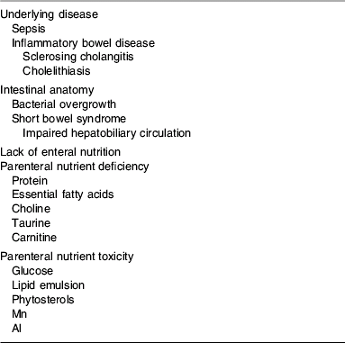
The aetiology of hepatic dysfunction in both adults and children receiving parenteral nutrition is complex and multifactorial. An understanding of the mechanisms behind the biochemical and histological changes observed in patients receiving parenteral nutrition is further complicated by differences in the patient populations studied and in the parenteral nutrition formulations used. In general, possible aetiological factors can be divided into those that are patient dependent, which include primary diagnosis and underlying disease state, presence of sepsis, intestinal anatomy and lack of enteral intake, and those resulting from either nutrient deficiencies or nutrient toxicities in the parenteral formulations employed (Table 1).
Table 1. Factors implicated in the aetiology of parenteral nutrition-associated liver disease

Underlying disease
Abnormalities of LFT may relate to underlying liver disease in patients receiving parenteral nutrition rather than the effects of the parenteral nutrition itself, although the effects of the latter may exacerbate the former. It is also important to be aware that treatment of the underlying disease may influence liver function as a result of drug hepatotoxicity. Patients with inflammatory bowel disease may have occult sclerosing cholangitis with histological evidence of steatosis or pericholangitis in the presence of normal LFT(Reference Nightingale16). Patients who have short-bowel syndrome as a result of mesenteric infarction may also have underlying insulin resistance, dyslipidaemia and non-alcoholic fatty liver disease. It has been shown that in adults receiving long-term parenteral nutrition the presence of pre-existing liver disease is associated with a threefold increased risk of chronic cholestasis(Reference Cavicchi, Beau, Crenn, Degott and Messing10).
Sepsis
As mentioned earlier, the presence of sepsis is an important precipitant of cholestasis in neonates and of abnormal LFT in adults receiving short-term parenteral nutrition. Bacterial overgrowth is relatively common in both children and adults with intestinal failure as a result of intestinal stasis. It has been proposed that bacterial overgrowth may be causative in the development of liver dysfunction in patients receiving parenteral nutrition because of bacterial translocation and the effects of bacterial endotoxin and also because of the generation of secondary bile salts such as lithocholic acid as a result of bacterial dehydroxylation of chenodeoxycholic acid(Reference Quigley, Marsh, Shaffer and Markin4). Recent human studies support the concept of bacterial translocation previously only seen in animal models(Reference Reddy, MacFie, Gatt, Macfarlane-Smith, Bitzopoulou and Snelling17), and experiments performed in animals suggest that the effects of bacterial endotoxins on the liver may be mediated by cytokines such as TNFα(Reference Pappo, Bercovier, Berry, Gallilly, Feigin and Freund18). The concept that bacterial overgrowth causes hepatic dysfunction via production of secondary bile acids has less support in human subjects, particularly since potentially-therapeutic exogenous bile salts such as ursodeoxycholic acid (UCDA) can also be metabolised to lithocholic acid(Reference Buchman, Iyer and Fryer19).
Intestinal anatomy
PNALD has been shown to be related to small intestinal length in a number of studies(Reference Luman and Shaffer6, Reference Ito and Shils8, Reference Cavicchi, Beau, Crenn, Degott and Messing10). One study has reported that a small intestinal length of <1 m is associated with abnormal LFT in adults receiving long-term parenteral nutrition(Reference Luman and Shaffer6), while another study has reported an increased risk of chronic cholestasis in adults with a small intestinal length of <0·5 m(Reference Cavicchi, Beau, Crenn, Degott and Messing10). Some authors have postulated that short-bowel syndrome predisposes to liver dysfunction as a result of impairment of enterohepatic bile salt circulation and abnormal bile acid metabolism(Reference Cavicchi, Beau, Crenn, Degott and Messing10). Others have argued that bowel length may simply be a surrogate marker of parenteral energy requirement(Reference Luman and Shaffer6). It is of interest that LFT have been noted to improve after isolated small intestinal transplantation, even in a small number of cases in which there was biopsy-proven hepatic fibrosis(Reference Langrehr, Reilly, Banner, Warty, Lee and Schraut20, Reference Lauro, Zanfi and Ercolani21).
Lack of enteral nutrition
Reduced enteral nutrition is intimately associated with an increased reliance on parenteral nutrition, and so it is very difficult to ascertain whether the increased risk of liver dysfunction seen in individuals with very little oral intake is a result of the lack of enteral nutrition, or the effects of parenteral feeding. Fasting coupled with total parenteral nutrition has been shown to reduce the secretion of a number of gastrointestinal hormones, including gastrin, motilin, pancreatic polypeptide, insulinotropic polypeptide and glucagon(Reference Greenberg, Wolman, Christofides, Bloom and Jeejeebhoy22). This reduction in secretion may reduce intestinal motility, promoting bacterial overgrowth, and may predispose to biliary stasis(Reference Greenberg, Wolman, Christofides, Bloom and Jeejeebhoy22). It has been suggested that total parenteral nutrition induces small intestinal mucosal atrophy, which in turn allows increased bacterial translocation across the gut mucosal barrier. However, although total parenteral nutrition induces marked mucosal atrophy in rodent models, the effect in human subjects is much less dramatic and the results of clinical studies are mixed(Reference Alpers23–Reference Sedman, MacFie, Palmer, Mitchell and Sagar25).
Nutrient deficiency
Individuals with kwashiorkor, resulting from severe protein–energy malnutrition, develop hepatic steatosis because there is insufficient protein for the manufacture of VLDL, which is needed for hepatic TAG export(Reference Cook and Hutt26). However, patients receiving parenteral nutrition should receive adequate amino acids for VLDL synthesis. Similarly, inadequate supply of linoleic acid may result in essential fatty acid deficiency, which is also associated with steatosis(Reference Richardson and Sgoutas27). Since the majority of parenteral lipid infusions are manufactured using soyabean oil, which is rich in linoleic acid, essential fatty acid deficiency is rare, unless fat-free parenteral regimens are employed in individuals with little or no enteral lipid intake.
There has been recent interest in the possibility that deficiencies of a number of methionine metabolites, such as carnitine, choline and taurine, may be responsible for both steatosis and cholestasis in patients receiving parenteral nutrition. Orally-ingested methionine can be converted to these metabolites via hepatic transulfuration pathways, although these pathways are underdeveloped in premature infants(Reference Vina, Vento, Garcia-Sala, Puertes, Gasco, Sastre, Asensi and Pallardo28). However, methionine administered parenterally to the systemic circulation rather than to the portal circulation is also transaminated to mercaptans, hence reducing the synthesis of carnitine, choline and taurine(Reference Chawla, Berry, Kutner and Rudman29). Carnitine, choline and taurine are not routinely administered as part of parenteral formulations and there is evidence that levels are low in individuals receiving parenteral nutrition(Reference Bowyer, Fleming, Ilstrup, Nelson, Reek and Burnes30–Reference Vinton, Laidlaw, Ament and Kopple34).
Carnitine is involved in the transport of long-chain fatty acids across the mitochondrial membrane so that they can undergo oxidation. In deficiency states in which carnitine levels are very low (<10% normal levels) hepatic steatosis can develop(Reference Karpati, Carpenter, Engel, Watters, Allen, Rothman, Klassen and Mamer35). Although plasma carnitine levels are low in patients receiving parenteral nutrition, they are about 50% normal levels and so considerably higher than levels in patients with congenital or acquired deficiency(Reference Bowyer, Fleming, Ilstrup, Nelson, Reek and Burnes30, Reference Moukarzel, Dahlstrom, Buchman and Ament31, Reference Karpati, Carpenter, Engel, Watters, Allen, Rothman, Klassen and Mamer35). There is limited evidence to suggest an inverse relationship between carnitine levels and ALP in patients receiving parenteral nutrition(Reference Berner, Larchian, Lowry, Nicroa, Brennan and Shike36). However, intervention studies have failed to show any benefit of parenteral carnitine supplementation in patients receiving long-term parenteral nutrition in relation to hepatic abnormalities(Reference Bowyer, Miles, Haymond and Fleming37).
Choline, like carnitine, is normally synthesised from methionine. Levels are low in >90% of patients receiving parenteral nutrition(Reference Buchman, Moukarzel, Jenden, Roch, Rice and Ament38) as a result of the abnormal metabolism described earlier and the lack of choline in standard parenteral formulations. Choline is required for the synthesis of VLDL and hence hepatic TAG export. Choline deficiency results in impaired hepatic TAG secretion and subsequent steatosis(Reference Buchman, Moukarzel, Jenden, Roch, Rice and Ament38). Choline deficiency in patients receiving parenteral nutrition has been shown to correlate with elevated transaminase levels and steatosis in both adults and children receiving parenteral nutrition(Reference Buchman, Moukarzel, Jenden, Roch, Rice and Ament38, Reference Buchman, Sohel and Moukarzel39). Small studies have shown that both parenteral choline supplementation and high-dose oral supplementation can reverse these abnormalities(Reference Buchman, Dubin, Jenden, Moukarzel, Roch, Rice, Gornbein, Ament and Eckhert40, Reference Buchman, Ament, Sohel, Dubin, Jenden, Roch, Pownall, Farley, Awal and Ahn41).
Taurine is important for bile salt conjugation, particularly in preterm infants. It promotes bile flow and attenuates the cholestatic effects of secondary bile salts such as lithocholate(Reference Belli, Roy, Fournier, Tuchweber, Giguere and Yousef42, Reference Dorvil, Yousef, Tuchweber and Roy43). Taurine deficiency in neonates is associated with cholestatic liver disease(Reference Cooper, Betts, Pereira and Ziegler44) and there is some evidence that this condition can be prevented by parenteral taurine supplementation(Reference Heird, Dell, Helms, Greene, Ament, Karna and Storm45, Reference Spencer, Yu and Tracy46). Studies in adults have shown that both plasma and biliary taurine levels are low(Reference Schneider, Joly, Gehrardt, Badran, Myara, Thuillier, Coudray-Lucas, Cynober, Trivin and Messing47), probably as a result of a combination of reduced synthesis via hepatic transulfuration pathways coupled with an increased loss of bile acids associated with ileo-colonic resection. Supplementation in adults with short-bowel syndrome has been shown to improve plasma but not biliary taurine levels, this change being accompanied by an improvement in plasma transaminase concentrations(Reference Schneider, Joly, Gehrardt, Badran, Myara, Thuillier, Coudray-Lucas, Cynober, Trivin and Messing47).
It has been proposed that individuals receiving parenteral nutrition may be deficient in antioxidants, in particular vitamin E and Se(Reference Nightingale16). Increased oxidative stress might predispose to lipid peroxidation of hepatic lipid stores resulting in inflammation and steatohepatitis(Reference Buchman, Iyer and Fryer19). Some experiments using animal models suggest that antioxidants such as glutathione may attenuate the hepatic dysfunction induced by parenteral feeding(Reference Hong, Wu and Cai48). However, although markers of oxidative stress are raised in adults receiving parenteral nutrition, there is little evidence that oxidative damage is increased despite infusion of soyabean-based lipid infusions rich in PUFA(Reference Schepens, Roelofs, Peters and Wanten49). Studies in children suggest that the occurrence of parenteral nutrition-associated cholestasis is independent of oxidant load(Reference Lavoie, Chessex, Gauthier, Levy, Alvarez, St-Louis and Rouleau50). In addition, there is little evidence that antioxidant levels are depleted in patients receiving parenteral nutrition, and vitamin E is commonly added to intravenous lipid emulsions(Reference Buchman, Iyer and Fryer19).
Nutrient toxicity
Early parenteral nutrition formulations contained large amounts of energy supplied as glucose, and the total glucose content often exceeded the maximum glucose oxidation rate. It is likely that this factor partly explains the high incidence of steatosis seen in these early studies(Reference Lindor, Fleming, Abrams and Hirschkorn2). Glucose infusion at rates of >5 mg/body weight (BW) per min have been demonstrated to result in steatosis in human subjects(Reference Burke, Wolfe, Mullany, Mathews and Bier51). High glucose infusion rates result in high plasma glucose concentrations, which in turn stimulate insulin release. High plasma insulin concentrations stimulate hepatic lipogenesis and the production of acylglycerol from glucose while concomitantly inhibiting mitochondrial fatty acid oxidation(Reference Li, Nussbaum, Teague, Gapen, Dayal and Fischer52). This process results in a build up of TAG within the hepatocytes. The adverse effects of insulin hypersecretion may also explain why continuous parenteral nutrition infusion is associated with a greater extent of hepatic dysfunction than cyclic infusion. Allowing ≥8 h each day without parenteral glucose infusion has been shown to lower insulin levels and improve LFT(Reference Hwang, Lue and Chen53). It was initially thought that the failure of the liver to secrete the excess TAG into the circulation was the result of inadequate amino acid content relative to carbohydrate content limiting adequate lipoprotein production(Reference Quigley, Marsh, Shaffer and Markin4). However, steatosis has been shown to result from excessive glucose infusion even if amino acid intake is adequate(Reference Guglielmi, Boggio-Bertinet and Federico54). Interestingly, in animal models infusion of glucagon along with parenteral glucose has been shown to stimulate hepatic fatty acid release and attenuate steatosis(Reference Li, Nussbaum, McFadden, Gapen, Dayal and Fischer55).
The replacement of a proportion of glucose energy with parenteral lipid has been shown to reduce the incidence of steatosis(Reference Meguid, Akahoshi, Jeffers, Hayashi and Hammond56). However, excess lipid has also been demonstrated to result in hepatic complications. Very high parenteral lipid intakes of >4 g/kg BW per d may result in a fat-overload syndrome because of the inability of the reticulo-endothelial system to clear such large amounts of PUFA and phospholipid(Reference Bigorgne, Le Tourneau, Messing, Rio, Giraud, Molina, Audouin and Diebold57). An association between more-modest parenteral lipid intake and the incidence of both chronic cholestasis and advanced liver disease has also been demonstrated in adults(Reference Cavicchi, Beau, Crenn, Degott and Messing10). In multivariate analysis parenteral intake of soyabean-based lipid emulsion of >1 g/kg BW per d has been shown to be associated with a relative risk of chronic cholestasis of 2·3 and a relative risk of advanced liver disease (fibrosis or cirrhosis on liver biopsy) of 5·5(Reference Cavicchi, Beau, Crenn, Degott and Messing10). The exact mechanism behind this effect is unclear. It has been postulated that lipid overloading of hepatic macrophages might impair phospholipid excretion into bile and cause intrahepatic cholestasis(Reference Cavicchi, Beau, Crenn, Degott and Messing10). Other authors have suggested that lipid infusion may inhibit hepatic TAG release leading to accumulation in the liver(Reference Luman and Shaffer6), or may cause accumulation of phytosterols(Reference Clayton, Whitfield and Iyer58). High plasma phytosterol concentrations have been documented in children but not in adults receiving parenteral lipid emulsions(Reference Clayton, Bowron, Mills, Massoud, Casteels and Milla59). There has been some suggestion that lipid infusions with a lower soyabean oil content may cause fewer hepatic complications, with studies showing a lower incidence of cholestasis in patients receiving lipid emulsions in which a proportion of the soyabean oil has been replaced with either medium-chain TAG(Reference Carpentier, Richelle, Haumont and Deckelbaum60) or MUFA(Reference Reimund, Arondel, Joly, Messing, Duclos and Baumann61). There are also case reports of parenteral fish oil supplementation improving hepatic function in children(Reference Gura, Duggan, Collier, Jennings, Folkman, Bistrian and Puder62), although similar studies in adults have not been published.
A number of other components of parenteral infusions have been suggested to cause abnormal LFT in patients receiving parenteral nutrition. High amino acid content has been proposed to promote cholestasis in neonates(Reference Vileisis, Inwood and Hunt63), although as mentioned earlier there may also be a relative deficiency of certain amino acids in these individuals. Historically, tryptophan degraded in parenteral solutions containing NaHSO4 to produce hepatotoxic metabolites(Reference Nightingale16). However, NaHSO4 is no longer used as a preservative in parenteral solutions. Similarly, Al has been shown to cause cholestasis in animal models but is not found in modern parenteral formulations in substantial quantities(Reference Klein64). Studies in children have shown an association between plasma Mn levels and cholestasis(Reference Fell, Reynolds, Meadows, Khan, Long, Quaghebeur, Taylor and Milla65). While it could be argued that since Mn is excreted via the biliary system this increase is simply a consequence of cholestasis rather than a cause, withdrawal of parenteral Mn improves hepatic function in children with high plasma levels(Reference Fell, Reynolds, Meadows, Khan, Long, Quaghebeur, Taylor and Milla65). Finally, Cu is also excreted via the biliary route and can accumulate in the liver if there is substantial cholestasis, which may exacerbate hepatic dysfunction(Reference Blaszyk, Wild, Oliveira, Kelly and Burgart66).
Management of parenteral nutrition-associated liver disease
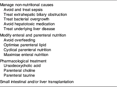
Strategies used in the management of PNALD are summarised in Table 2.
Table 2. Management of parenteral nutrition-associated liver disease

Treatment of non-nutritional causes
Given the importance of sepsis as a causative factor for PNALD, it is vital that every effort is made to reduce its incidence. Intra-abdominal sepsis should be treated using a procedure that is clinically appropriate, usually with a combination of antibiotics and/or minimally-invasive drainage procedures. It is also important to avoid central venous catheter-associated sepsis. To this end, studies in both adult and paediatric populations have demonstrated reduced rates of central venous catheter-associated sepsis in patients managed by specialist nutrition teams using strict aseptic techniques(Reference Kennedy and Nightingale67, Reference Beath, Booth, Murphy, Buckels, Mayer, McKiernan and Kelly68). Efforts to reduce bacterial overgrowth have been more successful in neonates than in adults, suggesting a greater pathophysiological role in the former. Several small studies have suggested that antibiotics such as metronidazole and gentamicin may reduce the incidence of cholestasis in neonates receiving parenteral nutrition(Reference Spurr, Grylack and Mehta69, Reference Kubota, Okada, Imura, Kawahara, Nezu, Kamata and Takagi70). Although a single study has suggested a benefit of metronidazole in adults, this study involved patients with active Crohn's disease and the reported effects of the antibiotic on ALP and γ-glutamyl transferase may well have been mediated via a beneficial effect on the activity of the Crohn's disease(Reference Capron, Gineston, Herve and Braillon71). Other approaches that might possibly reduce bacterial translocation, such as increased dietary fibre intake and parenteral glutamine supplementation, have not to date been shown to have a beneficial effect on PNALD in human subjects.
Cholelithiasis is common in both adults and children receiving parenteral nutrition because of a combination of factors including reduced oral intake, ileal resection, weight loss and drug treatment(Reference Quigley, Marsh, Shaffer and Markin4, Reference Nightingale16). The incidence of biliary sludge in adults receiving parenteral nutrition has been estimated to approach 100% after >6 weeks of treatment(Reference Messing, Bories, Kunstlinger and Bernier72) and gallstones have been demonstrated to form in 45% of patients with short-bowel syndrome(Reference Nightingale, Lennard-Jones, Gertner, Wood and Bartram73). Although often asymptomatic, gallstones may cause cholecystitis or extrahepatic biliary obstruction with resultant abnormalities of LFT. Cholelithiasis in patients receiving parenteral nutrition can be prevented by encouraging enteral feeding and administration of cholecystokinin but may necessitate prophylactic cholecystectomy(Reference Nightingale16).
Adjustments to parenteral nutrition
It is important not to overfeed patients receiving parenteral nutrition, given the deleterious effects of infusion of excess parenteral glucose and lipid described earlier. Both US and UK guidelines suggest a total daily energy intake of 105–146 kJ (25–35 kcal)/kg BW and a daily protein intake of 0·8–1·5 g/kg BW(Reference Buchman, Scolapio and Fryer74, 75), although more accurate assessment of BMR and energy requirements can be made by performing indirect calorimetry. Given that the majority of patients receiving long-term parenteral nutrition will continue to eat, it is also necessary to estimate enteral energy and protein absorption and reduce parenteral provision accordingly. In general, it is important to try to maximise enteral nutrition, although this approach may not always be possible. The oral route is preferable, although enteral-tube feeding can be employed and is often useful in infants. As well as countering the adverse effects of fasting described earlier and reducing parenteral requirements, enteral feeding promotes intestinal adaptation, which is of particular importance in infants for whom it may allow complete weaning from parenteral nutrition(Reference Wallis, Lloyd and Gabe76).
The optimal dose and type of parenteral lipid that should be provided to minimise hepatic dysfunction remains unclear. In patients who have no enteral intake some parenteral lipid is needed to prevent essential fatty acid deficiency(Reference Buchman, Scolapio and Fryer74). A linoleic acid intake of 2–4% total energy intake is adequate for this purpose(77) and, given the very high linoleic acid content of soyabean-based lipid emulsions, this level of intake is easily achievable. US guidelines suggest that parenteral lipid infusion should supply 20–30% total energy and daily intake should be <2·5 g/kg BW and ideally <1·5 g/kg BW(Reference Buchman, Scolapio and Fryer74). However, experimental data suggest that the intake of soyabean-based lipid should be <1·0 g/kg BW per d(Reference Cavicchi, Beau, Crenn, Degott and Messing10). As previously discussed there is growing evidence that lipid emulsions containing a mixture of long-chain and medium-chain TAG(Reference Chan, McCowen and Bistrian78) (e.g. Lipofundin MCT/LCT®; B Braun, Melsungen, Germany), emulsions with a high MUFA content(Reference Reimund, Arondel, Joly, Messing, Duclos and Baumann61, Reference Vahedi, Atlan, Joly, Le Brun, Evard, Perennec, Roux-Haguenau, Bereziat and Messing79) (e.g. Clinoleic®; Baxter, Maurepas, France) and emulsions containing fish oils(Reference Gura, Duggan, Collier, Jennings, Folkman, Bistrian and Puder62) (e.g. SMOF Lipid®; Fresenius Kabi, Bad Homburg, Germany) may be preferable to soyabean-based emulsions in relation to hepatic complications, but the latter remain in widespread use. Further clinical trials are necessary to explore this area further.
Finally, cyclical rather than continuous parenteral nutrition should be administered to minimise the adverse effects of prolonged insulin hypersecretion. Allowing an 8 h break from parenteral nutrition has been shown to improve LFT and reduce insulin levels(Reference Hwang, Lue and Chen53). Cyclical parenteral nutrition also allows greater patient freedom, which may be associated with an improved quality of life in patients receiving long-term parenteral nutrition(75).
Pharmacological treatment
UDCA is an exogenous bile acid used in the management of a number of cholestatic conditions, including primary biliary cirrhosis and cholelithiasis, with the aim of promoting biliary flow. There have been several small studies in neonates with parenteral nutrition-associated cholestasis that have shown a beneficial effect of orally-administered UDCA in doses of 10–30 mg/kg BW per d(Reference Chen, Tsao, Chen, Chou, Hsieh and Chang80–Reference Spagnuolo, Iorio, Vegnente and Guarino83). In this patient group UDCA has been shown to improve LFT and reduce the duration of cholestasis(Reference Chen, Tsao, Chen, Chou, Hsieh and Chang80–Reference Spagnuolo, Iorio, Vegnente and Guarino83). This outcome is achieved despite a very short residual intestine in some neonates. However, some studies have shown rebound cholestasis on withdrawal of UDCA in children in whom enteral nutrition had not been established(Reference De Marco, Sordino, Bruzzese, Di Caro, Mambretti, Tramontano, Colombo, Simoni and Guarino81). Evidence of a benefit of UDCA in adults is more limited, with a single study showing that treatment with an average of 11·2 mg oral UDCA/kg BW per d is associated with a reduction in γ-glutamyl transferase and alanine aminotransferase levels but not ALP, aspartate aminotransferase or bilirubin levels(Reference Beau, Labat-Labourdette, Ingrand and Beauchant84). UDCA may also worsen diarrhoea in some individuals(Reference Howard and Ashley85).
As mentioned earlier, there is evidence that levels of the S-containing amino acids choline and taurine are reduced in patients receiving parenteral nutrition(Reference Buchman32, Reference Cooper, Betts, Pereira and Ziegler44, Reference Schneider, Joly, Gehrardt, Badran, Myara, Thuillier, Coudray-Lucas, Cynober, Trivin and Messing47, Reference Buchman, Dubin, Moukarzel, Jenden, Roch, Rice, Gornbein and Ament86, Reference Misra, Ahn, Ament, Choi, Jenden, Roch and Buchman87). Taurine is especially low in neonates and is now often added to neonatal parenteral nutrition formulations, but not to those of adults. Multivariate analysis of data collected as part of a larger multi-centre trial investigating the management of cholestasis in neonates receiving parenteral nutrition has suggested that parenteral taurine reduces the incidence of cholestasis in severely-premature infants and those with necrotizing enterocolitis(Reference Spencer, Yu and Tracy46). There is considerably less evidence of a benefit of taurine supplementation in adults, with a reduction in aspartate aminotransferase levels being reported in a single small study of adults receiving long-term parenteral nutrition(Reference Schneider, Joly, Gehrardt, Badran, Myara, Thuillier, Coudray-Lucas, Cynober, Trivin and Messing47). Similarly, small-scale trials of choline supplementation in adults have demonstrated that both parenteral choline and high-dose oral lecithin can increase plasma choline concentrations(Reference Buchman, Dubin, Jenden, Moukarzel, Roch, Rice, Gornbein, Ament and Eckhert40, Reference Buchman, Ament, Sohel, Dubin, Jenden, Roch, Pownall, Farley, Awal and Ahn41, Reference Buchman, Dubin, Moukarzel, Jenden, Roch, Rice, Gornbein and Ament86). This outcome has been shown to be associated with a reduction in hepatic steatosis, assessed by computerised tomography scanning, and improvements in plasma ALP, alanine aminotransferase and aspartate aminotransferase concentrations(Reference Buchman, Ament, Sohel, Dubin, Jenden, Roch, Pownall, Farley, Awal and Ahn41). Further larger trials investigating the possible beneficial effects of both choline and taurine are ongoing.
Small intestine and liver transplantation
Impending or overt liver failure associated with PNALD is recognised as an indication for small intestinal transplantation(Reference Buchman, Scolapio and Fryer74), and PNALD is one of the commonest reasons for performing intestinal transplantation(Reference Grant, Abu-Elmagd, Reyes, Tzakis, Langnas, Fishbein, Goulet and Farmer88). The definition of impending and overt liver failure includes patients with ‘elevated serum bilirubin and/or liver enzymes, splenomegaly, thrombocytopenia, gastroesophageal varices, coagulopathy, stomal bleeding or hepatic fibrosis or cirrhosis’(Reference Buchman, Scolapio and Fryer74). This definition is very broad and would include patients ranging from those with relatively-mild LFT abnormalities to those with ESLD. UK transplant units have suggested consideration of transplantation only if there is portal hypertension, cirrhosis or bridging fibrosis(Reference Middleton and Jamieson89). Survival after intestinal transplantation is not as good as after other solid organ transplants(Reference Grant, Abu-Elmagd, Reyes, Tzakis, Langnas, Fishbein, Goulet and Farmer88). Survival is worst for individuals undergoing combined liver and small intestine transplantation, with a 1-year survival of only 60%; 1-year survival after transplantation of small intestine alone is better at 77%(Reference Grant, Abu-Elmagd, Reyes, Tzakis, Langnas, Fishbein, Goulet and Farmer88). There is evidence to suggest that for individuals who do not have irreversible PNALD small intestinal transplantation alone may be a viable option because of recovery of liver dysfunction after transplantation(Reference Lauro, Zanfi and Ercolani21). However, in adults with ESLD combined liver and small intestine transplantation will be required. In neonates and infants, for whom there is the possibility of an extent of intestinal adaptation and weaning from parenteral nutrition, transplantation of liver alone, which has a considerably better outcome than combined small intestine and liver transplantation, may be possible as a bridging measure until enteral nutrition is established.
Conclusions
Liver dysfunction is common in individuals receiving parenteral nutrition and particularly in neonates and infants. Abnormalities of LFT in patients receiving short-term parenteral nutrition are usually transient, but in individuals receiving long-term parenteral nutrition substantial liver damage and ultimately ESLD may occur. The aetiology of PNALD is complex, with a large number of patient-related and nutrition-related factors implicated. Effort should be made to prevent liver dysfunction by managing sepsis, avoiding parenteral overfeeding, employing cyclical parenteral feeding and encouraging enteral nutrition where possible. Intake of soyabean-based parenteral lipid emulsions should be reduced in individuals with established PNALD, but the role of more novel lipid emulsions containing medium-chain TAG, MUFA and fish oil remains unclear. Similarly, evidence supporting the widespread use of parenteral choline and taurine supplementation in the prevention or treatment of PNALD remains limited, although there are more data to support the use of oral antibiotics to treat bacterial overgrowth and oral UDCA in neonates. Ultimately, severe PNALD may necessitate referral for small intestine and/or liver transplantation.


