Hostname: page-component-848d4c4894-75dct Total loading time: 0 Render date: 2024-06-08T03:47:52.690Z Has data issue: false hasContentIssue false

Critical review evaluating the pig as a model for human nutritional physiology

Published online by Cambridge University Press:  13 May 2016

Eugeni Roura*
Affiliation:
The University of Queensland, Centre for Nutrition and Food Sciences, Queensland Alliance for Agriculture and Food Innovation, St. Lucia, QLD 4072, Australia
Sietse-Jan Koopmans
Affiliation:
Wageningen University and Research Centre, Animal Sciences Group, 6708 WD Wageningen, The Netherlands
Jean-Paul Lallès
Affiliation:
INRA, UR1341 ADNC, 35590 St Gilles, France
Isabelle Le Huerou-Luron
Affiliation:
INRA, UR1341 ADNC, 35590 St Gilles, France
Nadia de Jager
Affiliation:
The University of Queensland, Centre for Nutrition and Food Sciences, Queensland Alliance for Agriculture and Food Innovation, St. Lucia, QLD 4072, Australia
Teun Schuurman
Affiliation:
Wageningen University and Research Centre, Animal Sciences Group, 6708 WD Wageningen, The Netherlands
David Val-Laillet
Affiliation:
INRA, UR1341 ADNC, 35590 St Gilles, France
*
*Corresponding author: Eugeni Roura, email e.roura@uq.edu.au
Rights & Permissions [Opens in a new window]

Abstract

The present review examines the pig as a model for physiological studies in human subjects related to nutrient sensing, appetite regulation, gut barrier function, intestinal microbiota and nutritional neuroscience. The nutrient-sensing mechanisms regarding acids (sour), carbohydrates (sweet), glutamic acid (umami) and fatty acids are conserved between humans and pigs. In contrast, pigs show limited perception of high-intensity sweeteners and NaCl and sense a wider array of amino acids than humans. Differences on bitter taste may reflect the adaptation to ecosystems. In relation to appetite regulation, plasma concentrations of cholecystokinin and glucagon-like peptide-1 are similar in pigs and humans, while peptide YY in pigs is ten to twenty times higher and ghrelin two to five times lower than in humans. Pigs are an excellent model for human studies for vagal nerve function related to the hormonal regulation of food intake. Similarly, the study of gut barrier functions reveals conserved defence mechanisms between the two species particularly in functional permeability. However, human data are scant for some of the defence systems and nutritional programming. The pig model has been valuable for studying the changes in human microbiota following nutritional interventions. In particular, the use of human flora-associated pigs is a useful model for infants, but the long-term stability of the implanted human microbiota in pigs remains to be investigated. The similarity of the pig and human brain anatomy and development is paradigmatic. Brain explorations and therapies described in pig, when compared with available human data, highlight their value in nutritional neuroscience, particularly regarding functional neuroimaging techniques.

Type
Review Article
Copyright
Copyright © The Authors 2016 

Introduction

Rodents, mainly mice, have become the model for studies related to human physiology and genetics. However, the differences between rodents and humans in dietary habits, digestive strategies including coprophagy, nutrient requirements and nutrient–nutrient interactions suggest the pig may be a closer match than rodents for studying physiological functions relating to these research areas( Reference Baker 1 Reference Roura, Humphrey and Klasing 4 ). The suitability of the pig as a model for human research is becoming accepted, particularly related to physiological factors and biomedical applications( Reference Gandarillas and Bas 2 , Reference Spurlock and Gabler 5 Reference Bendixen, Danielsen and Larsen 7 ). Both humans and pigs are classified as omnivorous mammals and share similarities related to anatomical features of the gastrointestinal tract (GIT), such as the size of the compartments and the predominance of the colon rather than the caecum as the main fermentation site of plant/fibrous dietary components( Reference Gandarillas and Bas 2 , Reference Stephen, Correa-Matos and Donovan 8 ). Pigs have been used to study several areas relevant to human nutritional sciences( Reference Baker 1 , Reference Gandarillas and Bas 2 , Reference Labib, Erb and Kraus 9 Reference Nielsen, Hartvigsen and Hedemann 11 ), including metabolic syndromes, obesity, bariatric surgery( Reference Gandarillas and Bas 2 , Reference Spurlock and Gabler 5 ), neuroscience, brain imaging( Reference Clouard, Meunier-Salaun and Val-Laillet 3 , Reference Sauleau, Lapouble and Val-Laillet 12 ), food allergies( Reference McClain and Bannon 13 ), alcohol intake( Reference Casani, Segales and Vilahur 14 , Reference Kubotsu, Hu and Carnahan 15 ) and mother–offspring interactions( Reference Guilloteau, Zabielski and Hammon 16 ). In addition, the sequencing of the pig genome has widened support for using the pig as a model for humans by revealing the similarities and differences between pigs and humans at the gene and chromosomal levels( Reference Lunney 17 ).

The present review provides a critical evaluation of the pig as a model for studies related to humans in areas that encompass nutritional chemosensing and hormonal profiles relevant to feed/food intake, the gut barrier function, the host–microbiota interactions and the relationship between nutrition and brain development and metabolism. In addition, the present paper assesses, as quantitatively as possible, how appropriate it would be to use the pig as a model for humans for each of the areas discussed.

Nutritional chemosensing

The chemosensory system has evolved to allow animals to discriminate between foods in their environment, and is believed to reflect species-specific dietary needs( Reference Li and Zhang 18 ). For example, humans have developed trichromatic vision, which was a key event relevant to food recognition and may have contributed to decrease the reliance on olfaction( Reference Gilad, Przeworski and Lancet 19 ). In contrast, pigs appear to have the largest olfactory gene repertoire of any animal studied, with 1,301 potential olfactory receptor genes( Reference Groenen, Archibald and Uenishi 20 ). The potential for pigs to become a model for human research in olfaction is relevant in the case of maternal transfer of dietary cues( Reference Oostindjer, Bolhuis and van den Brand 21 Reference Langendijk, Bolhuis and Laurenssen 24 ).

Nutritional chemosensing is the science studying the perception of nutrients in biological systems which relates molecular mechanisms to changes in genomic, metabolic, physiological and behavioural parameters. Dietary nutrients are perceived in the oral cavity through the taste system in both pigs and humans( Reference Bachmanov and Beauchamp 25 , Reference Wellendorph, Johansen and Bräuner-Osborne 26 ).

Taste perception and the nutrient chemosensory system

Five basic tastes are accepted in humans: sweet, umami, salty, sour and bitter. In addition, fat perception and other nutrient sensing have been related to taste( Reference Bachmanov and Beauchamp 25 ). Sapid compounds solubilised in saliva are presented to the taste buds in the tongue papillae( Reference Matsuo 27 ). Taste perception involves a large set of taste receptors (referred hereinafter as TR for the receptors and TASR for the corresponding genes), which are expressed in the sensory cells of the taste buds and account for recognition of the tastants triggering a depolarisation of the cell and the subsequent signalling to the gustatory cortex of the brain through dedicated neuronal fibres( Reference Barretto, Gillis-Smith and Chandrashekar 28 ). Salty and sour sensing has been related to ligand-gated, transmembrane-channels: the epithelial Na channels (involving three genes ENaCα, β, γ) and the hydrogen-gated channels (involving two genes PKD1L3 and PKD2L1), respectively( Reference Bachmanov and Beauchamp 25 ). All other taste and nutrient receptors known to date are G-protein-coupled receptors (GPCR), including the family 1 (T1R), family 2 (T2R) and several other receptors related to amino acid (AA) and fatty acid (FA) sensing. In humans, the three genes of the taste receptor type 1 (TAS1R) subfamily have been related to umami and sweet tastes. They sense a limited range of l-AA (TAS1R1 and TAS1R3) and sugars (TAS1R2 and TAS1R3). The taste receptor type 2 (TAS2R) subfamily has evolved to identify potentially toxic compounds and elicits the bitter taste. In addition to the Tas1R family, AA and peptones are sensed in the oral cavity by five other GPCR receptors (mGluR1, mGluR4, GPRC6A, CaSR and GPR92). Medium- and long-chained FA are sensed by another set of five GPCR (GPR40, GPR41, GPR43, GPR84 and GPR120). Detailed reviews of the TR/TASR, including the standard nomenclature, are given for humans and rodents by Bachmanov & Beauchamp( Reference Bachmanov and Beauchamp 25 ), Wellendorph et al. ( Reference Wellendorph, Johansen and Bräuner-Osborne 26 ) and Foster et al. ( Reference Foster, Blank and See Hoe 29 , Reference Foster, Roura and Thomas 30 ).

The pig nutrient-sensing repertoire is less known but several in silico comparisons of taste receptors have been published in the last 3 years, after the appearance of the second annotation of the pig genome( Reference Li and Zhang 18 , Reference Groenen, Archibald and Uenishi 20 , Reference da Silva, de Jager and Burgos-Paz 31 Reference Herrero-Medrano, Megens and Groenen 33 ). Single gene sequencing in pigs has characterised the porcine TAS1R3( Reference Kiuchi, Yamada and Kiyokawa 34 ). The gene sequences for the porcine umami dimer, TAS1R1/TAS1R3, and the glutamate receptor, mGluR1, have also been published( Reference Roura, Humphrey and Klasing 4 , Reference Humphrey, Tedó and Klasing 35 ). More recently a study including allelic variation and oral expression of the porcine TAS2R family indicated the potential role of the bitter receptors in environmental adaptation( Reference da Silva, de Jager and Burgos-Paz 31 ).

Evidence for expression of TR in the GIT outside the oral cavity has emerged from both pig and human genome studies( Reference Wellendorph, Johansen and Bräuner-Osborne 26 , Reference Foster, Roura and Thomas 30 ). For example, expression of the Tas1r3 gene has been identified in the tongue, heart, lung, stomach, intestine, liver, kidney and testis( Reference Kiuchi, Yamada and Kiyokawa 34 ). Other nutrient receptors have been found along the GIT including T1R in the small intestine and stomach( Reference Moran, Al-Rammahi and Arora 36 Reference Zhang, Yin and Shu 38 ); AA and peptone receptors, GPRC6A, GPR92 and CaSR, in gastric mucosa( Reference Haid, Jordan-Biegger and Widmayer 39 ); and FA sensors, GPR40, 43 and 120, and seven TAS2R, in five different tissues of the GIT( Reference Colombo, Trevisi and Gandolfi 40 ).

Comparisons between pigs and humans in nutritional chemosensing

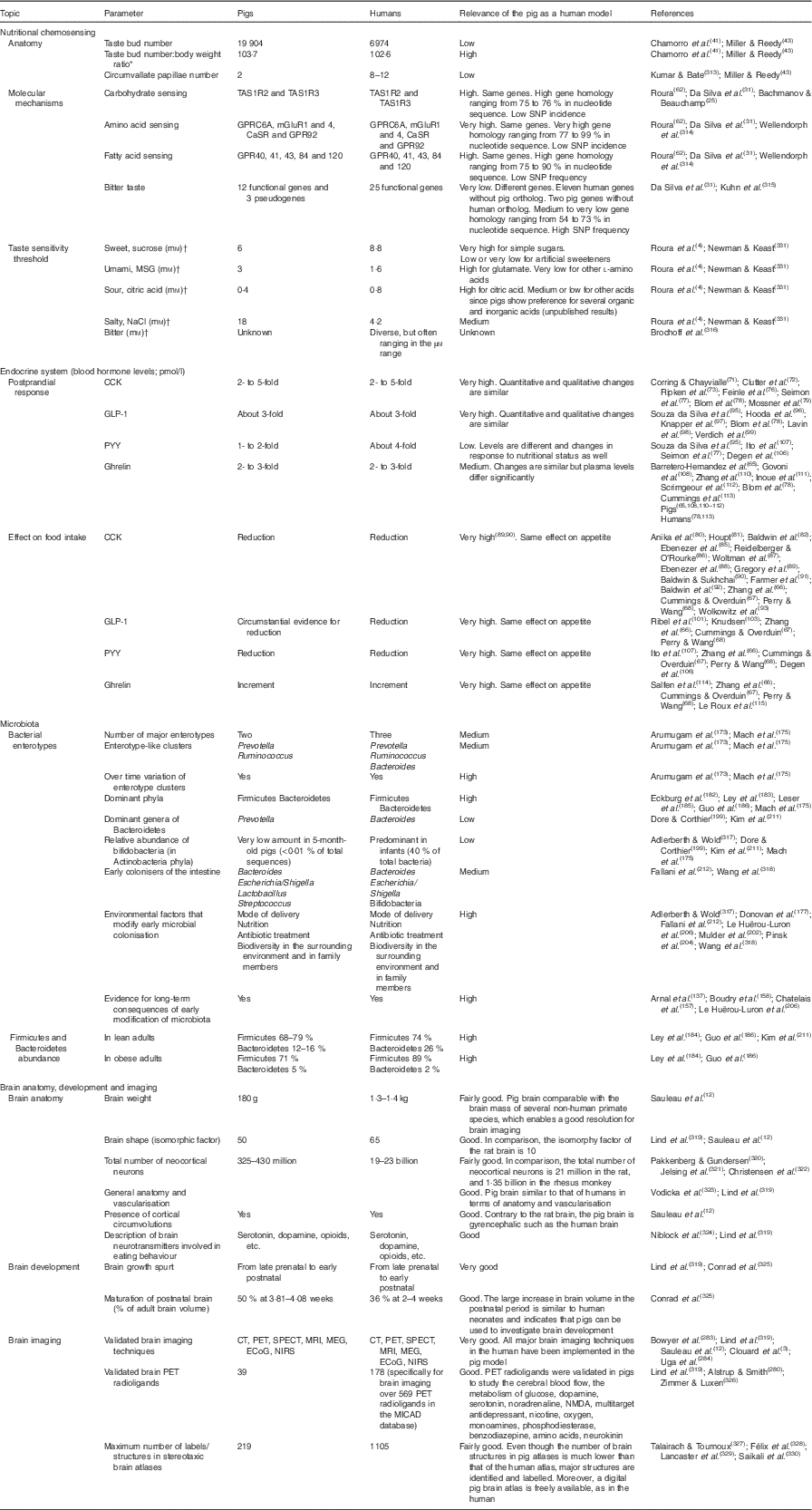
Pigs have a high number of taste buds (n 19 904) relative to other animal species. The number recorded for humans (n 6074) is about 30 % that for pigs. Most of these taste buds are on the tongue surface as part of the fungiform papillae or on the circumvallate papillae in pigs and humans( Reference Chamorro, de Paz and Fernandez 41 , Reference Roura, Humphrey and Tedo 42 ). Pigs have a larger number of fungiform papillae than humans and only two circumvallate papillae compared with eight to twelve in humans( Reference Miller and Reedy 43 ). However, the ratio of taste buds to adult body weight is similar for pigs and humans, as it is across many animal species( Reference Roura, Baldwin and Klasing 44 ).

Sensitivity of pigs compared with humans for the basic hedonic human tastes (sweet, umami, salty, sour and bitter) and homology between taste gene sequences are shown in Tables 1 and 2, respectively. Molecular mechanisms and taste sensitivity are generally similar between the two species for sweet, umami, sour and FA tastes. However, similarities are less for salty and low for bitter tastes.

Table 1 Efficacy of the pig model for humans in nutritional chemosensing; endocrine system; microbiota; and brain anatomy, development and imaging

TAS1R, taste receptor type 1; TAS2R, taste receptor type 2; MSG, monosodium glutamate; CCK, cholecystokinin; GLP-1, glucagon-like peptide-1; PYY, peptide YY; CT, computed tomography; PET, positron emission tomography; SPECT, single photon emission computed tomography; MEG, magnetoencephalography; ECoG, electrocorticography; NIRS, near-IR spectroscopy; NMDA, N-methyl-d-aspartate.

* The adult body weights for a pig and for a human used in the calculation were 192 and 68kg, respectively.

The preference tests in pigs consisted of double-choice tests including two solutions as described in Roura et al.( Reference Roura, Humphrey and Klasing 4 ). The threshold values for pigs refer to the lowest concentration of the taste active compound tested which resulted in significant (P<0·05) preference over water compared with 50 % (neutral value) (adapted from Roura et al. ( Reference Roura, Humphrey and Klasing 4 )). The threshold values for humans refer to the lowest concentration tested following the ascending forced choice triangle test method (Newman & Keast( Reference Newman and Keast 331 )), which resulted in significant (P<0·05) detection (E Roura, unpublished results).

Table 2 Studies on taste receptor and nutrient sensor genes in Sus scrofa compared with Homo sapiens Footnote *

TAS1R, taste receptor type 1; TAS2R, taste receptor type 2; IMP, inosine monophosphate; GMP, guanosine monophosphate; GLP-1, glucagon-like peptide-1.

* The homology percentage for pigs compared with humans was calculated between the amino acid sequences and between the nucleotide sequences for each gene.

Porcine genes that have been identified as pseudogenes according to Da Silva et al. ( Reference da Silva, de Jager and Burgos-Paz 31 ).

The porcine gene TAS2R134 has no known human homolog (Da Silva et al. ( Reference da Silva, de Jager and Burgos-Paz 31 )).

In relation to sweet tastes, the first studies in pigs from the 1950s were on appetite for sucrose (SUC)( Reference Lewis, Catron and Combs 45 , Reference Salmon-Legagneur and Fevrier 46 ). Kennedy & Baldwin( Reference Kennedy and Baldwin 47 ) also measured preferences by pigs for several sweet compounds and found similar recognition threshold concentrations in the diet for SUC and sodium saccharin (SAC) of about 5 to 10mm. The taste recognition threshold was higher for glucose (GLU) at between 10 and 30mm. Humans show a similar sensitivity to pigs for simple sugars and for two high-intensity sweeteners, sucralose and rebaudioside A( Reference Glaser, Wanner and Tinti 48 , Reference Roura, Shrestha and Diffey 49 ). However, pigs do not respond to aspartame, neohesperidin and thaumatin( Reference Glaser, Wanner and Tinti 48 , Reference Roura, Shrestha and Diffey 49 ), which are recognised as sweet by humans. SAC solutions above 100mm were rejected by pigs( Reference Lewis, Catron and Combs 45 ), suggesting an unpleasant sensation at high concentrations. This response is similar for humans, where high SAC concentrations elicit bitterness( Reference Galindo-Cuspinera, Winnig and Bufe 50 ).

Pigs have a preference for free l-AA including glutamic, aspartic, alanine, glutamine and lysine which have been related to the porcine umami taste receptor( Reference Roura, Humphrey and Klasing 4 ). In addition, preferences for serine, threonine, asparagine and hydroxyproline have also been reported in pigs( Reference Tinti, Glaser and Wanner 51 ). However, pig preferences for AA or sugars seem to increase with dietary deficiencies( Reference Guzmán-Pino, Sola-Oriol and Figueroa 52 , Reference Guzmán-Pino, Solà-Oriol and Figueroa 53 ). In contrast to the AA listed above, pigs have been shown to avoid branched-chained AA and tryptophan, presumably related to the onset of bitter taste( Reference Tinti, Glaser and Wanner 51 , Reference Tedo, Roura and Reina 54 , Reference Danilova, Hellekant and Jean-Marie 55 ). Compounds known to be bitter to humans such as pharmaceuticals, including antibiotics, caffeine, quinine HCl, among others, have also been shown to trigger avoidance in pigs( Reference Danilova, Roberts and Hellekant 56 , Reference Nelson and Sanregret 57 ).

Humans appear to be less sensitive to umami tastants than pigs. Umami taste in humans is stimulated by only two l-AA (glutamic and aspartic), whereas pigs recognise a wider array of l-AA( Reference Tinti, Glaser and Wanner 51 , Reference Tedo, Roura and Reina 54 ). The strong preference for non-glutamic AA in pigs is an important differential feature compared with humans. However, the umami receptor in laboratory rodents responds to an even wider array of l-AA compared with pigs( Reference Roura, Humphrey and Klasing 4 ).

Umami tastants in pigs, for example, monosodium glutamate, either alone or in combination with nucleotides, and some sweet tastants, stimulate nerve fibres identified with umami sensing( Reference Danilova, Roberts and Hellekant 56 ). Umami and sweet compounds are both perceived by dimeric taste receptors (TR) sharing one common receptor T1R3( Reference Li, Staszewski and Xu 58 ).

Characterisation of the umami dimer, TAS1R1/TAS1R3, and the glutamate receptor, mGluR1, revealed higher homologies of these porcine genes with the human orthologs compared with laboratory rodents( Reference Roura, Humphrey and Klasing 4 , Reference Humphrey, Tedó and Klasing 35 ). Studies of divergences and similarities in gene structure and expression between pigs and humans for the whole TASR repertoire showed high homology for the TAS1R subfamily coding for carbohydrates, AA and FA, but low homology for the TAS2R subfamily coding for bitter tastes( Reference da Silva, de Jager and Burgos-Paz 31 ). Recent publications indicate the pig has sixteen and the human twenty-five TAS2R genes( Reference Bachmanov and Beauchamp 25 , Reference da Silva, de Jager and Burgos-Paz 31 ). It may be speculated that the similarity in TAS1R gene structure between pigs and humans is consistent with the theory of parallel evolution, where both species are omnivorous and consumed diets with similar nutrient profiles. The lower number of TAS2R genes in pigs may indicate a higher resilience of pigs to bitter dietary compounds( Reference Groenen, Archibald and Uenishi 20 ). It has been suggested that the TAS2R receptors unique to humans tend to have a narrow specificity and may not have food-related significance particularly in light of their extra-oral expression( Reference Meyerhof, Batram and Kuhn 59 ). Da Silva et al. ( Reference da Silva, de Jager and Burgos-Paz 31 ) also reported a high incidence of non-synonymous polymorphisms in the porcine TAS2R repertoire when comparing fourteen different pig breeds across the globe. These findings are consistent with the view that bitter taste gene diversity is an adaptation within animal species( Reference Li and Zhang 18 , Reference Kosiol, Vinar and da Fonseca 60 ), including humans( Reference Campbell, Ranciaro and Zinshteyn 61 ), to their specific ecosystems. The porcine TAS2R is not a good model for the study of bitter agents in humans. However, the global widespread nature of pig breeds following human expansion may represent a unique model to study the role of taste in the adaptation to specific geo-ecosystems.

One limitation for using the pig as a model for humans to study taste sensitivity is a difference in methodology used between the two species. Threshold nutrient concentrations are determined in pigs using preference tests, often compared with water, whereas recognition thresholds are used in human studies. Compared with a recognition threshold, a preference threshold may require higher doses. Results from these two types of comparisons suggest that pigs are more sensitive to citric acid and SUC, but less sensitive to NaCl than humans( Reference Roura, Humphrey and Klasing 4 , Reference Roura 62 ). In addition, pigs and humans show strong discrepancies in the responses to most high-intensity sweeteners tested to date( Reference Glaser, Wanner and Tinti 48 , Reference Roura, Shrestha and Diffey 49 ).

Conclusion on using the pig as a model for human nutritional chemosensing studies

The chemosensing anatomy appears to be similar for pigs and humans with a similar ratio of taste buds in the mouth to mature body weight and the location of sensors throughout the GIT and other organs. The two species also show similarity in studies related to tasting simple sugars (sweet), glutamate (umami) and citric acid (sour). In contrast to humans, pigs do not have the same ability to taste some high-intensity sweeteners or non-glutamate AA and appear less sensitive to NaCl (salt). Overall, pigs are an excellent model for humans, when based on carbohydrate-, AA- and FA-sensing mechanisms. In contrast, the bitter sensory system of pigs and humans is diverse and characterised by species-specific features evolving from the adaptation to different ecosystems. Studying gene expression in human tissues can be difficult, particularly when using well-controlled nutrition intervention studies. The similarity in nutrient receptors and taste sensitivities between pigs and humans creates the opportunity to use the pig as a model in place of rodents for behavioural studies into chemosensing.

Endocrine regulation of food intake: gut–nutrient sensing and gut–brain communication

Sensory cells expressing TR in the GIT are part of the enteroendocrine system. Signalling of appetite or satiety in the gut can be both GIT hormone and nutrient specific( Reference Maljaars, Symersky and Kee 63 ). The release of GIT hormones is activated by fasting or food intake via intestinal receptors that respond to mechanical and/or chemical stimulation( Reference Ritter 64 ) including TR( Reference Barretero-Hernandez, Galyean and Vizcarra 65 ). Over forty GIT hormones have been identified with specific bioactivity( Reference Zhang, Ning and Handelsman 66 ) of which eight GIT hormones (cholecystokinin (CCK), glucagon-like peptide-1 (GLP-1), oxyntomodulin, peptide YY (PYY), apoA-IV, gastrin-releasing peptide and neuromedin B, gastric leptin and ghrelin) have been implicated in the regulation of food intake in mammals( Reference Cummings and Overduin 67 ). This section focuses on the following four hormones specifically released by the stomach and/or small and large intestine with published results in humans and pigs: CCK, GLP-1, PYY and ghrelin. The importance of these four GIT hormones in appetite regulation has been reviewed by Perry & Wang( Reference Perry and Wang 68 ). In the context of nutrient-induced release of satiety hormones it has been reported by Geraedts et al. ( Reference Geraedts, Troost and Tinnemans 69 ) that the release of CCK and GLP-1 in response to dietary proteins differs substantially between humans and rats, underlining the need for alternative, more human-like, animal models to study food intake regulation.

Once GIT hormones are released, they may exert their action via a neural or an endocrine route. The neural route involves binding of GIT hormones to local GIT receptors and subsequent signal transduction via afferent fibres of the abdominal vagal nerve to the brain. The endocrine route involves systemic transport of GIT hormones to the brain and by binding to brain receptors present in the area postrema, a brain structure serving as an interface between blood and brain, or by crossing the blood–brain barrier (BBB) and subsequent binding to receptors in specific brain regions. For proper translational research from pig to human, it is important that the kinetics of GIT hormones in blood follow a similar profile. For instance, the kinetics of circulating GLP-1 are similar in pigs and humans, but different in rats( Reference Larsen, Fledelius and Knudsen 70 ). This difference between the species is probably caused by the rapid inactivation of GLP-1 by circulating protease dipeptidyl peptidase-4 (DPP-4), which is more active in rats than in pigs and humans.

Similarities and differences of nutrient-induced release of satiety hormones and endocrine gut–brain communication in pigs and humans

Cholecystokinin

Depending on the amount and composition of the diet, CCK is secreted by the I cells in the duodenal and jejunal mucosa and also by neurons in the enteric nervous system. CCK contributes to satiation and satiety effects. The major effects of CCK are stimulation of gall bladder contraction, delay of gastric emptying and stimulation of pancreatic secretion( Reference Maljaars, Symersky and Kee 63 ). In pigs, CCK release is stimulated by a mixed meal from fasting to postprandial (30–120min) concentrations going from about 10pmol/l to about 30pmol/l, respectively, with a faster response when diets are enriched with starch than with fat( Reference Corring and Chayvialle 71 ). Clutter et al. ( Reference Clutter, Jiang and McCann 72 ) measured plasma CCK-8 in pigs, at fasting about 2pmol/l increasing to 10pmol/l at mixed meal feeding whereas Ripken et al. ( Reference Ripken, van der Wielen and van der Meulen 73 ) reported concentrations of 0·5pmol/l and 2pmol/l for fasting and postprandial, respectively. All experiments showed a 2- to 5-fold increase in plasma CCK-8 concentrations after mixed meal feeding. Another study in pigs( Reference Jakob, Mosenthin and Zabielski 74 ), using intraduodenal fat infusion, showed plasma CCK concentrations of approximately 5pmol/l both prior and following fat infusion. CCK release was higher to long-chain FA than to medium-chain TAG. In humans, CCK release is mostly stimulated by the presence of dietary protein, whereas carbohydrate provides a weaker stimulus( Reference Liddle, Goldfine and Rosen 75 ). To our knowledge, the effect of dietary protein on plasma CCK secretion has not been investigated to date in pigs. Plasma CCK concentrations are similar in pigs and humans, on average for humans at fasting 2pmol/l and at feeding 10pmol/l( Reference Feinle, Grundy and Otto 76 Reference Mossner, Grumann and Zeeh 79 ). In both species the magnitude of the CCK response is dependent on the composition of the meal. The effects of single macronutrients or combinations thereof on CCK release have not been investigated systematically in humans or pigs. The pig is a valid model to investigate these relationships.

Anika et al. ( Reference Anika, Houpt and Houpt 80 ) administered CCK intravenously at a rate of 0·2nmol/kg per min for total doses of 0·4, 0·8, 1·7 and 3·4nmol/kg to pigs. The lowest dose of CCK reduced food intake by 13 % and the highest dose by 94 %. Houpt( Reference Houpt 81 ) showed that intravenous (iv) CCK-8 (the synthetic and bioactive part of the CCK molecule) infusion to pigs of 14, 28 and 56 pmol/kg per min decreased meal size in a dose-dependent manner. A dose of 28pmol/kg per min reduced food intake by 30–40 %. Similar results were found by Baldwin et al. ( Reference Baldwin, Cooper and Parrott 82 ) who studied hungry pigs working for food, thirsty pigs responding for water, and non-deprived pigs working for SUC. In this study CCK produced significant dose-related decreases in response rates for all three conditions.

Nutrients exert much of their effects through the CCK-1 receptor( Reference Holzer, Turkelson and Solomon 83 ). There are two regions where CCK acts to produce satiety: the nucleus tractus solitarius in the hindbrain and the medial-basal hypothalamus( Reference Reidelberger, Hernandez and Fritzsch 84 ). CCK acts both at CCK-1 receptors beyond the BBB and by a CCK-1 receptor-mediated mechanism involving abdominal vagal nerves to inhibit food intake( Reference Reidelberger, Hernandez and Fritzsch 84 ). CCK-1 antagonists, such as devazepide (DVZ), are commonly used in pig appetite and satiety studies( Reference Ebenezer, Vellucci and Parrott 85 ). DVZ easily passes the BBB following iv administration inducing a dose-related increase in food intake at doses ranging from 17·5 to 140µg/kg with maximum increases occurring at about 70µg/kg( Reference Reidelberger and O’Rourke 86 Reference Ebenezer, de la Riva and Baldwin 88 ). Arterial injection of DVZ (0·1mg/kg) in pigs( Reference Gregory, McFadyen and Rayner 89 ) abolished the inhibition of food intake to duodenal infusion of emulsified fat and monoacylglycerols. However, it did not alter the inhibition of intake in response to oleic acid, to glycerol or to GLU, suggesting that monoacylglycerol-induced CCK secretion is mainly responsible for the satiety resulting from duodenal fat infusion in the pig. Baldwin & Sukhchai( Reference Baldwin and Sukhchai 90 ) reported that intracerebroventricular (icv) injection of DVZ (100µg) before the administration of 1µg CCK abolished the inhibition of GLU intake produced by CCK in pigs. However, DVZ itself had no effect on GLU intake. In a study by Farmer et al. ( Reference Farmer, Roy and Rushen 91 ), pigs were fasted for 24h, injected intravenously with DVZ at 70mg/kg and subsequently subjected to a feed motivation test (operant conditioning). The number of pushes, duration of eating and amount of feed eaten during the feed motivation test were all increased by fasting, and were further increased by DVZ injection, indicating that CCK induces satiation in pigs. An alternative CCK receptor antagonist used in pigs is 2-NAP (2-naphthalenesulfanyl-l-aspartyl-2-(phenethyl) amide), which does not cross the BBB. Baldwin et al. ( Reference Baldwin, de la Riva and Gerskowitch 92 ) revealed that iv administration (20 or 40mg/kg) of NAP injected before iv administration of CCK-8 (1µg/kg) abolished the inhibitory effects of CCK-8 on food intake in hungry pigs. Also, NAP abolished the inhibitory effects of CCK-8 on food intake in hungry pigs after icv injection of both compounds. Overall, these results indicate that endogenous CCK is involved in the regulation of satiety in pigs, in a way which is similar to what is known in humans( Reference Wolkowitz, Gertz and Weingartner 93 ), albeit the pig offers the possibility to conduct invasive mechanistic studies on CCK action.

Glucagon-like peptide-1

GLP-1 is mostly produced by the L cells in the distal small intestine and colon and contributes to satiety. It performs a variety of functions in the body such as inhibiting acid secretion and gastric emptying, while increasing insulin secretion from the pancreas in a GLU-dependent manner( Reference Holst 94 ). The secretion of GLP-1 is mediated by indirect, duodenal activated neurohumoral mechanisms, as well as by direct contact of nutrients with the distal small intestine( Reference Baldwin, Cooper and Parrott 82 ). Souza da Silva et al. ( Reference Souza da Silva, Haenen and Koopmans 95 ) showed that fasting plasma GLP-1 concentrations of 15pmol/l rose to 35pmol/l 1h after the beginning of a complete mash feed. Hooda et al. ( Reference Hooda, Matte and Vasanthan 96 ) showed that fasting plasma GLP-1 concentrations in pigs were approximately 8pmol/l and rose postprandial to 20pmol/l, 1–2h after initiation of the mixed-nutrient meal. The rise of plasma GLP-1 in response to intraduodenal infusion of GLU, fat and GLU–fat in pigs was modest (+5pmol/l) for GLU and intermediate (+10pmol/l) for fat, while the effect of combining GLU and fat was additive (+15pmol/l)( Reference Knapper, Morgan and Fletcher 97 ). To our knowledge, the effect of dietary protein on plasma GLP-1 secretion has not been investigated to date in pigs. In general, the GLP-1 responses to fasting/feeding in pigs are comparable with those in humans, with fasting concentrations of about 13pmol/l rising to 30–40pmol/l postprandial( Reference Blom, Lluch and Stafleu 78 , Reference Lavin, Wittert and Andrews 98 , Reference Verdich, Toubro and Buemann 99 ).

Although GLP-1 can cross the BBB, several studies suggest that peripheral GLP acts to reduce food intake primarily via activation of the vagal afferent nerve( Reference Asmar 100 ). Little information is available on the effect of GLP-1 on food intake in pigs. Ribel et al. ( Reference Ribel, Larsen and Rolin 101 ) showed that administration of a synthetic GLP-1 analogue reduced gastric emptying and therefore may induce satiation in pigs. In general, the pig is recognised as being a good model for studying human conditions( Reference Litten-Brown, Corson and Clarke 102 ), and in the case of GLP-1 analogues, it has found to be predictive of clinical findings in both reduction of hyperglycaemia (through improved insulin secretion and decreased glucagon secretion) and body-weight loss (through reduced gastric emptying and increased satiety)( Reference Knudsen 103 ). The icv administration of GLP-1 inhibited feeding in fasted rats and was counteracted by the icv injection of exendin 9–39, a GLP-1 receptor antagonist( Reference Turton, O’Shea and Gunn 104 ). No data are available in pigs.

Peptide YY

PYY is an anorexigenic hormone. It is a member of the pancreatic polypeptide (PP)-fold family of peptides. It is secreted by the L cells of the distal small-intestinal mucosa. The PYY-containing endocrine cells are found in highest numbers in the lower small intestine and colon. The gastrointestinal functions of PYY include inhibition of gastric acid and pepsin secretion, inhibition of pancreatic exocrine secretion, delay of gastric emptying and inhibition of jejunal and colonic motility( Reference Sheikh, Holst and Orskov 105 ). The secretion of PYY is stimulated by the presence of nutrients in the intestine. In humans, fats provide the strongest stimulus followed by carbohydrates and protein( Reference Degen, Oesch and Casanova 106 ). Little is known on the effects of intestinal nutrient loads on PYY secretion in pigs. Plasma PYY concentrations were higher (500pmol/l) in ad libitum-fed pigs compared with fasted pigs (200pmol/l)( Reference Ito, Thidarmyint and Murata 107 ). On the other hand, Souza da Silva et al. ( Reference Souza da Silva, Haenen and Koopmans 95 ) showed that fasting plasma PYY concentrations were approximately 700–800pmol/l, showing no response to a mixed-nutrient meal. Plasma PYY concentrations are lower in humans, ranging from 30pmol/l at fasting up to 116pmol/l after an intestinal nutrient load( Reference Seimon, Feltrin and Meyer 77 , Reference Degen, Oesch and Casanova 106 ). In the fast–refed condition, both single bolus injection (30mg/kg body weight) and iv infusion (0·25mg/kg per min) of PYY3–36 (the synthetic and bioactive part of the PYY molecule) suppressed feed intake in pigs( Reference Ito, Thidarmyint and Murata 107 ), suggesting that circulating PYY3–36 influences satiety and contributes to the termination of a meal such as in humans( Reference Perry and Wang 68 ).

Ghrelin

Ghrelin is an orexigenic intestinal hormone. In pigs, it is mostly produced in the oxyntic and cardiac gland and less commonly in the pyloric glands of the stomach( Reference Govoni, De Iasio and Cocco 108 ), with a similar pattern to that in humans( Reference Kojima and Kangawa 109 ). Ghrelin functions as a neuropeptide signal that reduces satiety and increases hunger. Govoni et al. ( Reference Govoni, De Iasio and Cocco 108 ) and Zhang et al. ( Reference Zhang, Yin and Li 110 ) showed that fasting increases plasma ghrelin concentrations from 10–25 to 50pmol/l in prepubertal gilts and weanling pigs, respectively. Ghrelin in pigs responded to changes in energy balance and the concentrations increased during fasting from approximately 25 to 75pmol/l( Reference Govoni, De Iasio and Cocco 108 , Reference Inoue, Watanuki and Myint 111 ). Barretero-Hernandez et al. ( Reference Barretero-Hernandez, Galyean and Vizcarra 65 ) reported postprandial plasma ghrelin concentrations of 6pmol/l, increasing to 10pmol/l at fasting in pigs. Scrimgeour et al. ( Reference Scrimgeour, Gresham and Giles 112 ) suggested that the dynamics in plasma ghrelin concentrations in the pig appear to be strongly influenced by prolonged fasting and change from 15pmol/l (feeding) to 100pmol/l (fasting). These ghrelin responses to fasting/feeding are comparable with those observed in humans( Reference Blom, Lluch and Stafleu 78 , Reference Cummings, Purnell and Frayo 113 ). However, the concentrations in humans are higher, being approximately 150pmol/l postprandial and 250pmol/l at fasting.

Salfen et al. ( Reference Salfen, Carroll and Keisler 114 ) showed that there was an increase in body weight of weanling pigs given exogenous ghrelin chronically, suggesting that feeding behaviour was influenced by the treatment. Ghrelin stimulates food intake in humans too. However, in vagotomised patients, ghrelin does not increase food intake, suggesting that an intact vagus nerve is required for exogenous ghrelin to increase appetite in humans( Reference Le Roux, Neary and Halsey 115 ). No pig studies have been conducted addressing the contribution of the vagus nerve to the orexogenic action of ghrelin.

Conclusions on nutrient-induced secretion of satiety hormones and on endocrine gut–brain communication in pigs and humans

Basal or fasting plasma concentrations of CCK and GLP-1 are similar in pigs and humans. The nutrient-induced increase of CCK and GLP-1 concentrations is also similar in both species. Plasma CCK increases postprandial 2- to 5-fold, whereas GLP-1 increases 3-fold in pigs and in humans. Therefore, pigs are a useful model for the investigation of the effects of single (macro) nutrients or combinations of nutrients on CCK- and GLP-1 release. Fasting and postprandial plasma concentrations of PYY in pigs are 10- to 20-fold higher compared with humans. The PYY response of pigs to feeding or intestinal infusion of nutrients (in terms of percentage increase of PYY concentration) seems to be smaller than in humans. For ghrelin the reverse is true; humans show 2- to 5-fold higher plasma concentrations than pigs. The responses of ghrelin to fasting or feeding are comparable in pigs and humans: 2- to 3-fold increases of ghrelin at fasting as compared with fed conditions. The relevance of the pig as a model for human GIT hormone dynamics seems therefore CCK and GLP-1>ghrelin>PYY.

Many pig studies support the human-like action of CCK on food intake regulation, but far fewer studies are available on the actions of GLP-1, PYY and ghrelin. Nevertheless, for GLP-1, PYY and ghrelin, human-like actions on food intake regulation have been reported in pigs( Reference Sternini, Anselmi and Rozengurt 116 ). Therefore, from an endocrine perspective, the pig is a suitable large animal model for the study of the humoral pathways of gut–brain communication as summarised in Table 1.

Gastrointestinal tract permeability and detoxification systems

Current outline of research in nutrition, gut-barrier and defence systems

An important aspect of the GIT function refers to metabolic disorders and obesity, which in humans are partially driven by excessive intake of high-energy diets and may be programmed early in life( Reference Vickers 117 , Reference Portha, Fournier and Kioon 118 ). Unbalanced, high-energy/fat and low-fibre diets may alter GIT permeability, allowing translocation of gut pro-inflammatory microbial-associated molecular patterns (MAMP) such as lipopolysaccharide (LPS) into the body and the development of metabolic inflammation, as demonstrated in mice( Reference Cani and Delzenne 119 ). These MAMP may then promote adipose tissue expansion, insulin resistance, metabolic disturbances and fat deposition.

The GIT is complex, comprising the mucus, the epithelial monolayer and the enteric immune system that includes intestinal epithelial cells( Reference Lallès 120 ). The intestinal epithelium participates in digestion and absorption, while tightly restricting body access of deleterious components. Passage of compounds across the epithelium is mainly regulated by tight paracellular junctions and by macromolecular uptake transcellular mechanisms( Reference Keita and Soderholm 121 ). LPS entry can be transcellular via the chylomicron pathway, following FA absorption, but also paracellular when the transcellular route is altered( Reference Mani, Weber and Baumgard 122 ). These mechanisms appear relevant to stress-related diseases (for example, gut chronic inflammation), the metabolic syndrome and obesity in humans( Reference Miele, Valenza and La Torre 123 Reference Horton, Wright and Smith 125 ).

Besides permeability, the intestinal mucosa is equipped with defence systems including epithelial intestinal alkaline phosphatase (IAP) and inducible heat shock proteins (HSP). IAP is produced by the enterocyte and acts as a major anti-inflammatory enzyme through two mechanisms: detoxification, by dephosphorylating pro-inflammatory MAMP (for example, LPS), and control of local (and systemic) inflammation through a down-regulation of the Toll-like receptor 4-triggered NF-κB activation and of inflammatory cytokine production( Reference Lallès 126 ). Intestinal epithelial cells are chronically exposed to a harsh environment and toxic substances and they have developed inducible HSP (HSP27, HSP70) as anti-inflammatory and antioxidant cytoprotection mechanisms( Reference Arnal and Lallès 127 ). Inducible HSP are involved in intracellular protein trafficking, with many functional implications, including protection against potentially invasive compounds and organisms( Reference Arnal and Lallès 127 ). Inducible HSP are produced in response to diverse microbial components and related metabolites (for example, LPS, butyrate) in vitro ( Reference Lallès 120 ). However, in vivo data are scarce.

Alterations in the function of the intestinal barrier and defence systems may lead to chronic inflammatory diseases( Reference Pastorelli, De Salvo and Mercado 128 ). Conversely, dietary approaches aimed at reducing intestinal permeability and/or stimulating IAP and inducible HSP may contribute to prevent or treat such diseases.

This section summarises comparisons between the pig and the human for intestinal physiology of permeability and its neuroimmune regulation, detoxification and defence systems, their dietary modulation and their early programming.

Similarities between pigs and humans

The pathophysiology, molecular basis and neuroimmune regulation of the intestinal barrier when under stress have been described for rodent models( Reference Keita and Soderholm 121 ). In summary, the mechanism involves hypothalamic corticotropin-releasing factor (CRF), central and peripheral CRF receptors, degranulation of mucosal mast cells and release of various bioactive mediators. Large quantitative inter-species differences exist for intestinal permeability of small, medium-size and large molecules. Notably, pigs are closer to humans than rodents for both trans- and para-cellular permeability values( Reference Nejdfors, Ekelund and Jeppsson 129 , Reference Wallon, Yang and Keita 130 ). Intestinal permeability regulation in pigs also involves enteric nerve activation, CRF, mast cells and released tryptase and TNF-α( Reference Lallès 120 , Reference Smith, Clark and Overman 131 , Reference Overman, Rivier and Moeser 132 ). Data are limited in humans, but they indicate essentially the same regulations( Reference Wallon, Yang and Keita 130 , Reference Vanuytsel, van Wanrooy and Vanheel 133 ).

MAMP detoxification function by IAP is conserved across species( Reference Yang, Wandler and Postlethwait 134 ). IAP is highly expressed along the villous epithelium of the small intestine in pigs( Reference Lackeyram, Yang and Archbold 135 ) and humans( Reference Tuin, Poelstra and de Jager-Krikken 136 ). IAP activity is 10- to 15-fold higher in the distal ileum compared with proximal colon in both species( Reference Tuin, Poelstra and de Jager-Krikken 136 , Reference Arnal, Zhang and Messori 137 ). In pigs, IAP activity is drastically reduced after weaning and may cause post-weaning intestinal alterations( Reference Lackeyram, Yang and Archbold 135 ). Depressed IAP is also suspected in various inflammatory diseases in humans( Reference Lallès 126 ). Administration of exogenous IAP has strong anti-inflammatory effects in both species( Reference Lallès 126 , Reference Tuin, Poelstra and de Jager-Krikken 136 , Reference Beumer, Wulferink and Raaben 138 , Reference Lukas, Drastich and Konecny 139 ). Plasma LPS is a marker of metabolic inflammation in humans( Reference Laugerette, Vors and Peretti 140 ). Intake of saturated fat is consistently reported to increase plasma LPS in humans( Reference Amar, Burcelin and Ruidavets 141 ) and pigs( Reference Mani, Hollis and Gabler 142 ), intestinal transport of LPS in pigs( Reference Mani, Hollis and Gabler 142 ) and plasma IAP in humans( Reference Domar, Karpe and Hamsten 143 ).

Differences between pigs and humans, or pig studies with no equivalent in humans

Differences exist between humans, pigs and rodents for IAP gene copies (n 1, 2 and 2, respectively) and their chromosome location( Reference Yang, Wandler and Postlethwait 134 ). IAP distribution along the small intestine is opposite between pigs and rodents, the former having higher IAP activity in the ileum and lower in the duodenum compared with rodents( Reference Lallès 126 , Reference Fan, Adeola and Asem 144 ). The distribution of IAP along the human intestine is yet to be elucidated. Human cell line Caco2 and porcine IPEC-I display inducible HSP (for example, HSP70)( Reference Lindemann, Grohs and Stange 145 , Reference Yi, Hou and Wang 146 ). However, data on intestinal HSP25 or HSP70 in human tissues are lacking. As a prototypic example of dietary modulation of intestinal defence systems, l-glutamine supplementation has been shown to improve the morphological integrity and barrier function of the intestines in humans( Reference Benjamin, Makharia and Ahuja 147 ) and pigs( Reference Ewaschuk, Murdoch and Johnson 148 , Reference Zhong, Zhang and Li 149 ). However, these studies are difficult to compare due to many differences in experimental conditions. Zn is a key element for intestinal and body homeostasis. One in vitro study with human (Caco2) and porcine (IPEC-J2) intestinal epithelial cells revealed cell line differences in permeability and Hsp70 responses to Zn, suggesting inter-species differences( Reference Lodemann, Einspanier and Scharfen 150 ). However, in vivo data in humans are lacking.

The concept of ‘developmental origin of health and disease’, linking early-life malnutrition (deficiency or excess) to metabolic diseases was formulated two decades ago( Reference Hales and Barker 151 ). The effects of nutrition in early life on gene expression and potential long-term effects have become a discipline of high interest in human( Reference Nehring, Kostka and von Kries 152 , Reference Mennella 153 ) and animal( Reference Oostindjer, Bolhuis and van den Brand 21 , Reference Hepper, Wells and Millsopp 154 ) models. However, data on how early-life gene programming may affect intestinal function and defence systems are limited in humans( Reference Lallès 155 , Reference Lallès, Michel and Theodorou 156 ). Various dietary components, including protein, fat, methyl donors and fibre, influence gene expression in the intestines( Reference Lallès 155 , Reference Lallès, Michel and Theodorou 156 ). The pig as a model for humans has a high potential value in this area of research, but comparative studies are limited( Reference Arnal, Zhang and Messori 137 , Reference Chatelais, Jamin and Gras-Le Guen 157 Reference Arnal, Zhang and Erridge 159 ). For example, high-protein milk formula transiently altered ex vivo ileal permeability in piglets and increased responses to LPS challenge in young adults( Reference Chatelais, Jamin and Gras-Le Guen 157 ). Neonatal dietary protein excess also led to long-term alterations in colonic barrier function under oxidant stress in female pigs( Reference Boudry, Jamin and Chatelais 158 ). Finally, alterations in mother-to-offspring transmission of GIT microbiota (for example, using antibiotics) had long-term consequences on IAP and iHSP along the GIT in pigs( Reference Arnal, Zhang and Messori 137 , Reference Arnal, Zhang and Erridge 159 ).

Intra-uterine growth retardation (IUGR) is a risk factor for the metabolic syndrome and obesity, possibly through low-grade inflammation( Reference Lakshmy 160 , Reference Salam, Das and Bhutta 161 ). For example, the prevalence of the metabolic syndrome was found to be 10-fold higher in human subjects weighing less than 3·0kg at birth compared with those with more than 4·3kg of birth weight( Reference Salam, Das and Bhutta 161 ). IUGR in the pig occurs naturally for some pigs in litters( Reference Ferenc, Pietrzak and Godlewski 162 ). IUGR piglets display immature gut, higher HSP70 both in utero and after birth, and altered pro-inflammatory NF-κB signalling pathway( Reference Zhong, Li and Huang 163 Reference D’Inca, Kloareg and Gras-Le Guen 165 ). The IUGR piglets fed high-protein milk formula had higher ileal permeability and altered neuronal regulation of the gut barrier function later in life( Reference Boudry, Morise and Seve 166 ). Finally, the GIT microbiota is an important modulator of gut development( Reference Lallès, Michel and Theodorou 156 , Reference Weng and Walker 167 ).

Conclusion

The available literature suggests that basic mechanisms of intestinal permeability and defence systems are conserved across species, including pigs and humans. Functional permeability studies ex vivo suggest the pig to be close to the human. However, data are scant for intestinal IAP and inducible HSP defence systems in humans, and only indirect evidence suggests some similarities and differences between pigs and humans. Data on pig nutrition and gut health are numerous( Reference Lallès, Bosi and Smidt 168 Reference Lallès and Guillou 170 ), making this species valuable to human nutrition research, due to anthropometric, dietary and GIT anatomical and physiological similarities. Moreover, nutritional programming of intestinal gene expression and long-term effects on growth and health can be assessed in this out-bred species and should be relevant to humans.

Host–microbiota interactions

One of the main aspects in the study of GIT function is the microbial population. Animals are associated with a diverse microbial community, primarily consisting of symbiotic and commensal bacteria. Mammalian bacterial diversity has been related to phylogeny and claimed to be influenced by the host diet, increasing in meat eaters compared with non-meat eaters( Reference Ley, Hamady and Lozupone 171 ). The GIT microbiota of modern humans is that of omnivorous primates. Original studies of the GIT microbiota focused on their role in inflammatory diseases, with the view that bacteria were pathogens only. However, the importance of the microbiota has been revisited in the past decade. It is now widely accepted that the GIT microbiota play a crucial role in maintaining homeostasis. The complex and intimate relationship between GIT microbial communities and its host is becoming clearer, due, in part, to large-scale microbial genome-sequencing programs( Reference Del Chierico, Vernocchi and Bonizzi 172 ). Metagenomic sequencing of total community DNA provides information about both the phylogenetic representation as well as functional genes. Interrogation of metagenomic information has revealed three distinct ‘enterotypes’ in the human microbiota that are identifiable by changes in the population of at least one of the three genera: Bacteroides, Prevotella and Ruminococcus ( Reference Arumugam, Raes and Pelletier 173 ). Enterotypes are not limited to humans, but also occur in mice( Reference Hildebrand, Nguyen and Brinkman 174 ) and pigs( Reference Mach, Berri and Estelle 175 ). Advancement in sequencing total RNA (metatranscriptomics), identifying total proteins (metaproteomics) and total metabolites (metametabolomics) has added further knowledge on the GIT ecosystem complexity both in humans and in pigs( Reference Lamendella, VerBerkmoes and Jansson 176 , Reference Donovan, Wang and Li 177 ). The use of an ecosystems biology approach, in association with ‘omics approaches, will lead to a more complete understanding of the complex interactions between the thousands of bacterial species in the GIT and the host( Reference Erickson, Cantarel and Lamendella 178 ).

The physiological similarity between humans and pigs in GIT development, digestive function, and gastrointestinal fermentation profiles (the colon being the main site of bacterial fermentation in pigs and humans) suggests that the pig is preferred over other non-primate models for digestive and metabolic disease studies relating to humans( Reference Guilloteau, Zabielski and Hammon 16 , Reference Le Bourgot, Ferret-Bernard and Le Normand 179 , Reference Heinritz, Mosenthin and Weiss 180 ). Moreover, the pig is a human-sized omnivorous species. The pig has been extensively used as a model for nutritional studies as its protein and lipid metabolism is comparable with humans( Reference Litten-Brown, Corson and Clarke 102 ). Increased knowledge of the pig microbiota composition and structure along the intestinal compartments being similar to humans further supports the pig as an ideal biomedical model for humans( Reference Mach, Berri and Estelle 175 , Reference Zhao, Wang and Liu 181 ). This section outlines the similarities and differences of the pig and human in host–microbiota interactions.

Similarities between pigs and humans in gut microbiota

Dominant phyla

The largest microbiota of the body is located in the GIT and the set of gene products provides a diverse range of biochemical and metabolic activities to complement host physiology. Hundreds of species are present in the GIT lumen, only belonging to a few microbial phyla. Firmicutes and Bacteroidetes are the two dominant bacterial phyla in the human and mouse gut, with the Proteobacteria, Actinobacteria, Fusobacteria and Verrucomicrobia phyla as subdominant phyla( Reference Ley, Hamady and Lozupone 171 , Reference Eckburg, Bik and Bernstein 182 Reference Ley, Turnbaugh and Klein 184 ). Similarly, the GIT microbiota in pigs, as well as in wild suidae, mainly consists of the Firmicutes and Bacteroidetes phyla( Reference Leser, Amenuvor and Jensen 185 , Reference Guo, Xia and Tang 186 )(Table 3). Recently two different enterotype-like clusters, primarily distinguished by unclassified Ruminococcus and Prevotella, have been identified in pig faeces( Reference Mach, Berri and Estelle 175 ). Their phylogenetic composition was highly similar to two of the enteroptypes described in humans( Reference Arumugam, Raes and Pelletier 173 ). Interestingly, in pigs, as in humans, enterotype-like clustering distribution can vary within an individual over time( Reference Knights, Ward and McKinlay 187 ).

Table 3 Mean values of the amount of total SCFA throughout life in pig and human faeces

* SCFA content in meconium; values adapted from Midtvedt & Midtvedt( Reference Midtvedt and Midtvedt 414 ).

Commercial pigs (Large White × Landrace × Pietrain breed) weaned 28d of age, data expressed per kg of faecal DM (Montagne et al. ( Reference Montagne, Le Floc’h and Arturo-Schaan 415 )).

5d post-weaning.

§ Post-pubescent (6·4 and 7·5 months-old Large White × Landrace × Pietrain breed) and gestating sows (1·5–2 years-old Large White × Landrace breed), data expressed per kg of faecal fresh matter (I Le Huerou-Luron, unpublished results).

Postnatal and early life microbial colonisation

During the first few hours after birth (postnatal), contact with environmental and colonising bacteria is essential for healthy intestinal and immune maturation. The major role of microbiota in the development of the neonatal GIT was confirmed in conditions where colonisation was modified early in life through exposure to micro-organisms of maternal and environmental origins, through nutrients consumed and through antibiotic treatments. During infancy, the composition of the intestinal microbiota is unstable and more variable than in older children and adults. Diet-induced adaptation of the microbiota may vary from the proximal to the distal parts of the intestine. The composition that is typically measured from faecal samples does not reflect the large bacterial diversity along the intestinal tract. Animal models, specifically cannulated pigs, are useful for obtaining a better understanding of the interactions between microbiota present in different niches of the intestine and physiology relevant to humans( Reference Saraoui, Parayre and Guernec 188 ). Studies with germ-free piglets clearly show that bacteria are essential for the growth and development of the digestive tract( Reference Chowdhury, King and Willing 189 ). The comparison of gene expression profiles in enterocytes of germ-free compared with conventional piglets has brought insight on the impact of microbiota on the GIT function( Reference Chowdhury, King and Willing 189 ). Bacterial colonisation induces the maturation and function of several components of the mucosal immune system and defence in order to prevent inflammatory responses that would compromise the barrier function( Reference Chowdhury, King and Willing 189 Reference Hooper 191 ). In humans, neonates compared with older individuals have a decreased innate defence, a low production of IgA and a defective interaction between dendritic cells, T lymphocytes and regulatory T cells( Reference El Aidy, Dinan and Cryan 192 ). Similarly, the mucosal immune system is essentially absent in the neonatal piglet, even though the systemic immune tissue is well developed( Reference Bailey, Haverson and Inman 193 ), and piglets begin to synthetise secretory IgA from the second week of age( Reference Le Huërou-Luron and Ferret-Bernard 194 ). The balance between T lymphocytes helper 1 and helper 2 responses in human and pig neonates is skewed toward the helper 2 profile, resulting in a high susceptibility to intracellular pathogen infection( Reference Le Bourgot, Ferret-Bernard and Apper-Bossard 195 , Reference Adkins, Leclerc and Marshall-Clarke 196 ). The impaired protection of the neonate against infections may be partly attributed to a deficient secretion of interferon( Reference Le Bourgot, Ferret-Bernard and Apper-Bossard 195 , Reference Wilson, Westall and Johnston 197 ).

It only takes a few hours for bacteria to appear in the faeces of mammalian neonates( Reference Thompson, Wang and Holmes 198 , Reference Dore and Corthier 199 ). Facultative anaerobic bacteria, such as Proteobacteria, are the first colonisers. These bacteria reduce oxygen concentration in the GIT and allow strict anaerobes, such as members from the genus Bacteroides and the phyla Actinobacteria and Firmicutes, to colonise the intestine. During the first year of life in humans and the first 6 months in pigs, the intestinal microbiota composition fluctuates widely between individuals and over time, before resembling the adult status (Table 4). The potential use of piglets as a model for human studies is reinforced by the early colonisers, Bacteroides and Escherichia/Shigella being similar in humans. However, the substantial presence of Lactobacillus and Streptococcus in pigs is unparalleled in humans.

Table 4 Comprehensive summary of the existing literature on the relationship between nutrition and brain composition/development in pig models

FA, fatty acids; ARA, arachidonic acid; DPA, docosapentaenoic acid; LA, linoleic acid; ALA, α-linolenic acid; MCT, medium-chain TAG; l-DOPA, l-3,4-dihydroxyphenylalanine; HC, high cholesterol; LC, low cholesterol; ppm, parts per million.

* Swainsonine is a plant toxin.

Quercetin is a dietary polyphenol with potential health benefits.

Early disturbances of the microbial colonisation process, such as induced by high-hygiene environments or by antibiotic treatment, have major consequences for the developmental sequence of the GIT microbiota and for host metabolism( Reference Cox and Blaser 200 ). An advantage of the porcine model is the flexibility to compare different early environmental-rearing conditions, using, for example, outdoor and indoor sow-reared piglets or isolator-reared neonates. Mulder et al. ( Reference Mulder, Schmidt and Stokes 201 ) showed large differences in composition of ileal-adherent microbiota between outdoor and indoor sow-reared animals, which corresponded to major differences in intestinal immune activation. Excessive hygiene appears to interfere with the normal processes of bacterial stabilisation and alters immune development. Mulder et al. ( Reference Mulder, Schmidt and Lewis 202 ) showed that the succession of events that lead to a stable adult microbiota depends on colonisation during the first 2d of life, and also on continuous exposure to highly diverse microbiota during the early development at least up to weaning at 4 weeks of age. The use of antibiotics, in combination with stressors in early life, was shown to affect adult pig microbiota and intestinal gene expression, including genes involved in immune-related processes( Reference Schokker, Zhang and Vastenhouw 203 ). This observation in pigs corroborates human studies indicating that changing the environmental conditions, and in particular microbial exposure, throughout early life affects the development of immune diseases( Reference Pinsk, Lemberg and Grewal 204 ). Whether inducing early change in immune homeostasis by modifying microbiota would lead to different sensitivity of pigs to infectious or inflammatory challenge, such as recently reported with early spray-dried supplementation( Reference Boyer, D’Costa and Edwards 205 ), warrants further investigation.

Many beneficial strategies have been suggested to strengthen the postnatal development of presumably beneficial microbiota and GIT functions, including: changing the composition of maternal food during gestation and lactation; changing the composition of infant formulas; and favouring breast-feeding over formula-feeding during the suckling period( Reference Heinritz, Mosenthin and Weiss 180 , Reference Le Huërou-Luron, Blat and Boudry 206 ). For example, feeding neonatal piglets with formula supplemented with prebiotics increased the bacterial numbers by 5-fold, the content of folic acid by 53 % and growth of the colon( Reference Aufreiter, Kim and O’Connor 207 ). Similarly, supplementation of the sow diet with prebiotics during gestation and lactation was associated with 50 % greater fermentative activity of the caecal microbiota, accelerated development of the intestinal immunity, and improved intestinal protection by increasing ileal Peyer’s patch production of secretory IgA in the offspring by 46 %( Reference Le Bourgot, Ferret-Bernard and Apper-Bossard 195 ). Faecal secretory IgA also increased by 170 % in healthy infants who receive a prebiotic-supplemented formula( Reference Scholtens, Alliet and Raes 208 ). These results in pigs and humans underline the key role of maternal nutrition during pregnancy in supporting neonatal development of the GIT immune system via modulation of microbiota.

The health benefits of breast-feeding have been recognised for a long time. Breast-feeding is associated with earlier colonisation with bifidobacteria, partly in relation to the presence of oligosaccharides in human maternal milk( Reference Donovan, Wang and Li 177 ). As in humans, breast-fed piglets showed lower intestinal growth and permeability compared with high protein formula-fed ones( Reference Boudry, Morise and Seve 166 , Reference Donovan, Wang and Li 177 ). One major issue in human studies on the effect of breast- v. formula-feeding on gut function is the great number of confounding factors which are difficult to circumvent, including quantification of food intake in breast-fed infants, variable length of exclusive breast-feeding, and variability of the composition of milk formulas. Animal models are used to help control these confounding factors, in particular, use of an automatic milk feeder that provides neonatal piglets with artificial milk as similar as possible to maternal milk( Reference Morise, Seve and Mace 209 ). These studies using a formula-fed piglet model provide strong support for the idea that short dietary changes before weaning associated with a modification of the early intestinal bacterial colonisation can have a long-term impact on the severity of inflammatory responses without changing the basal physiology of the intestinal barrier and cytokine profile in the intestine( Reference Chatelais, Jamin and Gras-Le Guen 157 , Reference Boudry, Jamin and Chatelais 158 ). No similar data are available from human studies due to the invasive procedure required for intestine functionality research. However, breast-feeding is clearly associated with lower incidence of necrotising enterocolitis and diarrhoea in both human and pig neonates( Reference Le Huërou-Luron, Blat and Boudry 206 , Reference Siggers, Siggers and Thymann 210 ).

Differences between pigs and humans in gut microbiota

Dominant phyla

Differences in the most abundant genera exist between the human and the pig intestinal microbiota( Reference Heinritz, Mosenthin and Weiss 180 ). Belonging to the Bacteroidetes phylum, the most abundant genus is Bacteroides in humans, averaging 9 to 42 % of total bacteria( Reference Dore and Corthier 199 ), while the most abundant genus is Prevotella in weaned pigs, accounting for more than 20 % of total bacteria( Reference Mach, Berri and Estelle 175 , Reference Kim, Borewicz and White 211 ). In adult humans, the phylum Actinobacteria may represent up to 15 % of total bacteria. It comprises bifidobacteria, the most predominant group detected in infants (40 % in average in faecal samples of 6-week-old European infants)( Reference Fallani, Young and Scott 212 ). The population of bifidobacteria present in the intestine of pigs is considerably lower, with less than 0·1 % of total sequences in the faecal samples of 22-week-old commercial pigs( Reference Kim, Borewicz and White 211 ). Dietary, environmental and behavioural (such as feeding habits) factors contribute to the species-specificity of the composition of microbiota.

Epigenetic mechanisms

The concept that early developmental dietary insults (poor or inadequate pre- or postnatal nutrition, for example) can have long-term consequences on health later in life has been termed developmental programming, or ‘developmental origins of health and disease’. The GIT microbiota appears to have an important role in the GIT programming as its initial composition creates distinct individuality during the lifespan( Reference Weng and Walker 167 , Reference Palmer, Bik and DiGiulio 213 , Reference Jakobsson, Jernberg and Andersson 214 ). A mechanism leading to these long-term effects may be due to epigenetically active fermentation metabolites such as in histone acetylation( Reference Strasak, Bartova and Harnicarova 215 , Reference Mischke and Plosch 216 ). However, no studies on epigenetic modifications underlying long-term effects on early microbial colonisation and gut function are available in pigs. Despite limited studies, use of the pig as a model for humans to assess the effects of early nutrition on the development of microbiota is gaining acceptance amongst the scientific community.

Conclusion

Although similarities exist between humans and pigs in terms of dynamics of postnatal maturation of microbiota diversity and structure, responses to environmental factors, including dietary factors, and phyla composition, differences in the most abundant genera exist. As reviewed by Heinritz et al. ( Reference Heinritz, Mosenthin and Weiss 180 ), understanding the crucial role and complexity of human microbiota could be improved by the use of human flora associations in pigs. This model has been successfully established taking advantage of the higher similarity between pigs and humans compared with the widely studied rodent models. In addition to the similarities in anatomy, physiology and metabolism between the pig and humans, the pig is more similar to humans than rodents with increased Bacteroides spp. and bifidibacteria( Reference Pang, Hua and Yang 217 , Reference Che, Pang and Hua 218 ). The pig has already successfully been used as a model for humans to study nutritional interventions( Reference Shen, Zhang and Wei 219 , Reference Wen, Tin and Wang 220 ). In addition, the human flora-associated pig can be considered as a useful model for human infants. However, stability of the implanted human microbiota in the gut of pigs during the lifespan remains to be investigated in studies regarding developmental programming.

The relationship between nutrition and the brain in the pig

Current outline on research in nutrition and neurosciences in pigs

Nutrient intake is driven by homeostatic and hedonic signals of peripheral, gastrointestinal, endocrinological and metabolic origin that convey in the central nervous system where they are integrated in a cognitive process referred to as the hunger–satiety cycle. Dietary nutrients also have an impact on brain development and function. Neuroscientific studies in pigs have progressed in recent years, partially to address scientific matters that cannot be studied in humans for ethical reasons.

This section summarises the pig studies, mostly in vivo and the minimally invasive neurocognitive explorations that are paralleled in human studies. Second, we will summarise the pig studies, mostly post-mortem explorations on brain tissues, which present differences or no equivalent in humans.

Similarities between pigs and humans in terms of brain functions

Brain responses to food signals

Describing brain responses to food signals is important to investigate food pleasure and motivation, or to decipher the brain networks underlying sensory and nutrient perception. An extensive literature is available on this topic in the human, describing mostly via functional MRI the brain responses to various food signals, according to different internal states (for example, hungry v. sated) or conditions (for example, lean v. obese), and many review papers are available on this topic (for example, Rolls( Reference Rolls 221 ); Stice et al. ( Reference Stice, Spoor and Ng 222 ); Carnell et al. ( Reference Carnell, Gibson and Benson 223 )). Studies using large animal models have not yet completely made use of the opportunities provided by in vivo brain imaging. The very first studies using functional imaging to describe food-induced brain responses in pigs used 99mTc-HMPAO (technetium hexamethylpropyleneamine oxime) SPECT (single photon emission computed tomography) and [18F]fluorodeoxyglucose positron emission tomography to map cerebral blood flow( Reference Boubaker, Val-Laillet and Guerin 224 Reference Gaultier, Meunier-Salaun and Malbert 226 ) and brain GLU metabolism( Reference Clouard, Jouhanneau and Meunier-Salaun 227 ), respectively. These studies addressed the use of the pig to study food conditioning, looking at specific modulations of the response of the brain reward circuit after exposure to flavours with positive or negative hedonic values( Reference Gaultier, Meunier-Salaun and Malbert 226 , Reference Clouard, Jouhanneau and Meunier-Salaun 227 ). These studies provide two major outcomes: (1) functional imaging in anaesthetised pigs can be implemented to explore brain responses to different food signals, as in humans; and (2) the brain circuits activated by the perception of food signals are similar to those described in the human (for example, frontostriatal areas, amygdala and insular cortex). Boubaker et al. ( Reference Boubaker, Val-Laillet and Guerin 224 ) showed in pigs that duodenal and portal GLU infusions led to different systemic and brain responses in areas regulating food intake and pleasure. Clouard et al. ( Reference Clouard, Meunier-Salaün and Meurice 225 ) compared congruent v. dissociated oral and duodenal SUC perception, and found different brain responses in the limbic and reward circuits. Studies in human subjects showed that brain responses to energy-providing sugars and sweeteners are not the same in the reward circuit (for reviews, see Low et al. ( Reference Low, Lacy and Keast 228 ) and Ochoa et al. ( Reference Ochoa, Lallès and Malbert 229 )), which resembles the results obtained by Clouard et al. ( Reference Clouard, Meunier-Salaün and Meurice 225 ) in pigs. These studies are important to understand how the human brain correlates sugar cravings, as well as the neurobehavioural changes that could emerge due to the chronic consumption of, for example; sugars or non-energy sweeteners.

Impact of diet on brain activity, neurotransmission and cognition

Minipigs have become a widely accepted model for studying obesity and the metabolic syndrome( Reference Johansen, Hansen and Richelsen 230 Reference Val-Laillet, Guerin and Malbert 234 ). They can be used to investigate the obesity-induced central modifications in humans, including decreased activity of the prefrontal cortex( Reference Le, Pannacciulli and Chen 235 Reference Volkow, Wang and Telang 237 ) and altered dopaminergic function( Reference Volkow, Wang and Telang 237 , Reference Wang, Volkow and Logan 238 ). Val-Laillet et al. ( Reference Val-Laillet, Layec and Guerin 239 ) demonstrated in the Göttingen minipig that brain alterations similar to those described in obese humans exist in this model and that they are an acquired feature of obesity correlated to weight gain. In Pitman–Moore minipigs, Val-Laillet et al. ( Reference Val-Laillet, Meurice and Lalles 240 ) also described the effects of three high-lipid diets differing in their lipid sources and found that the basal GLU metabolism of the anterior prefrontal cortex and nucleus accumbens was highest with a diet enriched with sunflower-seed oil, intermediary with a diet enriched with lard, and lowest with a diet enriched with fish oil. These results demonstrate that specific dietary nutrients can modify brain metabolism independently from body weight, and that specific nutrients in excess might favour the onset of brain metabolism anomalies.

In humans, cognitive test scores were positively related to breast milk DHA and negatively related to linoleic acid, suggesting that high levels of dietary linoleic acid may impair cognition( Reference Lassek and Gaulin 241 ). Individual consumption of dietary FA had an impact on cognitive measures in children( Reference Lassek and Gaulin 242 ), with n-3 FA being positively related to cognitive test scores in male and female children, while n-6 showed the reverse relationship. Higher scores in tests of neurodevelopment were found in infants fed formula with DHA than in infants fed formulas without DHA( Reference Willatts, Forsyth and DiModugno 243 ). DHA supplementation in young boys increased the prefrontal cortex activation during sustained attention( Reference McNamara, Able and Jandacek 244 ). These results are consistent with the hypothesis that dietary DHA is assimilated by the brain and has a positive influence on cognition. Autopsy data show lower DHA in brain of human infants fed formula rather than those who were breast-fed( Reference Farquharson, Cockburn and Patrick 245 , Reference Makrides, Neumann and Byard 246 ), which resembles the results obtained in pigs (Table 4).

In pigs, dietary FA significantly made an impact on the frontal cortex and striatum concentrations of neurotransmitters (for example, dopamine, serotonin)( Reference de la Presa Owens and Innis 247 , Reference de la Presa Owens and Innis 248 ). Another study showed fewer arm-entries in a maze in pigs receiving a low-PUFA diet compared with a high-PUFA diet, with the effect being probably dependent on central dopamine metabolism( Reference Ng and Innis 249 ). Epidemiological and clinical studies also suggest a relationship between dietary FA and altered functions of the nervous system, including neurocognitive disorders( Reference Grosso, Galvano and Marventano 250 ). Dietary n-3 FA could also protect against brain disorders on neurotransmission, neuroprotection and neurogenesis( Reference Denis, Potier and Heberden 251 ). However, data are lacking for pigs providing an opportunity to investigate the relationship between diet, brain activity and cognitive functions relevant to humans via in vivo imaging.

Peripheral neuromodulation to regulate eating behaviour in pigs

Vagal nerve stimulation (VNS) is a therapy for refractory epilepsy and psychiatric disorders( Reference Hotujac and Kuzman 252 , Reference Vonck, De Herdt and Boon 253 ), but it has also received attention as a way to modulate food intake. Animal models, including pigs, were used to investigate this question( Reference Val-Laillet, Biraben and Randuineau 254 ). Diaz-Guemes et al. ( Reference Diaz-Guemes, Sanchez and Luis 255 ) found that VNS increased central nervous activity, but no effect was observed on feeding behaviour. In contrast, other authors demonstrated a decreased weight gain, decreased fat gain and plasma insulin-like growth factor I( Reference Sobocki, Krolczyk and Herman 256 ), or decreased food intake and specific activations in several brain areas associated with altered gastric myoelectric activity( Reference Matyja, Thor and Sobocki 257 ) in growing pigs( Reference Biraben, Guérin and Bobillier 258 ). Another on-going study( Reference Malbert, Guérin and Bobillier 259 ) showed VNS-induced metabolism differences in the brain reward circuit only 7d after VNS onset, meaning that quick central neuroplasticity phenomena can be induced by VNS, possibly modulating homeostatic and cognitive processes. In Göttingen minipigs fed a Western diet, VNS prevented further weight gain, decreased food intake and sweet cravings( Reference Val-Laillet, Biraben and Randuineau 254 ), proving the therapeutic potential of this strategy. Similarly, some studies assessing the impact of VNS on eating behaviours and weight in individuals with other psychiatric and neurological disorders showed significant modulation of food cravings and body weight( Reference McClelland, Bozhilova and Campbell 260 , Reference Val-Laillet, Aarts and Weber 261 ), but there are significant discrepancies between studies. Overall, functional imaging in pigs has the potential to help validate and optimise therapies before their application to human patients.

Central neuromodulation to regulate eating behaviour in pigs

Recent development suggests that the deep-brain stimulation (DBS), a procedure for the treatment of Parkinson’s or depression, might also be used to combat obesity( Reference Halpern, Wolf and Bale 262 Reference Howland 265 ). The minipig has emerged as an ideal model for basic and preclinical studies on DBS( Reference Sorensen, Nielsen and Rosendal 266 ). Hypothalamic DBS was validated in the Göttingen minipig( Reference Bjarkam, Nielsen and Glud 267 , Reference Ettrup, Tornoe and Sorensen 268 ) and resulted in reduced weight gain( Reference Melega, Lacan and Gorgulho 269 ), as well as in behavioural and physiological changes that could be related to the activation of limbic and autonomic brain networks( Reference Ettrup, Sorensen and Rodell 270 ). These results with pigs are similar to those described in human studies (for reviewes, see McClelland et al. ( Reference McClelland, Bozhilova and Campbell 260 ) and Val-Laillet et al. ( Reference Val-Laillet, Aarts and Weber 261 )). Shon et al. ( Reference Shon, Lee and Goerss 271 ) showed that DBS of the subthalamic nuclei in pigs can stimulate striatal dopamine release, which is related to food motivation and obesity( Reference Volkow, Wang and Baler 272 , Reference Narayanaswami, Thompson and Cassis 273 ), whereas Sauleau et al. ( Reference Sauleau, Lapouble and Val-Laillet 12 ) managed to modify food motivation and learning. Knight et al. ( Reference Knight, Min and Hwang 274 ) demonstrated that DBS of the nucleus accumbens, a putative target to combat obesity( Reference Halpern, Wolf and Bale 262 ), modulated the activity of the prefrontal, cingulate and insular cortices, which are brain regions involved in eating behaviour. A similarly low metabolic activity of the prefrontal cortex was observed in obese humans( Reference Le, Pannacciulli and Chen 235 Reference Volkow, Wang and Telang 237 ) and minipigs( Reference Val-Laillet, Layec and Guerin 239 ), an anomaly that was normalised via DBS of the cortex. The combination of DBS and MRI has been explored in pigs, in terms of image-guided brain navigation( Reference White, Woolley and Bienemann 275 ), network activation( Reference Knight, Min and Hwang 274 , Reference Min, Hwang and Marsh 276 ) and safety( Reference Shrivastava, Abosch and Hanson 277 Reference Gorny, Presti and Goerss 279 ). These studies show the (mini) pig is a convenient model to study the impact of DBS on eating behaviour and nutritional diseases, and to test medical innovations in preclinical trials before being safely applied to humans.

New imaging approaches in pigs

In addition to the aforementioned neuromodulation studies, there are many innovative methods related to nutrition and brain activity used in humans that could potentially be investigated in pigs. Alstrup & Smith( Reference Alstrup and Smith 280 ) reviewed 10 years of positron emission tomography findings on neuromolecular processes in the living porcine brain and listed all the validated brain radio ligands including several molecules of interest for nutrition studies. Other methods can be used for molecular imaging in pigs, such as the wireless instantaneous neurotransmitter concentration system which allows the measure of neurotransmitter release in specific brain areas( Reference Agnesi, Tye and Bledsoe 281 , Reference Van Gompel, Chang and Goerss 282 ). The non-invasive magnetoencephalography and electrocorticography( Reference Bowyer, Okada and Papuashvili 283 ), as well as the functional near-IR spectroscopy and cortical imaging have been successfully implemented in the pig model( Reference Uga, Saito and Sano 284 ) and could be applied in the future to map brain responses to food and nutrient stimulations.

Differences between pigs and humans, or pig studies with no equivalent in humans

The most important corpus of literature investigating the impact of nutrition on the pig brain has been focused on brain composition and development, and especially on the role of dietary FA. The health consequences of dietary deficits or supplements of n-3 and n-6 FA are still controversial areas of human nutrition due to conflicting results. However, animal models such as the pig have the potential to bring new insight through better-controlled experimental designs( Reference Innis 285 , Reference Innis 286 ). Table 4 provides a comprehensive summary of this literature. Most of these results have no equivalent in human studies (other than exceptional post-mortem case studies), because it is not possible to assess, non-invasively, human brain composition or the administration of specific toxicants.

The BBB is important for the regulation of food intake and the blood-to-brain transport of dietary compounds( Reference Banks 287 ). The passage of xenobiotics( Reference Milbury and Kalt 288 ) or bismuth( Reference Pollet, Albouz and Le Saux 289 ) into the brain has been investigated in pigs via post-mortem tissue analyses. However, the emergence of in vitro models of pig BBB( Reference Goti, Balazs and Panzenboeck 290 Reference Patabendige, Skinner and Morgan 293 ) have significant advantages for investigating the transport mechanisms of compounds into the brain. BBB transport of glutamate( Reference Kim, Virella and Braunberg 294 ), alkaloids( Reference Mulac, Huwel and Galla 295 ), mycotoxins( Reference Weidner, Huwel and Ebert 296 ) and central nervous system-active drugs( Reference Campbell, Regina and Kharasch 297 ) was assessed with this model, which might help to understand the neural toxicology of dietary compounds and the effectiveness of medicines. An in vivo imaging study in obese minipigs showed increased BBB permeability with a diet enriched with fish oil characterised by an excessive amount of n-3 FA( Reference Val-Laillet, Meurice and Lalles 240 ). Transport of nutrients and their impact on the BBB integrity should receive more attention in the future to understand how the gut–brain axis is altered by nutritional diseases( Reference Buckman, Thompson and Moreno 298 ), and to provide nutritional recommendations in humans. If the increased BBB permeability induced by high doses of n-3 FA is confirmed in humans, this could also have unexpected beneficial outcomes, for example to improve drug delivery to the brain in Alzheimer’s disease( Reference Banks 299 ) or other neuropsychiatric disorders.

In pigs, central concentration of serotonin can modulate operant food intake behaviour( Reference Ebenezer, Vellucci and Parrott 85 ). In addition, brain DHA, which depends on dietary DHA, promotes central dopamine metabolism( Reference Ng and Innis 249 ). Dietary AA can also affect brain neurotransmitters in the hypothalamus( Reference Adeola and Ball 300 Reference Shen, Voilque and Kim 303 ), suggesting that dietary manipulation of AA precursors of neurotransmitters may offer a practical means of reducing stress responses. Further studies are needed to verify whether these results may be of benefit in human nutrition, for example to help patients with stress disorders. Elmquist et al. ( Reference Elmquist, Ross and Hsu 304 ) suggested that central CCK increases with time in piglets in parallel to the ability to assimilate nutrients from a solid diet. Kenk et al. ( Reference Kenk, Thomas and Lortie 305 ) demonstrated via positron emission tomography imaging of cAMP, a strong region-specific signal in the brain, as well as an impaired cAMP-mediated signalling in obese pigs, giving some insight into pathological progression with potential for directing therapy in humans. Mycotoxin( Reference Prelusky 306 , Reference Swamy, Smith and Karrow 307 ) and feed additives( Reference Poletto, Cheng and Meisel 308 ) were also found to alter behaviour, neurotransmitter activity and metabolism in pigs. The decreased feed intake and increased aggressive behaviour observed in subjects contaminated with mycotoxins might be related to anomalies in brain monoamines, including dopamine and serotonin. Gbore( Reference Gbore 309 ) demonstrated that the acetylcholinesterase activity in the hypophyses, hypothalamus and amygdala decreased with increased mycotoxin concentrations in the diet. These results are important to identify the risk associated with mycotoxin contamination and possible therapeutic interventions in humans.

Pig studies exploring the brain responses to specific diets relying on post-mortem methods have no equivalent human data. Kanitz et al. ( Reference Kanitz, Otten and Tuchscherer 310 ) showed that low protein:carbohydrate dietary ratio during gestation may alter the brain expression of genes encoding key determinants of glucocorticoid activity in the fetus, with potential long-lasting consequences for stress adaptation and health. Also, increased c-Fos (a transcription factor) immunoreactivity in several brain structures was described after oral administration of fungi extracts in pigs( Reference Gaige, Bonnet and Tardivel 311 ). Madsen et al. ( Reference Madsen, Birck and Fredholm 312 ) found that expression of the fat mass and obesity associated gene (FTO) transcript was detected at high levels in brain tissues and that these levels varied through the development and between specific brain areas. These results demonstrate a relationship between the genetic propensity to develop obesity and dietary habits at the cerebral level. Since the FTO gene has recently been associated with increased BMI in several human populations, the pig model might be used to investigate the epigenetic mechanisms that could lead to obesity-related brain anomalies in humans.

Conclusions on the relationship between nutrition and the brain

The general comparison between the brain of pigs and humans (Table 1) shows that, even if there are some differences in terms of size and structure, the overall brain anatomy and development in pigs are similar to those of humans. In addition, similar functional neuroimaging approaches have been transferred from humans to pigs. Understandably, many pig studies do not have any equivalent in humans (especially the invasive and terminal experiments). However, most of the in vivo functional brain explorations and therapies described in pig models are echoed in human studies, which highlight the fantastic potential of pig models for translational research in nutrition and neurosciences. In addition, the pig model is of high value to perform mechanistic, toxicological and epigenetics studies that could not be performed in humans for practical and ethical reasons.

Acknowledgements

The authors would like to thank Dr John L. Black and Dr Anton Pluschke for reviewing the manuscript and for their sound comments.

There are no conflicts of interest.

References

1. Baker, DH (2008) Animal models in nutrition research. J Nutr 138, 391396.Google Scholar
2. Gandarillas, M & Bas, F (2009) The domestic pig (Sus scrofa domestica) as a model for evaluating nutritional and metabolic consequences of bariatric surgery practiced on morbid obese humans. Cienc Investig Agrar 36, 163176.Google Scholar
3. Clouard, C, Meunier-Salaun, MC & Val-Laillet, D (2012) Food preferences and aversions in human health and nutrition: how can pigs help the biomedical research? Animal 6, 118136.Google Scholar
4. Roura, E, Humphrey, B, Klasing, K, et al. (2011) Is the pig a good umami sensing model for humans? A comparative taste receptor study. Flavour Frag J 26, 282285.Google Scholar
5. Spurlock, ME & Gabler, NK (2008) The development of porcine models of obesity and the metabolic syndrome. J Nutr 138, 397402.CrossRefGoogle ScholarPubMed
6. Verma, N, Rettenmeier, AW & Schmitz-Spanke, S (2011) Recent advances in the use of Sus scrofa (pig) as a model system for proteomic studies. Proteomics 11, 776793.Google Scholar
7. Bendixen, E, Danielsen, M, Larsen, K, et al. (2010) Advances in porcine genomics and proteomics – a toolbox for developing the pig as a model organism for molecular biomedical research. Brief Funct Genomics 9, 208219.CrossRefGoogle Scholar
8. Stephen, RM, Correa-Matos, NJ, Donovan, SM, et al. (2004) The effect of fermentable fibers on intestinal function and structure following Salmonella typhimurium infection. Gastroenterology 126, A517A517.Google Scholar
9. Labib, S, Erb, A, Kraus, M, et al. (2004) The pig caecum model: a suitable tool to study the intestinal metabolism of flavonoids. Mol Nutr Food Res 48, 326332.CrossRefGoogle Scholar
10. Mickelson, BD, Greer, FR & Benevenga, NJ (2005) The contribution of body protein to the supply of energy in starved newborn piglets is not preferentially suppressed by intravenous provision of glucose and fat. J Nutr 135, 26092615.Google Scholar
11. Nielsen, KL, Hartvigsen, ML, Hedemann, MS, et al. (2014) Similar metabolic responses in pigs and humans to breads with different contents and compositions of dietary fibers: a metabolomics study. Am J Clin Nutr 99, 941949.Google Scholar
12. Sauleau, P, Lapouble, E, Val-Laillet, D, et al. (2009) The pig model in brain imaging and neurosurgery. Animal 3, 11381151.CrossRefGoogle ScholarPubMed
13. McClain, S & Bannon, GA (2006) Animal models of food allergy: opportunities and barriers. Curr Allergy Asthma Rep 6, 141144.Google Scholar
14. Casani, L, Segales, E, Vilahur, G, et al. (2004) Moderate daily intake of red wine inhibits mural thrombosis and monocyte tissue factor expression in an experimental porcine model. Circulation 110, 460465.Google Scholar
15. Kubotsu, SL, Hu, J, Carnahan, KG, et al. (2003) The effects of chronic ethanol consumption during early pregnancy on conceptus health and uterine function in pigs. Alcohol Clin Exp Res 27, 712719.Google Scholar
16. Guilloteau, P, Zabielski, R, Hammon, HM, et al. (2010) Nutritional programming of gastrointestinal tract development. Is the pig a good model for man? Nutr Res Rev 23, 422.Google Scholar
17. Lunney, JK (2007) Advances in swine biomedical model genomics. Int J Biol Sci 3, 179184.Google Scholar
18. Li, D & Zhang, J (2013) Diet shapes the evolution of the vertebrate bitter taste receptor gene repertoire. Mol Biol Evol 31, 303309.Google Scholar
19. Gilad, Y, Przeworski, M & Lancet, D (2007) Loss of olfactory receptor genes coincides with the acquisition of full trichromatic vision in primates. PLoS Biol 5, e148.Google Scholar
20. Groenen, MA, Archibald, AL, Uenishi, H, et al. (2012) Analyses of pig genomes provide insight into porcine demography and evolution. Nature 491, 393398.Google Scholar
21. Oostindjer, M, Bolhuis, JE, van den Brand, H, et al. (2010) Prenatal flavor exposure affects growth, health and behavior of newly weaned piglets. Physiol Behav 99, 579586.Google Scholar
22. Bolhuis, JE, Oostindjer, M, Van den Brand, H, et al. (2009) Voluntary feed intake in piglets: potential impact of early experience with flavours derived from maternal diet. In Voluntary Feed Intake in Pigs, pp. 3752 [D Torrallardona and E Roura, editors]. Wageningen: Wageningen Academic Publishers.Google Scholar
23. Oostindjer, M, Bolhuis, JE, Simon, K, et al. (2011) Perinatal flavour learning and adaptation to being weaned: all the pig needs is smell. PLoS One 6, e25318.CrossRefGoogle ScholarPubMed
24. Langendijk, P, Bolhuis, JE & Laurenssen, BFA (2007) Effects of pre- and postnatal exposure to garlic and aniseed flavour on pre- and postweaning feed intake in pigs. Livest Sci 108, 284287.CrossRefGoogle Scholar
25. Bachmanov, AA & Beauchamp, GK (2007) Taste receptor genes. Annu Rev Nutr 27, 389414.Google Scholar
26. Wellendorph, P, Johansen, LD & Bräuner-Osborne, H (2010) The emerging role of promiscuous 7TM receptors as chemosensors for food intake. In Vitamins and Hormones, pp. 151184 [L Gerald, editor]. London: Academic Press.Google Scholar
27. Matsuo, R (2000) Role of saliva in the maintenance of taste sensitivity. Crit Rev Oral Biol Med 11, 216229.Google Scholar
28. Barretto, RPJ, Gillis-Smith, S, Chandrashekar, J, et al. (2015) The neural representation of taste quality at the periphery. Nature 517, 373376.Google Scholar
29. Foster, SR, Blank, K, See Hoe, LE, et al. (2014) Bitter taste receptor agonists elicit G-protein-dependent negative inotropy in the murine heart. FASEB J 28, 44974508.CrossRefGoogle ScholarPubMed
30. Foster, SR, Roura, E & Thomas, WG (2014) Extrasensory perception: odorant and taste receptors beyond the nose and mouth. Pharmacol Ther 142, 4161.CrossRefGoogle ScholarPubMed
31. da Silva, EC, de Jager, N, Burgos-Paz, W, et al. (2014) Characterization of the porcine nutrient and taste receptor gene repertoire in domestic and wild populations across the globe. BMC Genomics 15, 1057.Google Scholar
32. Feng, P & Zhao, H (2013) Complex evolutionary history of the vertebrate sweet/umami taste receptor genes. Chin Sci Bull 58, 21982204.Google Scholar
33. Herrero-Medrano, JM, Megens, HJ, Groenen, MA, et al. (2014) Whole-genome sequence analysis reveals differences in population management and selection of European low-input pig breeds. BMC Genomics 15, 601.CrossRefGoogle ScholarPubMed
34. Kiuchi, S, Yamada, T, Kiyokawa, N, et al. (2006) Genomic structure of swine taste receptor family 1 member 3, TAS1R3, and its expression in tissues. Cytogenet Genome Res 115, 5161.Google Scholar
35. Humphrey, B, Tedó, G, Klasing, KC, et al. (2009) Characterization of Porcine Umami Taste Receptors (pT1r1 and pT1r3). 41èmes Journées de la Recherche Porcine; Paris, France, pp. 165–166.Google Scholar
36. Moran, AW, Al-Rammahi, MA, Arora, DK, et al. (2010) Expression of Na+/glucose co-transporter 1 (SGLT1) in the intestine of piglets weaned to different concentrations of dietary carbohydrate. Br J Nutr 104, 647655.Google Scholar
37. Widmayer, P, Breer, H & Hass, N (2011) Candidate chemosensory cells in the porcine stomach. Histochem Cell Biol 136, 3745.Google Scholar
38. Zhang, J, Yin, YL, Shu, XG, et al. (2013) Oral administration of MSG increases expression of glutamate receptors and transporters in the gastrointestinal tract of young piglets. Amino Acids 45, 11691177.Google Scholar
39. Haid, DC, Jordan-Biegger, C, Widmayer, P, et al. (2012) Receptors responsive to protein breakdown products in G-cells and D-cells of mouse, swine and human. Front Physiol 3, 65.Google Scholar
40. Colombo, M, Trevisi, P, Gandolfi, G, et al. (2012) Assessment of the presence of chemosensing receptors based on bitter and fat taste in the gastrointestinal tract of young pig. J Anim Sci 90, Suppl. 4, 128130.Google Scholar
41. Chamorro, CA, de Paz, P, Fernandez, JG, et al. (1993) Fungiform papillae of the pig and the wild boar analyzed by scanning electron microscopy. Scanning Microsc 7, 313322.Google Scholar
42. Roura, E, Humphrey, B, Tedo, G, et al. (2008) Unfolding the codes of short-term feed appetence in farm and companion animals. A comparative oronasal nutrient sensing biology review. Can J Anim Sci 88, 535558.Google Scholar
43. Miller, IJ Jr & Reedy, FE Jr (1990) Variations in human taste bud density and taste intensity perception. Physiol Behav 47, 12131219.CrossRefGoogle ScholarPubMed
44. Roura, E, Baldwin, MW & Klasing, KC (2013) The avian taste system: potential implications in poultry nutrition. Anim Feed Sci Technol 180, 19.CrossRefGoogle Scholar
45. Lewis, CJ, Catron, DV, Combs, GE, et al. (1955) Sugar in pig starters. J Anim Sci 14, 11031115.Google Scholar
46. Salmon-Legagneur, E & Fevrier, R (1956) Feed preferences in young pigs. 2. Sugar in rations for young pigs. Ann Zootech 5, 7379.Google Scholar
47. Kennedy, JM & Baldwin, BA (1972) Taste preferences in pigs for nutritive and non-nutritive sweet solutions. Anim Behav 20, 706718.Google Scholar
48. Glaser, D, Wanner, M, Tinti, JM, et al. (2000) Gustatory responses of pigs to various natural and artificial compounds known to be sweet in man. Food Chem 68, 375385.CrossRefGoogle Scholar
49. Roura, E, Shrestha, B & Diffey, S (2013) Preference thresholds and sensory-motivated intake for four high intensity sweeteners in piglets. In Manipulating Pig Production XIV: Proceedings of the 14th Biennial Conference of the Australasian Pig Science Association. Melbourne, Australia; 24–27 November 2013, p. 44. Werribee, VIC: Australasian Pig Science Association (Inc.).Google Scholar
50. Galindo-Cuspinera, V, Winnig, M, Bufe, B, et al. (2006) A TAS1R receptor-based explanation of sweet ‘water-taste’. Nature 441, 354357.Google Scholar
51. Tinti, JM, Glaser, D, Wanner, M, et al. (2000) Comparison of gustatory responses to amino acids in pigs and in humans. Lebensm Wiss Technol 33, 578583.Google Scholar
52. Guzmán-Pino, SA, Sola-Oriol, D, Figueroa, J, et al. (2012) Dietary energy density affects the preference for protein or carbohydrate solutions and piglet performance after weaning. J Anim Sci 90, Suppl. 4, 7173.Google Scholar
53. Guzmán-Pino, SA, Solà-Oriol, D, Figueroa, J, et al. (2014) Influence of the protein status of piglets on their ability to select and prefer protein sources. Physiol Behav 129, 4349.Google Scholar
54. Tedo, G, Roura, E, Reina, M, et al. (2010) Well-fed piglets prefer amino acids that elicit umami taste. J Anim Sci 88, e.suppl. 2, 211.Google Scholar
55. Danilova, V, Hellekant, G, Jean-Marie, T, et al. (1998) Gustatory responses of the hamster Mesocricetus auratus to various compounds considered sweet by humans. J Neurophysiol 80, 21022112.CrossRefGoogle ScholarPubMed
56. Danilova, V, Roberts, T & Hellekant, G (1999) Responses of single taste fibers and whole chorda tympani and glossopharyngeal nerve in the domestic pig. Sus scrofa. Chem Senses 24, 301316.Google Scholar
57. Nelson, SL & Sanregret, JD (1997) Response of pigs to bitter-tasting compounds. Chem Senses 22, 129132.Google Scholar
58. Li, X, Staszewski, L, Xu, H, et al. (2002) Human receptors for sweet and umami taste. Proc Natl Acad Sci U S A 99, 46924696.Google Scholar
59. Meyerhof, W, Batram, C, Kuhn, C, et al. (2010) The molecular receptive ranges of human TAS2R bitter taste receptors. Chem Senses 35, 157170.Google Scholar
60. Kosiol, C, Vinar, T, da Fonseca, RR, et al. (2008) Patterns of positive selection in six mammalian genomes. PLoS Genet 4, e1000144.Google Scholar
61. Campbell, MC, Ranciaro, A, Zinshteyn, D, et al. (2014) Origin and differential selection of allelic variation at tas2r16 associated with salicin bitter taste sensitivity in Africa. Mol Biol Evol 31, 288302.Google Scholar
62. Roura, E (2011) Taste beyond taste. In Manipulating Pig Production XIII: Proceedings of the 13th Biennial Conference of the Australasian Pig Science Association. Adelaide, Australia; 27–30 November 2011, pp. 106–117. Werribee, VIC: Australasian Pig Science Association (Inc.).Google Scholar
63. Maljaars, PW, Symersky, T, Kee, BC, et al. (2008) Effect of ileal fat perfusion on satiety and hormone release in healthy volunteers. Int J Obes 32, 16331639.CrossRefGoogle ScholarPubMed
64. Ritter, RC (2004) Gastrointestinal mechanisms of satiation for food. Physiol Behav 81, 249273.CrossRefGoogle ScholarPubMed
65. Barretero-Hernandez, R, Galyean, ML & Vizcarra, JA (2010) The effect of feed restriction on plasma ghrelin, growth hormone, insulin, and glucose tolerance in pigs. Prof Anim Sci 26, 2634.Google Scholar
66. Zhang, Y, Ning, G, Handelsman, Y, et al. (2010) Gut hormones and the brain. J Diabetes 2, 138145.Google Scholar
67. Cummings, DE & Overduin, J (2007) Gastrointestinal regulation of food intake. J Clin Invest 117, 1323.Google Scholar
68. Perry, B & Wang, Y (2012) Appetite regulation and weight control: the role of gut hormones. Nutr Diabetes 2, e26.CrossRefGoogle ScholarPubMed
69. Geraedts, MC, Troost, FJ, Tinnemans, R, et al. (2010) Release of satiety hormones in response to specific dietary proteins is different between human and murine small intestinal mucosa. Ann Nutr Metab 56, 308313.Google Scholar
70. Larsen, PJ, Fledelius, C, Knudsen, LB, et al. (2001) Systemic administration of the long-acting GLP-1 derivative NN2211 induces lasting and reversible weight loss in both normal and obese rats. Diabetes 50, 25302539.Google Scholar
71. Corring, T & Chayvialle, JA (1987) Diet composition and the plasma levels of some peptides regulating pancreatic secretion in the pig. Reprod Nutr Dev 27, 967977.Google Scholar
72. Clutter, AC, Jiang, R, McCann, JP, et al. (1998) Plasma cholecystokinin-8 in pigs with divergent genetic potential for feed intake and growth. Domest Anim Endocrinol 15, 921.Google Scholar
73. Ripken, D, van der Wielen, N, van der Meulen, J, et al. (2015) Cholecystokinin regulates satiation independently of the abdominal vagal nerve in a pig model of total subdiaphragmatic vagotomy. Physiol Behav 139, 167176.Google Scholar
74. Jakob, S, Mosenthin, R, Zabielski, R, et al. (2000) Fats infused intraduodenally affect the postprandial secretion of the exocrine pancreas and the plasma concentration of cholecystokinin but not of peptide YY in growing pigs. J Nutr 130, 24502455.Google Scholar
75. Liddle, RA, Goldfine, ID, Rosen, MS, et al. (1985) Cholecystokinin bioactivity in human plasma. Molecular forms, responses to feeding, and relationship to gallbladder contraction. J Clin Invest 75, 11441152.Google Scholar
76. Feinle, C, Grundy, D, Otto, B, et al. (2000) Relationship between increasing duodenal lipid doses, gastric perception, and plasma hormone levels in humans. Am J Physiol Regul Integr Comp Physiol 278, R1217R1223.Google Scholar
77. Seimon, RV, Feltrin, KL, Meyer, JH, et al. (2009) Effects of varying combinations of intraduodenal lipid and carbohydrate on antropyloroduodenal motility, hormone release, and appetite in healthy males. Am J Physiol Regul Integr Comp Physiol 296, R912R920.Google Scholar
78. Blom, WA, Lluch, A, Stafleu, A, et al. (2006) Effect of a high-protein breakfast on the postprandial ghrelin response. Am J Clin Nutr 83, 211220.Google Scholar
79. Mossner, J, Grumann, M, Zeeh, J, et al. (1992) Influence of various nutrients and their mode of application on plasma cholecystokinin (CCK) bioactivity. Clin Invest 70, 125129.Google Scholar
80. Anika, SM, Houpt, TR & Houpt, KA (1981) Cholecystokinin and satiety in pigs. Am J Physiol 240, R310R318.Google Scholar
81. Houpt, TR (1983) The sites of action of cholecystokinin in decreasing meal size in pigs. Physiol Behav 31, 693698.Google Scholar
82. Baldwin, BA, Cooper, TR & Parrott, RF (1983) Intravenous cholecystokinin octapeptide in pigs reduces operant responding for food, water, sucrose solution or radiant heat. Physiol Behav 30, 399403.Google Scholar
83. Holzer, HH, Turkelson, CM, Solomon, TE, et al. (1994) Intestinal lipid inhibits gastric emptying via CCK and a vagal capsaicin-sensitive afferent pathway in rats. Am J Physiol 267, G625G629.Google Scholar
84. Reidelberger, RD, Hernandez, J, Fritzsch, B, et al. (2004) Abdominal vagal mediation of the satiety effects of CCK in rats. Am J Physiol Regul Integr Comp Physiol 286, R1005R1012.Google Scholar
85. Ebenezer, IS, Vellucci, SV & Parrott, RF (2001) The differential effects of intravenously administered 8-OH-DPAT on operant food intake in satiated and food-deprived pigs are mediated by central 5-HT1A receptors. Physiol Behav 73, 223227.Google Scholar
86. Reidelberger, RD & O’Rourke, MF (1989) Potent cholecystokinin antagonist L 364718 stimulates food intake in rats. Am J Physiol 257, R1512R1518.Google ScholarPubMed
87. Woltman, TA, Hulce, M & Reidelberger, RD (1999) Relative blood–brain barrier permeabilities of the cholecystokinin receptor antagonists devazepide and A-65186 in rats. J Pharm Pharmacol 51, 917920.Google Scholar
88. Ebenezer, IS, de la Riva, C & Baldwin, BA (1990) Effects of the CCK receptor antagonist MK-329 on food intake in pigs. Physiol Behav 47, 145148.Google Scholar
89. Gregory, PC, McFadyen, M & Rayner, DV (1989) Duodenal infusion of fat, cholecystokinin secretion and satiety in the pig. Physiol Behav 45, 10211024.Google Scholar
90. Baldwin, BA & Sukhchai, S (1996) Intracerebroventricular injection of CCK reduces operant sugar intake in pigs. Physiol Behav 60, 231233.Google Scholar
91. Farmer, C, Roy, N, Rushen, J, et al. (2001) Feeding motivation in swine: relation with insulin, glucose & free fatty acids in portal and jugular blood, and involvement of cholecystokinin. Can J Anim Sci 81, 7582.Google Scholar
92. Baldwin, BA, de la Riva, C & Gerskowitch, VP (1994) Effect of a novel CCKA receptor antagonist (2-NAP) on the reduction in food intake produced by CCK in pigs. Physiol Behav 55, 175179.Google Scholar
93. Wolkowitz, OM, Gertz, B, Weingartner, H, et al. (1990) Hunger in humans induced by MK-329, a specific peripheral-type cholecystokinin receptor antagonist. Biol Psychiatry 28, 169173.Google Scholar
94. Holst, JJ (2007) The physiology of glucagon-like peptide 1. Physiol Rev 87, 14091439.Google Scholar
95. Souza da Silva, C, Haenen, D, Koopmans, SJ, et al. (2014) Effects of resistant starch on behaviour, satiety-related hormones and metabolites in growing pigs. Animal 8, 14021411.Google Scholar
96. Hooda, S, Matte, JJ, Vasanthan, T, et al. (2010) Dietary oat β-glucan reduces peak net glucose flux and insulin production and modulates plasma incretin in portal-vein catheterized grower pigs. J Nutr 140, 15641569.Google Scholar
97. Knapper, JM, Morgan, LM, Fletcher, JM, et al. (1995) Plasma and intestinal concentrations of GIP and GLP-1 (7–36) amide during suckling and after weaning in pigs. Horm Metab Res 27, 485490.Google Scholar
98. Lavin, JH, Wittert, GA, Andrews, J, et al. (1998) Interaction of insulin, glucagon-like peptide 1, gastric inhibitory polypeptide, and appetite in response to intraduodenal carbohydrate. Am J Clin Nutr 68, 591598.Google Scholar
99. Verdich, C, Toubro, S, Buemann, B, et al. (2001) The role of postprandial releases of insulin and incretin hormones in meal-induced satiety – effect of obesity and weight reduction. Int J Obes Relat Metab Disord 25, 12061214.Google Scholar
100. Asmar, M (2011) New physiological effects of the incretin hormones GLP-1 and GIP. Dan Med Bull 58, B4248.Google Scholar
101. Ribel, U, Larsen, MO, Rolin, B, et al. (2002) NN2211: a long-acting glucagon-like peptide-1 derivative with anti-diabetic effects in glucose-intolerant pigs. Eur J Pharmacol 451, 217225.Google Scholar
102. Litten-Brown, JC, Corson, AM & Clarke, L (2010) Porcine models for the metabolic syndrome, digestive and bone disorders: a general overview. Animal 4, 899920.CrossRefGoogle ScholarPubMed
103. Knudsen, LB (2010) Liraglutide: the therapeutic promise from animal models. Int J Clin Pract Suppl 167, 411.Google Scholar
104. Turton, MD, O’Shea, D, Gunn, I, et al. (1996) A role for glucagon-like peptide-1 in the central regulation of feeding. Nature 379, 6972.Google Scholar
105. Sheikh, SP, Holst, JJ, Orskov, C, et al. (1989) Release of PYY from pig intestinal mucosa; luminal and neural regulation. Regul Pept 26, 253266.Google Scholar
106. Degen, L, Oesch, S, Casanova, M, et al. (2005) Effect of peptide YY3-36 on food intake in humans. Gastroenterology 129, 14301436.CrossRefGoogle ScholarPubMed
107. Ito, T, Thidarmyint, H, Murata, T, et al. (2006) Effects of peripheral administration of PYY3-36 on feed intake and plasma acyl-ghrelin levels in pigs. J Endocrinol 191, 113119.Google Scholar
108. Govoni, N, De Iasio, R, Cocco, C, et al. (2005) Gastric immunolocalization and plasma profiles of acyl-ghrelin in fasted and fasted-refed prepuberal gilts. J Endocrinol 186, 505513.Google Scholar
109. Kojima, M & Kangawa, K (2005) Ghrelin: structure and function. Physiol Rev 85, 495522.Google Scholar
110. Zhang, H, Yin, J, Li, D, et al. (2007) Tryptophan enhances ghrelin expression and secretion associated with increased food intake and weight gain in weanling pigs. Domest Anim Endocrinol 33, 4761.Google Scholar
111. Inoue, H, Watanuki, M, Myint, HT, et al. (2005) Effects of fasting and refeeding on plasma concentrations of leptin, ghrelin, insulin, growth hormone and metabolites in swine. Anim Sci J 76, 367374.Google Scholar
112. Scrimgeour, K, Gresham, MJ, Giles, LR, et al. (2008) Ghrelin secretion is more closely aligned to energy balance than with feeding behaviour in the grower pig. J Endocrinol 198, 135145.Google Scholar
113. Cummings, DE, Purnell, JQ, Frayo, RS, et al. (2001) A preprandial rise in plasma ghrelin levels suggests a role in meal initiation in humans. Diabetes 50, 17141719.Google Scholar
114. Salfen, BE, Carroll, JA, Keisler, DH, et al. (2004) Effects of exogenous ghrelin on feed intake, weight gain, behavior, and endocrine responses in weanling pigs. J Anim Sci 82, 19571966.Google Scholar
115. Le Roux, CW, Neary, NM, Halsey, TJ, et al. (2005) Ghrelin does not stimulate food intake in patients with surgical procedures involving vagotomy. J Clin Endocrinol Metab 90, 45214524.CrossRefGoogle ScholarPubMed
116. Sternini, C, Anselmi, L & Rozengurt, E (2008) Enteroendocrine cells: a site of ‘taste’ in gastrointestinal chemosensing. Curr Opin Endocrinol Diabetes Obes 15, 7378.Google Scholar
117. Vickers, MH (2014) Early life nutrition, epigenetics and programming of later life disease. Nutrients 6, 21652178.Google Scholar
118. Portha, B, Fournier, A, Kioon, MD, et al. (2014) Early environmental factors, alteration of epigenetic marks and metabolic disease susceptibility. Biochimie 97, 115.Google Scholar
119. Cani, PD & Delzenne, NM (2009) The role of the gut microbiota in energy metabolism and metabolic disease. Curr Pharm Des 15, 15461558.Google Scholar
120. Lallès, JP (2009) Basis and regulation of gut barrier function and epithelial cell proliferation – applications to the weaned pig. In Dynamics in Animal Nutrition, pp. 3151 [J Doppenberg and PJ Van der Aar, editors]. Wageningen: Wageningen Academic Publishers.Google Scholar
121. Keita, AV & Soderholm, JD (2010) The intestinal barrier and its regulation by neuroimmune factors. Neurogastroenterol Motil 22, 718733.Google Scholar
122. Mani, V, Weber, TE, Baumgard, LH, et al. (2012) Growth and Development Symposium: Endotoxin, inflammation, and intestinal function in livestock. J Anim Sci 90, 14521465.Google Scholar
123. Miele, L, Valenza, V, La Torre, G, et al. (2009) Increased intestinal permeability and tight junction alterations in nonalcoholic fatty liver disease. Hepatology 49, 18771887.Google Scholar
124. Leber, B, Tripolt, NJ, Blattl, D, et al. (2012) The influence of probiotic supplementation on gut permeability in patients with metabolic syndrome: an open label, randomized pilot study. Eur J Clin Nutr 66, 11101115.Google Scholar
125. Horton, F, Wright, J, Smith, L, et al. (2014) Increased intestinal permeability to oral chromium (51Cr)-EDTA in human type 2 diabetes. Diabet Med 31, 559563.Google Scholar
126. Lallès, JP (2014) Intestinal alkaline phosphatase: novel functions and protective effects. Nutr Rev 72, 8294.Google Scholar
127. Arnal, ME & Lallès, JP (2016) Gut epithelial inducible heat shock proteins: protective properties and modulation by the microbiota and the diet. Nutr Rev 74, 181197.Google Scholar
128. Pastorelli, L, De Salvo, C, Mercado, JR, et al. (2013) Central role of the gut epithelial barrier in the pathogenesis of chronic intestinal inflammation: lessons learned from animal models and human genetics. Front Immunol 4, 280.Google Scholar
129. Nejdfors, P, Ekelund, M, Jeppsson, B, et al. (2000) Mucosal in vitro permeability in the intestinal tract of the pig, the rat, and man: species- and region-related differences. Scand J Gastroenterol 35, 501507.Google Scholar
130. Wallon, C, Yang, PC, Keita, AV, et al. (2008) Corticotropin-releasing hormone (CRH) regulates macromolecular permeability via mast cells in normal human colonic biopsies in vitro . Gut 57, 5058.CrossRefGoogle ScholarPubMed
131. Smith, F, Clark, JE, Overman, BL, et al. (2010) Early weaning stress impairs development of mucosal barrier function in the porcine intestine. Am J Physiol Gastrointest Liver Physiol 298, G352G363.Google Scholar
132. Overman, EL, Rivier, JE & Moeser, AJ (2012) CRF induces intestinal epithelial barrier injury via the release of mast cell proteases and TNF-α. PLOS ONE 7, e39935.Google Scholar
133. Vanuytsel, T, van Wanrooy, S, Vanheel, H, et al. (2014) Psychological stress and corticotropin-releasing hormone increase intestinal permeability in humans by a mast cell-dependent mechanism. Gut 63, 12931299.CrossRefGoogle ScholarPubMed
134. Yang, Y, Wandler, AM, Postlethwait, JH, et al. (2012) Dynamic evolution of the LPS-detoxifying enzyme intestinal alkaline phosphatase in zebrafish and other vertebrates. Front Immunol 3, 314.Google Scholar
135. Lackeyram, D, Yang, C, Archbold, T, et al. (2010) Early weaning reduces small intestinal alkaline phosphatase expression in pigs. J Nutr 140, 461468.Google Scholar
136. Tuin, A, Poelstra, K, de Jager-Krikken, A, et al. (2009) Role of alkaline phosphatase in colitis in man and rats. Gut 58, 379387.Google Scholar
137. Arnal, ME, Zhang, J, Messori, S, et al. (2014) Early changes in microbial colonization selectively modulate intestinal enzymes, but not inducible heat shock proteins in young adult Swine. PLOS ONE 9, e98730.Google Scholar
138. Beumer, C, Wulferink, M, Raaben, W, et al. (2003) Calf intestinal alkaline phosphatase, a novel therapeutic drug for lipopolysaccharide (LPS)-mediated diseases, attenuates LPS toxicity in mice and piglets. J Pharmacol Exp Ther 307, 737744.Google Scholar
139. Lukas, M, Drastich, P, Konecny, M, et al. (2010) Exogenous alkaline phosphatase for the treatment of patients with moderate to severe ulcerative colitis. Inflamm Bowel Dis 16, 11801186.Google Scholar
140. Laugerette, F, Vors, C, Peretti, N, et al. (2011) Complex links between dietary lipids, endogenous endotoxins and metabolic inflammation. Biochimie 93, 3945.Google Scholar
141. Amar, J, Burcelin, R, Ruidavets, JB, et al. (2008) Energy intake is associated with endotoxemia in apparently healthy men. Am J Clin Nutr 87, 12191223.Google Scholar
142. Mani, V, Hollis, JH & Gabler, NK (2013) Dietary oil composition differentially modulates intestinal endotoxin transport and postprandial endotoxemia. Nutr Metab 10, 6.Google Scholar
143. Domar, U, Karpe, F, Hamsten, A, et al. (1993) Human intestinal alkaline phosphatase – release to the blood is linked to lipid absorption, but removal from the blood is not linked to lipoprotein clearance. Eur J Clin Invest 23, 753760.Google Scholar
144. Fan, MZ, Adeola, O & Asem, EK (1999) Characterization of brush border membrane-bound alkaline phosphatase activity in different segments of the porcine small intestine. J Nutr Biochem 10, 299305.Google Scholar
145. Lindemann, G, Grohs, M, Stange, EF, et al. (2001) Limited heat-shock protein 72 induction in Caco-2 cells by l-glutamine. Digestion 64, 8186.Google Scholar
146. Yi, D, Hou, Y, Wang, L, et al. (2015) l-Glutamine enhances enterocyte growth via activation of the mTOR signaling pathway independently of AMPK. Amino Acids 47, 6578.CrossRefGoogle ScholarPubMed
147. Benjamin, J, Makharia, G, Ahuja, V, et al. (2012) Glutamine and whey protein improve intestinal permeability and morphology in patients with Crohn’s disease: a randomized controlled trial. Dig Dis Sci 57, 10001012.Google Scholar
148. Ewaschuk, JB, Murdoch, GK, Johnson, IR, et al. (2011) Glutamine supplementation improves intestinal barrier function in a weaned piglet model of Escherichia coli infection. Br J Nutr 106, 870877.Google Scholar
149. Zhong, X, Zhang, XH, Li, XM, et al. (2011) Intestinal growth and morphology is associated with the increase in heat shock protein 70 expression in weaning piglets through supplementation with glutamine. J Anim Sci 89, 36343642.Google Scholar
150. Lodemann, U, Einspanier, R, Scharfen, F, et al. (2013) Effects of zinc on epithelial barrier properties and viability in a human and a porcine intestinal cell culture model. Toxicol In Vitro 27, 834843.Google Scholar
151. Hales, CN & Barker, DJ (1992) Type 2 (non-insulin-dependent) diabetes mellitus: the thrifty phenotype hypothesis. Diabetologia 35, 595601.Google Scholar
152. Nehring, I, Kostka, T, von Kries, R, et al. (2015) Impacts of in utero and early infant taste experiences on later taste acceptance: a systematic review. J Nutr 145, 12711279.Google Scholar
153. Mennella, JA (2014) Ontogeny of taste preferences: basic biology and implications for health. Am J Clin Nutr 99, 704S711S.Google Scholar
154. Hepper, PG, Wells, DL, Millsopp, S, et al. (2012) Prenatal and early sucking influences on dietary preference in newborn, weaning, and young adult cats. Chem Senses 37, 755766.Google Scholar
155. Lallès, JP (2012) Long term effects of pre- and early postnatal nutrition and environment on the gut. J Anim Sci 90, Suppl. 4, 421429.Google Scholar
156. Lallès, JP, Michel, C, Theodorou, V, et al. (2015) Epigenetic regulation of gastrointestinal epithelial barrier and developmental origins of health and disease. In The Epigenome and Developmental Origins of Health and Disease, pp. 337360 [C Rosenfeld, editor]. London: Academic Press.Google Scholar
157. Chatelais, L, Jamin, A, Gras-Le Guen, C, et al. (2011) The level of protein in milk formula modifies ileal sensitivity to LPS later in life in a piglet model. PLoS One 6, e19594.Google Scholar
158. Boudry, G, Jamin, A, Chatelais, L, et al. (2013) Dietary protein excess during neonatal life alters colonic microbiota and mucosal response to inflammatory mediators later in life in female pigs. J Nutr 143, 12251232.Google Scholar
159. Arnal, ME, Zhang, J, Erridge, C, et al. (2015) Maternal antibiotic-induced early changes in microbial colonization selectively modulate colonic permeability and inducible heat shock proteins, and digesta concentrations of alkaline phosphatase and TLR-stimulants in swine offspring. PLoS One 10, e0118092.Google Scholar
160. Lakshmy, R (2013) Metabolic syndrome: role of maternal undernutrition and fetal programming. Rev Endocr Metab Disord 14, 229240.Google Scholar
161. Salam, RA, Das, JK & Bhutta, ZA (2014) Impact of intrauterine growth restriction on long-term health. Curr Opin Clin Nutr Metab Care 17, 249254.Google Scholar
162. Ferenc, K, Pietrzak, P, Godlewski, MM, et al. (2014) Intrauterine growth retarded piglet as a model for humans – studies on the perinatal development of the gut structure and function. Reprod Biol 14, 5160.Google Scholar
163. Zhong, X, Li, W, Huang, X, et al. (2012) Impairment of cellular immunity is associated with overexpression of heat shock protein 70 in neonatal pigs with intrauterine growth retardation. Cell Stress Chaperon 17, 495505.Google Scholar
164. Wang, X, Lin, G, Liu, C, et al. (2014) Temporal proteomic analysis reveals defects in small-intestinal development of porcine fetuses with intrauterine growth restriction. J Nutr Biochem 25, 785795.Google Scholar
165. D’Inca, R, Kloareg, M, Gras-Le Guen, C, et al. (2010) Intrauterine growth restriction modifies the developmental pattern of intestinal structure, transcriptomic profile, and bacterial colonization in neonatal pigs. J Nutr 140, 925931.Google Scholar
166. Boudry, G, Morise, A, Seve, B, et al. (2011) Effect of milk formula protein content on intestinal barrier function in a porcine model of LBW neonates. Pediatr Res 69, 49.Google Scholar
167. Weng, M & Walker, WA (2013) The role of gut microbiota in programming the immune phenotype. J Dev Orig Health Dis 4, 203214.Google Scholar
168. Lallès, JP, Bosi, P, Smidt, H, et al. (2007) Nutritional management of gut health in pigs around weaning. Proc Nutr Soc 66, 260268.Google Scholar
169. Lallès, JP, Bosi, P, Janczyk, P, et al. (2009) Impact of bioactive substances on the gastrointestinal tract and performance of weaned piglets: a review. Animal 3, 16251643.Google Scholar
170. Lallès, JP & Guillou, D (2015) Pig intestine, weaning and dietary interventions. In Intestinal Health, Key to Optimise Production, pp. 139168 [T Niewold, editor]. Wageningen: Wageningen Academic Publishers.Google Scholar
171. Ley, RE, Hamady, M, Lozupone, C, et al. (2008) Evolution of mammals and their gut microbes. Science 320, 16471651.Google Scholar
172. Del Chierico, F, Vernocchi, P, Bonizzi, L, et al. (2012) Early-life gut microbiota under physiological and pathological conditions: the central role of combined meta-omics-based approaches. J Proteomics 75, 45804587.Google Scholar
173. Arumugam, M, Raes, J, Pelletier, E, et al. (2011) Enterotypes of the human gut microbiome. Nature 473, 174180.Google Scholar
174. Hildebrand, F, Nguyen, TL, Brinkman, B, et al. (2013) Inflammation-associated enterotypes, host genotype, cage and inter-individual effects drive gut microbiota variation in common laboratory mice. Genome Biol 14, R4.Google Scholar
175. Mach, N, Berri, M, Estelle, J, et al. (2015) Early life establishment of the swine gut microbiome and impact on host phenotypes. Environ Microbiol Rep 7, 554569.Google Scholar
176. Lamendella, R, VerBerkmoes, N & Jansson, JK (2012) ‘Omics’ of the mammalian gut – new insights into function. Curr Opin Biotechnol 23, 491500.Google Scholar
177. Donovan, SM, Wang, M, Li, M, et al. (2012) Host–microbe interactions in the neonatal intestine: role of human milk oligosaccharides. Adv Nutr 3, 450S455S.Google Scholar
178. Erickson, AR, Cantarel, BL, Lamendella, R, et al. (2012) Integrated metagenomics/metaproteomics reveals human host–microbiota signatures of Crohn’s disease. PLoS One 7, e49138.Google Scholar
179. Le Bourgot, C, Ferret-Bernard, S, Le Normand, L, et al. (2014) Maternal short-chain fructooligosaccharide supplementation influences intestinal immune system maturation in piglets. PLoS One 9, e107508.Google Scholar
180. Heinritz, SN, Mosenthin, R & Weiss, E (2013) Use of pigs as a potential model for research into dietary modulation of the human gut microbiota. Nutr Res Rev 26, 191209.Google Scholar
181. Zhao, W, Wang, Y, Liu, S, et al. (2015) The dynamic distribution of porcine microbiota across different ages and gastrointestinal tract segments. PLoS One 10, e0117441.Google Scholar
182. Eckburg, PB, Bik, EM, Bernstein, CN, et al. (2005) Diversity of the human intestinal microbial flora. Science 308, 16351638.Google Scholar
183. Ley, RE, Backhed, F, Turnbaugh, P, et al. (2005) Obesity alters gut microbial ecology. Proc Natl Acad Sci U S A 102, 1107011075.Google Scholar
184. Ley, RE, Turnbaugh, PJ, Klein, S, et al. (2006) Microbial ecology: human gut microbes associated with obesity. Nature 444, 10221023.Google Scholar
185. Leser, TD, Amenuvor, JZ, Jensen, TK, et al. (2002) Culture-independent analysis of gut bacteria: the pig gastrointestinal tract microbiota revisited. Appl Environ Microbiol 68, 673690.Google Scholar
186. Guo, X, Xia, X, Tang, R, et al. (2008) Development of a real-time PCR method for Firmicutes and Bacteroidetes in faeces and its application to quantify intestinal population of obese and lean pigs. Lett Appl Microbiol 47, 367373.Google Scholar
187. Knights, D, Ward, TL, McKinlay, CE, et al. (2014) Rethinking “enterotypes”. Cell Host Microbe 16, 433437.Google Scholar
188. Saraoui, T, Parayre, S, Guernec, G, et al. (2013) A unique in vivo experimental approach reveals metabolic adaptation of the probiotic Propionibacterium freudenreichii to the colon environment. BMC Genomics 14, 911.Google Scholar
189. Chowdhury, SR, King, DE, Willing, BP, et al. (2007) Transcriptome profiling of the small intestinal epithelium in germfree versus conventional piglets. BMC Genomics 8, 215.Google Scholar
190. Backhed, F, Ding, H, Wang, T, et al. (2004) The gut microbiota as an environmental factor that regulates fat storage. Proc Natl Acad Sci U S A 101, 1571815723.Google Scholar
191. Hooper, LV (2004) Bacterial contributions to mammalian gut development. Trends Microbiol 12, 129134.Google Scholar
192. El Aidy, S, Dinan, TG & Cryan, JF (2015) Gut microbiota: the conductor in the orchestra of immune–neuroendocrine communication. Clin Ther 37, 954967.Google Scholar
193. Bailey, M, Haverson, K, Inman, C, et al. (2005) The development of the mucosal immune system pre- and post-weaning: balancing regulatory and effector function. Proc Nutr Soc 64, 451457.Google Scholar
194. Le Huërou-Luron, I & Ferret-Bernard, S (2015) Development of gut and gut-associated lymphoid tissues in piglets: role of maternal environment. In The Gestating and Lactating Sow, pp. 335356 [C Farmer, editor]. Wageningen: Wageningen Academic Publishers.Google Scholar
195. Le Bourgot, C, Ferret-Bernard, S, Apper-Bossard, E, et al. (2013) A maternal scFOS supplementation modulates maturation of the immune system of piglets. In Proceedings of the 64th Annual Meeting of the European Federation of Animal Science. Nantes, France. 20–30 August 2013, p. 552. Wageningen: Wageningen Academic Publishers.Google Scholar
196. Adkins, B, Leclerc, C & Marshall-Clarke, S (2004) Neonatal adaptive immunity comes of age. Nat Rev Immunol 4, 553564.Google Scholar
197. Wilson, CB, Westall, J, Johnston, L, et al. (1986) Decreased production of interferon-γ by human neonatal cells. Intrinsic and regulatory deficiencies. J Clin Invest 77, 860867.Google Scholar
198. Thompson, CL, Wang, B & Holmes, AJ (2008) The immediate environment during postnatal development has long-term impact on gut community structure in pigs. ISME J 2, 739748.Google Scholar
199. Dore, J & Corthier, G (2010) The human intestinal microbiota. Gastroenterol Clin Biol 34, Suppl. 1, S7S15.Google Scholar
200. Cox, LM & Blaser, MJ (2015) Antibiotics in early life and obesity. Nat Rev Endocrinol 11, 182190.Google Scholar
201. Mulder, IE, Schmidt, B, Stokes, CR, et al. (2009) Environmentally-acquired bacteria influence microbial diversity and natural innate immune responses at gut surfaces. BMC Biol 7, 7999.Google Scholar
202. Mulder, IE, Schmidt, B, Lewis, M, et al. (2011) Restricting microbial exposure in early life negates the immune benefits associated with gut colonization in environments of high microbial diversity. PLoS One 6, e28279.Google Scholar
203. Schokker, D, Zhang, J, Vastenhouw, SA, et al. (2015) Long-lasting effects of early-life antibiotic treatment and routine animal handling on gut microbiota composition and immune system in pigs. PLoS One 10, e0116523.Google Scholar
204. Pinsk, V, Lemberg, DA, Grewal, K, et al. (2007) Inflammatory bowel disease in the South Asian pediatric population of British Columbia. Am J Gastroenterol 102, 10771083.Google Scholar
205. Boyer, PE, D’Costa, S, Edwards, LL, et al. (2015) Early-life dietary spray-dried plasma influences immunological and intestinal injury responses to later-life Salmonella typhimurium challenge. Br J Nutr 113, 783793.Google Scholar
206. Le Huërou-Luron, I, Blat, S & Boudry, G (2010) Breast- v. formula-feeding: impacts on the digestive tract and immediate and long-term health effects. Nutr Res Rev 23, 2336.Google Scholar
207. Aufreiter, S, Kim, JH & O’Connor, DL (2011) Dietary oligosaccharides increase colonic weight and the amount but not concentration of bacterially synthesized folate in the colon of piglets. J Nutr 141, 366372.Google Scholar
208. Scholtens, PA, Alliet, P, Raes, M, et al. (2008) Fecal secretory immunoglobulin A is increased in healthy infants who receive a formula with short-chain galacto-oligosaccharides and long-chain fructo-oligosaccharides. J Nutr 138, 11411147.Google Scholar
209. Morise, A, Seve, B, Mace, K, et al. (2009) Impact of intrauterine growth retardation and early protein intake on growth, adipose tissue, and the insulin-like growth factor system in piglets. Pediatr Res 65, 4550.Google Scholar
210. Siggers, RH, Siggers, J, Thymann, T, et al. (2011) Nutritional modulation of the gut microbiota and immune system in preterm neonates susceptible to necrotizing enterocolitis. J Nutr Biochem 22, 511521.Google Scholar
211. Kim, HB, Borewicz, K, White, BA, et al. (2011) Longitudinal investigation of the age-related bacterial diversity in the feces of commercial pigs. Vet Microbiol 153, 124133.Google Scholar
212. Fallani, M, Young, D, Scott, J, et al. (2010) Intestinal microbiota of 6-week-old infants across Europe: geographic influence beyond delivery mode, breast-feeding, and antibiotics. J Pediatr Gastroenterol Nutr 51, 7784.Google Scholar
213. Palmer, C, Bik, EM, DiGiulio, DB, et al. (2007) Development of the human infant intestinal microbiota. PLoS Biol 5, e177.Google Scholar
214. Jakobsson, HE, Jernberg, C, Andersson, AF, et al. (2010) Short-term antibiotic treatment has differing long-term impacts on the human throat and gut microbiome. PLoS One 5, e9836.Google Scholar
215. Strasak, L, Bartova, E, Harnicarova, A, et al. (2009) H3K9 acetylation and radial chromatin positioning. J Cell Physiol 220, 91101.Google Scholar
216. Mischke, M & Plosch, T (2013) More than just a gut instinct – the potential interplay between a baby’s nutrition, its gut microbiome, and the epigenome. Am J Physiol Regul Integr Comp Physiol 304, R1065R1069.Google Scholar
217. Pang, X, Hua, X, Yang, Q, et al. (2007) Inter-species transplantation of gut microbiota from human to pigs. ISME J 1, 156162.Google Scholar
218. Che, C, Pang, X, Hua, X, et al. (2009) Effects of human fecal flora on intestinal morphology and mucosal immunity in human flora-associated piglet. Scand J Immunol 69, 223233.Google Scholar
219. Shen, J, Zhang, B, Wei, H, et al. (2010) Assessment of the modulating effects of fructo-oligosaccharides on fecal microbiota using human flora-associated piglets. Arch Microbiol 192, 959968.Google Scholar
220. Wen, K, Tin, C, Wang, H, et al. (2014) Probiotic Lactobacillus rhamnosus GG enhanced Th1 cellular immunity but did not affect antibody responses in a human gut microbiota transplanted neonatal gnotobiotic pig model. PLoS One 9, e94504.Google Scholar
221. Rolls, ET (2006) Brain mechanisms underlying flavour and appetite. Philos Trans R Soc Lond B Biol Sci 361, 11231136.Google Scholar
222. Stice, E, Spoor, S, Ng, J, et al. (2009) Relation of obesity to consummatory and anticipatory food reward. Physiol Behav 97, 551560.Google Scholar
223. Carnell, S, Gibson, C, Benson, L, et al. (2012) Neuroimaging and obesity: current knowledge and future directions. Obes Rev 13, 4356.Google Scholar
224. Boubaker, J, Val-Laillet, D, Guerin, S, et al. (2012) Brain processing of duodenal and portal glucose sensing. J Neuroendocrinol 24, 10961105.Google Scholar
225. Clouard, C, Meunier-Salaün, M-C, Meurice, P, et al. (2014) Combined compared to dissociated oral and intestinal sucrose stimuli induce different brain hedonic processes. Front Psychol 5, 861.Google Scholar
226. Gaultier, A, Meunier-Salaun, MC, Malbert, CH, et al. (2011) Flavour exposures after conditioned aversion or preference trigger different brain processes in anaesthetised pigs. Eur J Neurosci 34, 15001511.Google Scholar
227. Clouard, C, Jouhanneau, M, Meunier-Salaun, MC, et al. (2012) Exposures to conditioned flavours with different hedonic values induce contrasted behavioural and brain responses in pigs. PLOS ONE 7, e37968.Google Scholar
228. Low, YQ, Lacy, K & Keast, R (2014) The role of sweet taste in satiation and satiety. Nutrients 6, 34313450.Google Scholar
229. Ochoa, M, Lallès, J-P, Malbert, C-H, et al. (2015) Dietary sugars: their detection by the gut–brain axis and their peripheral and central effects in health and diseases. Eur J Nutr 54, 124.Google Scholar
230. Johansen, T, Hansen, HS, Richelsen, B, et al. (2001) The obese Göttingen minipig as a model of the metabolic syndrome: dietary effects on obesity, insulin sensitivity, and growth hormone profile. Comp Med 51, 150155.Google Scholar
231. Clarke, IJ (2010) Whatever way weight goes, inflammation shows. Endocrinology 151, 846848.Google Scholar
232. Neeb, ZP, Edwards, JM, Alloosh, M, et al. (2010) Metabolic syndrome and coronary artery disease in Ossabaw compared with Yucatan swine. Comp Med 60, 300315.Google Scholar
233. Val-Laillet, D, Blat, S, Louveau, I, et al. (2010) A computed tomography scan application to evaluate adiposity in a minipig model of human obesity. Br J Nutr 104, 17191728.Google Scholar
234. Val-Laillet, D, Guerin, S & Malbert, CH (2010) Slower eating rate is independent to gastric emptying in obese minipigs. Physiol Behav 101, 462468.Google Scholar
235. Le, DS, Pannacciulli, N, Chen, K, et al. (2006) Less activation of the left dorsolateral prefrontal cortex in response to a meal: a feature of obesity. Am J Clin Nutr 84, 725731.CrossRefGoogle Scholar
236. Le, DS, Pannacciulli, N, Chen, K, et al. (2007) Less activation in the left dorsolateral prefrontal cortex in the reanalysis of the response to a meal in obese than in lean women and its association with successful weight loss. Am J Clin Nutr 86, 573579.Google Scholar
237. Volkow, ND, Wang, GJ, Telang, F, et al. (2009) Inverse association between BMI and prefrontal metabolic activity in healthy adults. Obesity (Silver Spring) 17, 6065.Google Scholar
238. Wang, GJ, Volkow, ND, Logan, J, et al. (2001) Brain dopamine and obesity. Lancet 357, 354357.Google Scholar
239. Val-Laillet, D, Layec, S, Guerin, S, et al. (2011) Changes in brain activity after a diet-induced obesity. Obesity (Silver Spring) 19, 749756.Google Scholar
240. Val-Laillet, D, Meurice, P, Lalles, JP, et al. (2013) Central functions altered by chronic high-lipids diets enriched with omega 3, omega 6 or saturated fat. Gastroenterology 144, S837S837.Google Scholar
241. Lassek, WD & Gaulin, SJC (2014) Linoleic and docosahexaenoic acids in human milk have opposite relationships with cognitive test performance in a sample of 28 countries. Prostaglandins Leukot Essent Fatty Acids 91, 195201.Google Scholar
242. Lassek, WD & Gaulin, SJ (2011) Sex differences in the relationship of dietary fatty acids to cognitive measures in American children. Front Evol Neurosci 3, 5.Google Scholar
243. Willatts, P, Forsyth, JS, DiModugno, MK, et al. (1998) Effect of long-chain polyunsaturated fatty acids in infant formula on problem solving at 10 months of age. Lancet 352, 688691.Google Scholar
244. McNamara, RK, Able, J, Jandacek, R, et al. (2010) Docosahexaenoic acid supplementation increases prefrontal cortex activation during sustained attention in healthy boys: a placebo-controlled, dose-ranging, functional magnetic resonance imaging study. Am J Clin Nutr 91, 10601067.Google Scholar
245. Farquharson, J, Cockburn, F, Patrick, WA, et al. (1992) Infant cerebral cortex phospholipid fatty-acid composition and diet. Lancet 340, 810813.Google Scholar
246. Makrides, M, Neumann, MA, Byard, RW, et al. (1994) Fatty acid composition of brain, retina, and erythrocytes in breast- and formula-fed infants. Am J Clin Nutr 60, 189194.Google Scholar
247. de la Presa Owens, S & Innis, SM (1999) Docosahexaenoic and arachidonic acid prevent a decrease in dopaminergic and serotoninergic neurotransmitters in frontal cortex caused by a linoleic and α-linolenic acid deficient diet in formula-fed piglets. J Nutr 129, 20882093.Google Scholar
248. de la Presa Owens, S & Innis, SM (2000) Diverse, region-specific effects of addition of arachidonic and docosahexanoic acids to formula with low or adequate linoleic and α-linolenic acids on piglet brain monoaminergic neurotransmitters. Pediatr Res 48, 125130.Google Scholar
249. Ng, KF & Innis, SM (2003) Behavioral responses are altered in piglets with decreased frontal cortex docosahexaenoic acid. J Nutr 133, 32223227.Google Scholar
250. Grosso, G, Galvano, F, Marventano, S, et al. (2014) Omega-3 fatty acids and depression: scientific evidence and biological mechanisms. Oxid Med Cell Longev 2014, 313570.Google Scholar
251. Denis, I, Potier, B, Heberden, C, et al. (2015) Omega-3 polyunsaturated fatty acids and brain aging. Curr Opin Clin Nutr Metab Care 18, 139146.Google Scholar
252. Hotujac, L & Kuzman, MR (2008) Vagus nerve stimulation in the treatment of pharmacoresistant depression. Neuro Endocrinol Lett 29, Suppl, 1, 133146.Google Scholar
253. Vonck, K, De Herdt, V & Boon, P (2009) Vagal nerve stimulation – a 15-year survey of an established treatment modality in epilepsy surgery. Adv Tech Stand Neurosurg 34, 111146.Google Scholar
254. Val-Laillet, D, Biraben, A, Randuineau, G, et al. (2010) Chronic vagus nerve stimulation decreased weight gain, food consumption and sweet craving in adult obese minipigs. Appetite 55, 245252.Google Scholar
255. Diaz-Guemes, I, Sanchez, FM, Luis, L, et al. (2007) Continuous vagus nerve stimulation effects on the gut–brain axis in swine. Neuromodulation 10, 5258.Google Scholar
256. Sobocki, J, Krolczyk, G, Herman, RM, et al. (2005) Influence of vagal nerve stimulation on food intake and body weight – results of experimental studies. J Physiol Pharmacol 56, Suppl. 6, 2733.Google Scholar
257. Matyja, A, Thor, PJ, Sobocki, J, et al. (2004) Effects of vagal pacing on food intake and body mass in pigs. Folia Med Cracov 45, 5562.Google Scholar
258. Biraben, A, Guérin, S, Bobillier, E, et al. (2008) Central activation after chronic vagus nerve stimulation in pigs: contribution of functional imaging. Bull Acad Vet France 161, 441448.Google Scholar
259. Malbert, CH, Guérin, S, Bobillier, E, et al. (2014) Early changes in brain metabolism following vagal stimulation. In 2nd Nuclear Technologies for Health Symposium. Nantes, France, 12–14 February 2014. Nantes, France: Labex IRON and Nucsan.Google Scholar
260. McClelland, J, Bozhilova, N, Campbell, I, et al. (2013) A systematic review of the effects of neuromodulation on eating and body weight: evidence from human and animal studies. Eur Eat Disord Rev 21, 436455.Google Scholar
261. Val-Laillet, D, Aarts, E, Weber, B, et al. (2015) Neuroimaging and neuromodulation approaches to study eating behavior and prevent and treat eating disorders and obesity. NeuroImage Clin 8, 131.Google Scholar
262. Halpern, CH, Wolf, JA, Bale, TL, et al. (2008) Deep brain stimulation in the treatment of obesity. J Neurosurg 109, 625634.Google Scholar
263. Sankar, T, Tierney, TS & Hamani, C (2012) Novel applications of deep brain stimulation. Surg Neurol Int 3, S26S33.Google Scholar
264. Whiting, DM, Tomycz, ND, Bailes, J, et al. (2013) Lateral hypothalamic area deep brain stimulation for refractory obesity: a pilot study with preliminary data on safety, body weight, and energy metabolism. J Neurosurg 119, 5663.Google Scholar
265. Howland, RH (2014) Update on deep brain stimulation. J Psychosoc Nurs Ment Health Serv 52, 2326.Google ScholarPubMed
266. Sorensen, JC, Nielsen, MS, Rosendal, F, et al. (2011) Development of neuromodulation treatments in a large animal model – do neurosurgeons dream of electric pigs? Prog Brain Res 194, 97103.Google Scholar
267. Bjarkam, CR, Nielsen, MS, Glud, AN, et al. (2008) Neuromodulation in a minipig MPTP model of Parkinson disease. Br J Neurosurg 22, Suppl. 1, S9S12.Google Scholar
268. Ettrup, KS, Tornoe, J, Sorensen, JC, et al. (2011) A surgical device for minimally invasive implantation of experimental deep brain stimulation leads in large research animals. J Neurosci Methods 200, 4146.Google Scholar
269. Melega, WP, Lacan, G, Gorgulho, AA, et al. (2012) Hypothalamic deep brain stimulation reduces weight gain in an obesity-animal model. PLOS ONE 7, e30672.Google Scholar
270. Ettrup, KS, Sorensen, JC, Rodell, A, et al. (2012) Hypothalamic deep brain stimulation influences autonomic and limbic circuitry involved in the regulation of aggression and cardiocerebrovascular control in the Göttingen minipig. Stereotact Funct Neurosurg 90, 281291.Google Scholar
271. Shon, YM, Lee, KH, Goerss, SJ, et al. (2010) High frequency stimulation of the subthalamic nucleus evokes striatal dopamine release in a large animal model of human DBS neurosurgery. Neurosci Lett 475, 136140.Google Scholar
272. Volkow, ND, Wang, GJ & Baler, RD (2011) Reward, dopamine and the control of food intake: implications for obesity. Trends Cogn Sci 15, 3746.Google Scholar
273. Narayanaswami, V, Thompson, AC, Cassis, LA, et al. (2013) Diet-induced obesity: dopamine transporter function, impulsivity and motivation. Int J Obes (Lond) 37, 10951103.Google Scholar
274. Knight, EJ, Min, HK, Hwang, SC, et al. (2013) Nucleus accumbens deep brain stimulation results in insula and prefrontal activation: a large animal fMRI study. PLoS One 8, e56640.Google Scholar
275. White, E, Woolley, M, Bienemann, A, et al. (2011) A robust MRI-compatible system to facilitate highly accurate stereotactic administration of therapeutic agents to targets within the brain of a large animal model. J Neurosci Methods 195, 7887.Google Scholar
276. Min, HK, Hwang, SC, Marsh, MP, et al. (2012) Deep brain stimulation induces BOLD activation in motor and non-motor networks: an fMRI comparison study of STN and EN/GPi DBS in large animals. NeuroImage 63, 14081420.Google Scholar
277. Shrivastava, D, Abosch, A, Hanson, T, et al. (2010) Effect of the extracranial deep brain stimulation lead on radiofrequency heating at 9.4 Tesla (400.2 MHz). J Magn Reson Imaging 32, 600607.Google Scholar
278. Shrivastava, D, Abosch, A, Hughes, J, et al. (2012) Heating induced near deep brain stimulation lead electrodes during magnetic resonance imaging with a 3 T transceive volume head coil. Phys Med Biol 57, 56515665.Google Scholar
279. Gorny, KR, Presti, MF, Goerss, SJ, et al. (2013) Measurements of RF heating during 3.0-T MRI of a pig implanted with deep brain stimulator. Magn Reson Imaging 31, 783788.Google Scholar
280. Alstrup, AKO & Smith, DF (2012) PET neuroimaging in pigs. Scand J Lab Anim Sci 39, 2545.Google Scholar
281. Agnesi, F, Tye, SJ, Bledsoe, JM, et al. (2009) Wireless Instantaneous Neurotransmitter Concentration System-based amperometric detection of dopamine, adenosine, and glutamate for intraoperative neurochemical monitoring. J Neurosurg 111, 701711.Google Scholar
282. Van Gompel, JJ, Chang, SY, Goerss, SJ, et al. (2010) Development of intraoperative electrochemical detection: wireless instantaneous neurochemical concentration sensor for deep brain stimulation feedback. Neurosurg Focus 29, E6.Google Scholar
283. Bowyer, SM, Okada, YC, Papuashvili, N, et al. (1999) Analysis of MEG signals of spreading cortical depression with propagation constrained to a rectangular cortical strip. I. Lissencephalic rabbit model. Brain Res 843, 7178.Google Scholar
284. Uga, M, Saito, T, Sano, T, et al. (2014) Direct cortical hemodynamic mapping of somatotopy of pig nostril sensation by functional near-infrared cortical imaging (fNCI). NeuroImage 91, 138145.Google Scholar
285. Innis, SM (2000) Essential fatty acids in infant nutrition: lessons and limitations from animal studies in relation to studies on infant fatty acid requirements. Am J Clin Nutr 71, 238S244S.Google Scholar
286. Innis, SM (2000) The role of dietary n-6 and n-3 fatty acids in the developing brain. Dev Neurosci 22, 474480.Google Scholar
287. Banks, WA (2012) Role of the blood–brain barrier in the evolution of feeding and cognition. Ann N Y Acad Sci 1264, 1319.Google Scholar
288. Milbury, PE & Kalt, W (2010) Xenobiotic metabolism and berry flavonoid transport across the blood–brain barrier. J Agric Food Chem 58, 39503956.Google Scholar
289. Pollet, S, Albouz, S, Le Saux, F, et al. (1979) Bismuth intoxication: bismuth level in pig brain lipids and in subcellular fractions. Toxicol Eur Res 2, 123125.Google Scholar
290. Goti, D, Balazs, Z, Panzenboeck, U, et al. (2002) Effects of lipoprotein lipase on uptake and transcytosis of low density lipoprotein (LDL) and LDL-associated α-tocopherol in a porcine in vitro blood–brain barrier model. J Biol Chem 277, 2853728544.Google Scholar
291. Patabendige, A (2012) The value of in vitro models of the blood–brain barrier and their uses. Altern Lab Anim 40, 335338.Google Scholar
292. Patabendige, A, Skinner, RA & Abbott, NJ (2013) Establishment of a simplified in vitro porcine blood–brain barrier model with high transendothelial electrical resistance. Brain Res 1521, 115.Google Scholar
293. Patabendige, A, Skinner, RA, Morgan, L, et al. (2013) A detailed method for preparation of a functional and flexible blood–brain barrier model using porcine brain endothelial cells. Brain Res 1521, 1630.Google Scholar
294. Kim, CS, Virella, A, Braunberg, RC, et al. (1996) Kinetic analysis of glutamate transport by the miniswine choroid plexus in vitro . Brain Res 709, 5964.Google Scholar
295. Mulac, D, Huwel, S, Galla, HJ, et al. (2012) Permeability of ergot alkaloids across the blood–brain barrier in vitro and influence on the barrier integrity. Mol Nutr Food Res 56, 475485.Google Scholar
296. Weidner, M, Huwel, S, Ebert, F, et al. (2013) Influence of T-2 and HT-2 toxin on the blood–brain barrier in vitro: new experimental hints for neurotoxic effects. PLoS One 8, e60484.Google Scholar
297. Campbell, SD, Regina, KJ & Kharasch, ED (2014) Significance of lipid composition in a blood–brain barrier-mimetic PAMPA assay. J Biomol Screen 19, 437444.Google Scholar
298. Buckman, LB, Thompson, MM, Moreno, HN, et al. (2013) Regional astrogliosis in the mouse hypothalamus in response to obesity. J Comp Neurol 521, 13221333.Google Scholar
299. Banks, WA (2012) Drug delivery to the brain in Alzheimer’s disease: consideration of the blood–brain barrier. Adv Drug Deliv Rev 64, 629639.Google Scholar
300. Adeola, O & Ball, RO (1992) Hypothalamic neurotransmitter concentrations and meat quality in stressed pigs offered excess dietary tryptophan and tyrosine. J Anim Sci 70, 18881894.Google Scholar
301. Henry, Y, Seve, B, Colleaux, Y, et al. (1992) Interactive effects of dietary levels of tryptophan and protein on voluntary feed intake and growth performance in pigs, in relation to plasma free amino acids and hypothalamic serotonin. J Anim Sci 70, 18731887.Google Scholar
302. Henry, Y, Seve, B, Mounier, A, et al. (1996) Growth performance and brain neurotransmitters in pigs as affected by tryptophan, protein, and sex. J Anim Sci 74, 27002710.Google Scholar
303. Shen, YB, Voilque, G, Kim, JD, et al. (2012) Effects of increasing tryptophan intake on growth and physiological changes in nursery pigs. J Anim Sci 90, 22642275.Google Scholar
304. Elmquist, JK, Ross, LR, Hsu, W, et al. (1993) Cholecystokinin like immunoreactivity in the brains of young Meishan and Duroc pigs. J Anim Breed Genet 110, 473479.Google Scholar
305. Kenk, M, Thomas, A, Lortie, M, et al. (2011) PET measurements of cAMP-mediated phosphodiesterase-4 with (R)-[11C]rolipram. Curr Radiopharm 4, 4458.Google Scholar
306. Prelusky, DB (1993) The effect of low-level deoxynivalenol on neurotransmitter levels measured in pig cerebral spinal fluid. J Environ Sci Health B 28, 731761.Google Scholar
307. Swamy, HV, Smith, TK, Karrow, NA, et al. (2004) Effects of feeding blends of grains naturally contaminated with Fusarium mycotoxins on growth and immunological parameters of broiler chickens. Poult Sci 83, 533543.Google Scholar
308. Poletto, R, Cheng, HW, Meisel, RL, et al. (2010) Aggressiveness and brain amine concentration in dominant and subordinate finishing pigs fed the β-adrenoreceptor agonist ractopamine. J Anim Sci 88, 31073120.Google Scholar
309. Gbore, FA (2010) Brain and hypophyseal acetylcholinesterase activity of pubertal boars fed dietary fumonisin B1. J Anim Physiol Anim Nutr (Berl) 94, e123e129.Google Scholar
310. Kanitz, E, Otten, W, Tuchscherer, M, et al. (2012) High and low protein: carbohydrate dietary ratios during gestation alter maternal–fetal cortisol regulation in pigs. PLoS One 7, e52748.Google Scholar
311. Gaige, S, Bonnet, MS, Tardivel, C, et al. (2013) c-Fos immunoreactivity in the pig brain following deoxynivalenol intoxication: focus on NUCB2/nesfatin-1 expressing neurons. Neurotoxicology 34, 135149.Google Scholar
312. Madsen, MB, Birck, MM, Fredholm, M, et al. (2010) Expression studies of the obesity candidate gene FTO in pig. Anim Biotechnol 21, 5163.Google Scholar
313. Kumar, S & Bate, LA (2004) Scanning electron microscopy of the tongue papillae in the pig (Sus scrofa). Microsc Res Tech 63, 253258.Google Scholar
314. Wellendorph, P, Johansen, LD & Bräuner-Osborne, H (2009) Molecular pharmacology of promiscuous seven transmembrane receptors sensing organic nutrients. Mol Pharmacol 76, 453465.Google Scholar
315. Kuhn, C, Bufe, B, Batram, C, et al. (2010) Oligomerization of TAS2R bitter taste receptors. Chem Senses 35, 395406.Google Scholar
316. Brockhoff, A, Behrens, M, Roudnitzky, N, et al. (2011) Receptor agonism and antagonism of dietary bitter compounds. J Neurosci 31, 1477514782.Google Scholar
317. Adlerberth, I & Wold, AE (2009) Establishment of the gut microbiota in Western infants. Acta Paediatr 98, 229238.Google Scholar
318. Wang, M, Radlowski, EC, Monaco, MH, et al. (2013) Mode of delivery and early nutrition modulate microbial colonization and fermentation products in neonatal piglets. J Nutr 143, 795803.Google Scholar
319. Lind, NM, Moustgaard, A, Jelsing, J, et al. (2007) The use of pigs in neuroscience: modeling brain disorders. Neurosci Biobehav Rev 31, 728751.Google Scholar
320. Pakkenberg, B & Gundersen, HJ (1997) Neocortical neuron number in humans: effect of sex and age. J Comp Neurol 384, 312320.Google Scholar
321. Jelsing, J, Nielsen, R, Olsen, AK, et al. (2006) The postnatal development of neocortical neurons and glial cells in the Göttingen minipig and the domestic pig brain. J Exp Biol 209, 14541462.Google Scholar
322. Christensen, JR, Larsen, KB, Lisanby, SH, et al. (2007) Neocortical and hippocampal neuron and glial cell numbers in the rhesus monkey. Anat Rec (Hoboken) 290, 330340.Google Scholar
323. Vodicka, P, Smetana, K Jr, Dvorankova, B, et al. (2005) The miniature pig as an animal model in biomedical research. Ann N Y Acad Sci 1049, 161171.Google Scholar
324. Niblock, MM, Luce, CJ, Belliveau, RA, et al. (2005) Comparative anatomical assessment of the piglet as a model for the developing human medullary serotonergic system. Brain Res Rev 50, 169183.Google Scholar
325. Conrad, MS, Dilger, RN & Johnson, RW (2012) Brain growth of the domestic pig (Sus scrofa) from 2 to 24 weeks of age: a longitudinal MRI study. Dev Neurosci 34, 291298.Google Scholar
326. Zimmer, L & Luxen, A (2012) PET radiotracers for molecular imaging in the brain: past, present and future. NeuroImage 61, 363370.Google Scholar
327. Talairach, J & Tournoux, P (1988) Co-planar Stereotaxic Atlas of the Human Brain . New York: Thieme.Google Scholar
328. Félix, B, Leger, ME, Albe-Fessard, D, et al. (1999) Stereotaxic atlas of the pig brain. Brain Res Bull 49, 1137.Google Scholar
329. Lancaster, JL, Woldorff, MG, Parsons, LM, et al. (2000) Automated Talairach atlas labels for functional brain mapping. Hum Brain Mapp 10, 120131.Google Scholar
330. Saikali, S, Meurice, P, Sauleau, P, et al. (2010) A three-dimensional digital segmented and deformable brain atlas of the domestic pig. J Neurosci Methods 192, 102109.Google Scholar
331. Newman, L & Keast, RJ (2013) The test–retest reliability of fatty acid taste thresholds. Chemosens Percept 6, 7077.Google Scholar
332. Conigrave, AD & Hampson, DR (2006) Broad-spectrum l-amino acid sensing by class 3 G-protein-coupled receptors. Trends Endocrinol Metab 17, 398407.Google Scholar
333. Zucker, CS, Ryba, NJP, Feng, L, et al. (2002) An amino-acid taste receptor. Nature 416, 191194.Google Scholar
334. De Jager, N, Zhan, M, Rzepus, M, et al. (2013) Towards defining the taste receptor repertoire in the pig. In Manipulating Pig Production XIV: Proceedings of the 14th Biennial Conference of the Australasian Pig Science Association: Melbourne Australia; 24–27 November 2013, p. 47. Werribee, VIC: Australasian Pig Science Association (Inc.).Google Scholar
335. Rzepus, M, De Jager, N, Preston, M, et al. (2013) Isoenergetic diets differing in arabinoxylans or β glucans show similar taste receptor expression profile in pig tongue. In Manipulating Pig Production XIV: Proceedings of the 14th Biennial Conference of the Australasian Pig Science Association: Melbourne Australia; 24–27 November 2013, p. 46. Werribee, VIC: Australasian Pig Science Association (Inc.).Google Scholar
336. Meyer, D, Voigt, A, Widmayer, P, et al. (2012) Expression of Tas1 taste receptors in mammalian spermatozoa: functional role of Tas1r1 in regulating basal Ca2+ and cAMP concentrations in spermatozoa. PLOS ONE 7, e32354.Google Scholar
337. Bezencon, C, le Coutre, J & Damak, S (2007) Taste-signaling proteins are coexpressed in solitary intestinal epithelial cells. Chem Senses 32, 4149.Google Scholar
338. Toyono, T, Seta, Y, Kataoka, S, et al. (2007) CCAAT/enhancer-binding protein β regulates expression of human T1R3 taste receptor gene in the bile duct carcinoma cell line, HuCCT1. Biochim Biophys Acta 1769, 641648.Google Scholar
339. Wauson, EM, Zaganjor, E, Lee, AY, et al. (2012) The G protein-coupled taste receptor T1R1/T1R3 regulates mTORC1 and autophagy. Mol Cell 28, 851862.Google Scholar
340. Raliou, M, Boucher, Y, Wiencis, A, et al. (2009) Tas1R1-Tas1R3 taste receptor variants in human fungiform papillae. Neurosci Lett 451, 217221.Google Scholar
341. Flegel, C, Manteniotis, S, Osthold, S, et al. (2013) Expression profile of ectopic olfactory receptors determined by deep sequencing. PLOS ONE 8, e55368.Google Scholar
342. Symonds, EL, Peiris, M, Page, AJ, et al. (2015) Mechanisms of activation of mouse and human enteroendocrine cells by nutrients. Gut 64, 618626.Google Scholar
343. Taniguchi, K (2004) Expression of the sweet receptor protein, T1R3, in the human liver and pancreas. J Vet Med Sci 66, 13111314.Google Scholar
344. Rozengurt, E & Sternini, C (2007) Taste receptor signaling in the mammalian gut. Curr Opin Pharmacol 7, 557562.Google Scholar
345. Jang, HJ, Kokrashvili, Z, Theodorakis, MJ, et al. (2007) Gut-expressed gustducin and taste receptors regulate secretion of glucagon-like peptide-1. Proc Natl Acad Sci U S A 104, 1506915074.Google Scholar
346. Nelson, G, Hoon, MA, Chandrashekar, J, et al. (2001) Mammalian sweet taste receptors. Cell 106, 381390.Google Scholar
347. Blad, CC, Tang, C & Offermanns, S (2012) G protein-coupled receptors for energy metabolites as new therapeutic targets. Nat Rev Drug Discov 11, 603619.Google Scholar
348. Moran, AW, Al-Rammahi, MA, Arora, DK, et al. (2010) Expression of Na+/glucose co-transporter 1 (SGLT1) is enhanced by supplementation of the diet of weaning piglets with artificial sweeteners. Br J Nutr 104, 637646.Google Scholar
349. Young, RL, Sutherland, K, Pezos, N, et al. (2009) Expression of taste molecules in the upper gastrointestinal tract in humans with and without type 2 diabetes. Gut 58, 337346.Google Scholar
350. Zhang, X, Bedigian, AV, Wang, W, et al. (2012) G protein-coupled receptors participate in cytokinesis. Cytoskeleton (Hoboken) 69, 810818.Google Scholar
351. Elliott, RA, Kapoor, S & Tincello, DG (2011) Expression and distribution of the sweet taste receptor isoforms T1R2 and T1R3 in human and rat bladders. J Urol 186, 24552462.Google Scholar
352. Montmayeur, JP & Matsunami, H (2002) Receptors for bitter and sweet taste. Curr Opin Neurobiol 12, 366371.Google Scholar
353. Tordoff, MG, Alarcon, LK, Valmeki, S, et al. (2012) T1R3: a human calcium taste receptor. Sci Rep 2, 496.Google Scholar
354. Van der Wielen, N, Van Avesaat, M, De Wit, NJ, et al. (2014) Cross-species comparison of genes related to nutrient sensing mechanisms expressed along the intestine. PLOS ONE 9, e107531.Google Scholar
355. Dotson, CD, Zhang, L, Xu, H, et al. (2008) Bitter taste receptors influence glucose homeostasis. PLoS One 3, e3974.Google Scholar
356. Rozengurt, E (2006) Taste receptors in the gastrointestinal tract. I. Bitter taste receptors and α-gustducin in the mammalian gut. Am J Physiol Gastrointest Liver Physiol 291, G171G177.Google Scholar
357. Foster, SR, Porrello, ER, Purdue, B, et al. (2013) Expression, regulation and putative nutrient-sensing function of taste GPCRs in the heart. PLoS One 8, e64579.Google Scholar
358. Max, M, Shanker, YG, Huang, L, et al. (2001) Tas1r3, encoding a new candidate taste receptor, is allelic to the sweet responsiveness locus Sac . Nat Genet 28, 5863.Google Scholar
359. Behrens, M, Foerster, S, Staehler, F, et al. (2007) Gustatory expression pattern of the human TAS2R bitter receptor gene family reveals a heterogenous population of bitter responsive taste receptor cells. J Neurosci 27, 1263012640.Google Scholar
360. Kaji, I, Karaki, S, Fukami, Y, et al. (2009) Secretory effects of a luminal bitter tastant and expressions of bitter taste receptors, T2Rs, in the human and rat large intestine. Am J Physiol Gastrointest Liver Physiol 296, G971G981.Google Scholar
361. Shah, AS, Ben-Shahar, Y, Moninger, TO, et al. (2009) Motile cilia of human airway epithelia are chemosensory. Science 325, 11311134.Google Scholar
362. Deshpande, DA, Wang, WC, McIlmoyle, EL, et al. (2010) Bitter taste receptors on airway smooth muscle bronchodilate by localized calcium signaling and reverse obstruction. Nat Med 16, 12991304.Google Scholar
363. Robinett, KS, Golding, A, Lockatell, V, et al. (2014) Differential expression and suppressive function of bitter taste receptors in Th1 and Th2 lymphocytes. B101. Asthma Pathogenesis, p. A3683. http://www.atsjournals.org/doi/abs/10.1164/ajrccm-conference.2014.189.1_MeetingAbstracts.A3683 (accessed April 2016).Google Scholar
364. Gerspach, AC, Steinert, RE, Schonenberger, L, et al. (2011) The role of the gut sweet taste receptor in regulating GLP-1, PYY, and CCK release in humans. Am J Physiol Endocrinol Metab 301, E317E325.Google Scholar
365. Li, F (2013) Taste perception: from the tongue to the testis. Mol Hum Reprod 19, 349360.Google Scholar
366. Soares, S, Kohl, S, Thalmann, S, et al. (2013) Different phenolic compounds activate distinct human bitter taste receptors. J Agric Food Chem 61, 15251533.Google Scholar
367. Orsmark-Pietras, C, James, A, Konradsen, JR, et al. (2013) Transcriptome analysis reveals upregulation of bitter taste receptors in severe asthmatics. Eur Respir J 42, 6578.Google Scholar
368. Pydi, SP, Sobotkiewicz, T, Billakanti, R, et al. (2014) Amino acid derivatives as bitter taste receptor (T2R) blockers. J Biol Chem 289, 2505425066.Google Scholar
369. Garcia-Esparcia, P, Schluter, A, Carmona, M, et al. (2013) Functional genomics reveals dysregulation of cortical olfactory receptors in Parkinson disease: novel putative chemoreceptors in the human brain. J Neuropathol Exp Neurol 72, 524539.Google Scholar
370. Behrens, M & Meyerhof, W (2011) Gustatory and extragustatory functions of mammalian taste receptors. Physiol Behav 105, 413.Google Scholar
371. Cohen, SP, Buckley, BK, Kosloff, M, et al. (2012) Regulator of G-protein signaling-21 (RGS21) is an inhibitor of bitter gustatory signaling found in lingual and airway epithelia. J Biol Chem 287, 4170641719.Google Scholar
372. Lee, RJ, Xiong, G, Kofonow, JM, et al. (2012) T2R38 taste receptor polymorphisms underlie susceptibility to upper respiratory infection. J Clin Invest 122, 41454159.Google Scholar
373. Thalmann, S, Behrens, M & Meyerhof, W (2013) Major haplotypes of the human bitter taste receptor TAS2R41 encode functional receptors for chloramphenicol. Biochem Biophys Res Commun 435, 267273.Google Scholar
374. Hirasawa, A, Tsumaya, K, Awaji, T, et al. (2005) Free fatty acids regulate gut incretin glucagon-like peptide-1 secretion through GPR120. Nat Med 11, 9094.Google Scholar
375. Ichimura, A, Hirasawa, A, Poulain-Godefroy, O, et al. (2012) Dysfunction of lipid sensor GPR120 leads to obesity in both mouse and human. Nature 483, 350354.Google Scholar
376. Fontanesi, L, Bertolini, F, Scotti, E, et al. (2015) Next generation semiconductor based-sequencing of a nutrigenetics target gene (GPR120) and association with average growth rate in Italian large white pigs. Anim Biotechnol 26, 9297.Google Scholar
377. Briscoe, CP, Tadayyon, M, Andrews, JL, et al. (2003) The orphan G protein-coupled receptor GPR40 is activated by medium and long chain fatty acids. J Biol Chem 278, 1130311311.Google Scholar
378. Itoh, Y, Kawamata, Y, Harada, M, et al. (2003) Free fatty acids regulate insulin secretion from pancreatic β cells through GPR40. Nature 422, 173176.Google Scholar
379. Del Guerra, S, Bugliani, M, D’Aleo, V, et al. (2010) G-protein-coupled receptor 40 (GPR40) expression and its regulation in human pancreatic islets: the role of type 2 diabetes and fatty acids. Nutr Metab Cardiovasc Dis 20, 2225.Google Scholar
380. Brown, AJ, Goldsworthy, SM, Barnes, AA, et al. (2003) The orphan G protein-coupled receptors GPR41 and GPR43 are activated by propionate and other short chain carboxylic acids. J Biol Chem 278, 1131211319.Google Scholar
381. Le Poul, E, Loison, C, Struyf, S, et al. (2003) Functional characterization of human receptors for short chain fatty acids and their role in polymorphonuclear cell activation. J Biol Chem 278, 2548125489.Google Scholar
382. Haenen, D, Zhang, J, Souza da Silva, C, et al. (2013) A diet high in resistant starch modulates microbiota composition, SCFA concentrations, and gene expression in pig intestine. J Nutr 143, 274283.Google Scholar
383. Regard, JB, Kataoka, H, Cano, DA, et al. (2007) Probing cell type-specific functions of Gi in vivo identifies GPCR regulators of insulin secretion. J Clin Invest 117, 40344043.Google Scholar
384. Karaki, S, Tazoe, H, Hayashi, H, et al. (2008) Expression of the short-chain fatty acid receptor, GPR43, in the human colon. J Mol Histol 39, 135142.Google Scholar
385. Tazoe, H, Otomo, Y, Karaki, S, et al. (2009) Expression of short-chain fatty acid receptor GPR41 in the human colon. Biomed Res 30, 149156.Google Scholar
386. Wang, J, Wu, X, Simonavicius, N, et al. (2006) Medium-chain fatty acids as ligands for orphan G protein-coupled receptor GPR84. J Biol Chem 281, 3445734464.Google Scholar
387. Laugerette, F, Passilly-Degrace, P, Patris, B, et al. (2005) CD36 involvement in orosensory detection of dietary lipids, spontaneous fat preference, and digestive secretions. J Clin Invest 115, 31773184.Google Scholar
388. Simons, PJ, Kummer, JA, Luiken, JJ, et al. (2011) Apical CD36 immunolocalization in human and porcine taste buds from circumvallate and foliate papillae. Acta Histochem 113, 839843.Google Scholar
389. Simons, PJ & Boon, L (2011) Lingual CD36 and obesity: a matter of fat taste? Acta Histochem 113, 765767 (author reply 768–769.Google Scholar
390. Fairbairn, L, Kapetanovic, R, Beraldi, D, et al. (2013) Comparative analysis of monocyte subsets in the pig. J Immunol 190, 63896396.Google Scholar
391. Wellendorph, P, Hansen, KB, Balsgaard, A, et al. (2005) Deorphanization of GPRC6A: a promiscuous l-α-amino acid receptor with preference for basic amino acids. Mol Pharmacol 67, 589597.Google Scholar
392. Wellendorph, P & Brauner-Osborne, H (2004) Molecular cloning, expression, and sequence analysis of GPRC6A, a novel family C G-protein-coupled receptor. Gene 335, 3746.Google Scholar
393. San Gabriel, A, Uneyama, H, Yoshie, S, et al. (2005) Cloning and characterization of a novel mGluR1 variant from vallate papillae that functions as a receptor for l-glutamate stimuli. Chem Senses 30, 2526.Google Scholar
394. Li, S & Huang, Y (2014) In vivo imaging of the metabotropic glutamate receptor 1 (mGluR1) with positron emission tomography: recent advance and perspective. Curr Med Chem 21, 113123.Google Scholar
395. Wangari-Talbot, J, Wall, BA, Goydos, JS, et al. (2012) Functional effects of GRM1 suppression in human melanoma cells. Mol Cancer Res 10, 14401450.CrossRefGoogle ScholarPubMed
396. Lee, HJ, Wall, BA, Wangari-Talbot, J, et al. (2012) Regulation of mGluR1 expression in human melanocytes and melanoma cells. Biochim Biophys Acta 1819, 11231131.Google Scholar
397. Hong, S-P, Liu, KG, Ma, G, et al. (2011) Tricyclic thiazolopyrazole derivatives as metabotropic glutamate receptor 4 positive allosteric modulators. J Med Chem 54, 50705081.Google Scholar
398. Conigrave, AD, Quinn, SJ & Brown, EM (2000) l-Amino acid sensing by the extracellular Ca2+-sensing receptor. Proc Natl Acad Sci U S A 97, 48144819.Google Scholar
399. Conigrave, AD, Mun, HC, Delbridge, L, et al. (2004) l-Amino acids regulate parathyroid hormone secretion. J Biol Chem 279, 3815138159.Google Scholar
400. Garrett, JE, Tamir, H, Kifor, O, et al. (1995) Calcitonin-secreting cells of the thyroid express an extracellular calcium receptor gene. Endocrinology 136, 52025211.Google Scholar
401. Garrett, JE, Capuano, IV, Hammerland, LG, et al. (1995) Molecular cloning and functional expression of human parathyroid calcium receptor cDNAs. J Biol Chem 270, 1291912925.Google Scholar
402. Chattopadhyay, N, Ye, C, Singh, DP, et al. (1997) Expression of extracellular calcium-sensing receptor by human lens epithelial cells. Biochem Biophys Res Commun 233, 801805.Google Scholar
403. Mihai, R, Stevens, J, McKinney, C, et al. (2006) Expression of the calcium receptor in human breast cancer – a potential new marker predicting the risk of bone metastases. Eur J Surg Oncol 32, 511515.Google Scholar
404. Ray, JM, Squires, PE, Curtis, SB, et al. (1997) Expression of the calcium-sensing receptor on human antral gastrin cells in culture. J Clin Invest 99, 23282333.Google Scholar
405. Racz, GZ, Kittel, A, Riccardi, D, et al. (2002) Extracellular calcium sensing receptor in human pancreatic cells. Gut 51, 705711.Google Scholar
406. Topala, CN, Schoeber, JP, Searchfield, LE, et al. (2009) Activation of the Ca2+-sensing receptor stimulates the activity of the epithelial Ca2+ channel TRPV5. Cell Calcium 45, 331339.Google Scholar
407. Riccardi, D & Brown, EM (2010) Physiology and pathophysiology of the calcium-sensing receptor in the kidney. Am J Physiol Renal Physiol 298, F485F499.Google Scholar
408. Tfelt-Hansen, J & Brown, EM (2005) The calcium-sensing receptor in normal physiology and pathophysiology: a review. Crit Rev Clin Lab Sci 42, 3570.Google Scholar
409. Buchan, AM, Squires, PE, Ring, M, et al. (2001) Mechanism of action of the calcium-sensing receptor in human antral gastrin cells. Gastroenterology 120, 11281139.Google Scholar
410. Justinich, CJ, Mak, N, Pacheco, I, et al. (2008) The extracellular calcium-sensing receptor (CaSR) on human esophagus and evidence of expression of the CaSR on the esophageal epithelial cell line (HET-1A). Am J Physiol Gastrointest Liver Physiol 294, G120G129.Google Scholar
411. Maeda, H, Nakano, T, Tomokiyo, A, et al. (2010) Mineral trioxide aggregate induces bone morphogenetic protein-2 expression and calcification in human periodontal ligament cells. J Endod 36, 647652.Google Scholar
412. Lundequist, A & Boyce, JA (2011) LPA5 is abundantly expressed by human mast cells and important for lysophosphatidic acid induced MIP-1β release. PLoS One 6, e18192.Google Scholar
413. Lund, TC, Kobs, AJ, Kramer, A, et al. (2013) Bone marrow stromal and vascular smooth muscle cells have chemosensory capacity via bitter taste receptor expression. PLoS One 8, e58945.Google Scholar
414. Midtvedt, AC & Midtvedt, T (1992) Production of short chain fatty acids by the intestinal microflora during the first 2 years of human life. J Pediatr Gastroenterol Nutr 15, 395403.Google Scholar
415. Montagne, L, Le Floc’h, N, Arturo-Schaan, M, et al. (2012) Comparative effects of level of dietary fiber and sanitary conditions on the growth and health of weanling pigs. J Anim Sci 90, 25562569.Google Scholar
416. Arbuckle, LD & Innis, SM (1993) Docosahexaenoic acid is transferred through maternal diet to milk and to tissues of natural milk-fed piglets. J Nutr 123, 16681675.Google Scholar
417. Rooke, JA, Bland, IM & Edwards, SA (1999) Relationships between fatty acid status of sow plasma and that of umbilical cord, plasma and tissues of newborn piglets when sows were fed on diets containing tuna oil or soyabean oil in late pregnancy. Br J Nutr 82, 213221.Google Scholar
418. de Quelen, F, Boudry, G & Mourot, J (2010) Linseed oil in the maternal diet increases long chain-PUFA status of the foetus and the newborn during the suckling period in pigs. Br J Nutr 104, 533543.Google Scholar
419. Sampels, S, Pickova, J, Hogberg, A, et al. (2011) Fatty acid transfer from sow to piglet differs for different polyunsaturated fatty acids (PUFA). Physiol Res 60, 113124.Google Scholar
420. Wall, KM, Diersen-Schade, D & Innis, SM (1994) Plasma and tissue lipids of piglets fed formula containing saturated fatty acids from medium-chain triglycerides with or without fish oil. Am J Clin Nutr 59, 13171324.Google Scholar
421. Alessandri, JM, Goustard, B, Guesnet, P, et al. (1996) Polyunsaturated fatty acids status in blood, heart, liver, intestine, retina and brain of newborn piglets fed either sow milk or a milk replacer diet. Reprod Nutr Dev 36, 95109.Google Scholar
422. Goustard-Langelier, B, Guesnet, P, Durand, G, et al. (1999) n-3 and n-6 fatty acid enrichment by dietary fish oil and phospholipid sources in brain cortical areas and nonneural tissues of formula-fed piglets. Lipids 34, 516.Google Scholar
423. Morris, SA, Simmer, KN, van Barneveld, R, et al. (1999) Developmental sensitivity of the piglet brain to docosahexanoic acid. Pediatr Res 46, 401405.Google Scholar
424. Novak, EM, Dyer, RA & Innis, SM (2008) High dietary omega-6 fatty acids contribute to reduced docosahexaenoic acid in the developing brain and inhibit secondary neurite growth. Brain Res 1237, 136145.Google Scholar
425. Li, P, Kim, SW, Li, X, et al. (2009) Dietary supplementation with cholesterol and docosahexaenoic acid affects concentrations of amino acids in tissues of young pigs. Amino Acids 37, 709716.Google Scholar
426. Tyburczy, C, Brenna, ME, DeMari, JA, et al. (2011) Evaluation of bioequivalency and toxicological effects of three sources of arachidonic acid (ARA) in domestic piglets. Food Chem Toxicol 49, 23202327.Google Scholar
427. Rytych, JL, Elmore, MR, Burton, MD, et al. (2012) Early life iron deficiency impairs spatial cognition in neonatal piglets. J Nutr 142, 20502056.Google Scholar
428. Pierzynowski, S, Ushakova, G, Kovalenko, T, et al. (2014) Impact of colostrum and plasma immunoglobulin intake on hippocampus structure during early postnatal development in pigs. Int J Dev Neurosci 35, 6471.Google Scholar
429. Pettersen, J & Opstvedt, J (1988) Trans fatty acids. 2. Fatty acid composition of the brain and other organs in the mature female pig. Lipids 23, 720726.Google Scholar
430. Harris, KB, Cross, HR, Pond, WG, et al. (1993) Effect of dietary fat and cholesterol level on tissue cholesterol concentrations of growing pigs selected for high or low serum cholesterol. J Anim Sci 71, 807810.Google Scholar
431. Dullemeijer, C, Zock, PL, Coronel, R, et al. (2008) Differences in fatty acid composition between cerebral brain lobes in juvenile pigs after fish oil feeding. Br J Nutr 100, 794800.Google Scholar
432. Pond, WG, Mersmann, HJ, Su, D, et al. (2008) Neonatal dietary cholesterol and alleles of cholesterol 7-α hydroxylase affect piglet cerebrum weight, cholesterol concentration, and behavior. J Nutr 138, 282286.Google Scholar
433. Lin, X, Bo, J, Oliver, SA, et al. (2011) Dietary conjugated linoleic acid alters long chain polyunsaturated fatty acid metabolism in brain and liver of neonatal pigs. J Nutr Biochem 22, 10471054.Google Scholar
434. Hanhineva, K, Barri, T, Kolehmainen, M, et al. (2013) Comparative nontargeted profiling of metabolic changes in tissues and biofluids in high-fat diet-fed Ossabaw pig. J Proteome Res 12, 39803992.Google Scholar
435. Castellano, CA, Plourde, M, Briand, SI, et al. (2014) Safety of dietary conjugated α-linolenic acid (CLNA) in a neonatal pig model. Food Chem Toxicol 64, 119125.Google Scholar
436. Jericho, KW, Strausz, KI & Martin, PJ (1973) Observations on diseased pigs with high sulfate intake and normal tissue copper levels. Can J Comp Med 37, 228238.Google Scholar
437. Klinghardt, GW, Fredman, P & Svennerholm, L (1981) Chloroquine intoxication induces ganglioside storage in nervous tissue: a chemical and histopathological study of brain, spinal cord, dorsal root ganglia, and retinal in the miniature pig. J Neurochem 37, 897908.Google Scholar
438. Tulsiani, DR, Broquist, HP, James, LF, et al. (1988) Production of hybrid glycoproteins and accumulation of oligosaccharides in the brain of sheep and pigs administered swainsonine or locoweed. Arch Biochem Biophys 264, 607617.Google Scholar
439. Rambeck, WA, Brehm, HW & Kollmer, WE (1991) The effect of increased copper supplements in feed on the development of cadmium residues in swine [article in German]. Z Ernahrungswiss 30, 298306.Google Scholar
440. Pond, WG, Ellis, KJ, Mersmann, HJ, et al. (1996) Severe protein deficiency and repletion alter body and brain composition and organ weights in infant pigs. J Nutr 126, 290302.Google Scholar
441. de Boer, VC, Dihal, AA, van der Woude, H, et al. (2005) Tissue distribution of quercetin in rats and pigs. J Nutr 135, 17181725.Google Scholar
Figure 0

Table 1 Efficacy of the pig model for humans in nutritional chemosensing; endocrine system; microbiota; and brain anatomy, development and imaging

Figure 1

Table 2 Studies on taste receptor and nutrient sensor genes in Sus scrofa compared with Homo sapiens*

Figure 2

Table 3 Mean values of the amount of total SCFA throughout life in pig and human faeces

Figure 3

Table 4 Comprehensive summary of the existing literature on the relationship between nutrition and brain composition/development in pig models