


Introduction
Within the last two decades, reports of abnormalities in elasmobranch embryos have increased worldwide, and a wide variety of abnormal morphological conditions have been described. The reports come from a range of seas and oceans, including the Atlantic Ocean (Clark, Reference Clark2002; Coelho & Erzini, Reference Coelho and Erzini2006; Mancini et al., Reference Mancini, Casas and Amorim2006; Delpiani et al., Reference Delpiani, Deli Antoni, Barbini and Figueroa2011; Zaera & Johnsen, Reference Zaera and Johnsen2011; Wagner et al., Reference Wagner, Rice and Pease2013; Dos Santos & Gadig, Reference Dos Santos and Gadig2014; Afonso et al., Reference Afonso, Niella, Cavalcanti, Andrade, Afonso, Pinto and Hazin2016; Lamarca et al., Reference Lamarca, Ribeiro, Galheigo and Vianna2017; Ramírez-Amaro et al., Reference Ramirez-Amaro, Fernández-Peralta, Serna and Puerto2019; Prado et al., Reference Prado, Leite, Koerbel, Bornatowski, Padilha and Wosnick2020), Pacific Ocean (Goto et al., Reference Goto, Taniuchi, Kuga and Iwata1981; Clark, Reference Clark2002; Bejarano-Álvarez et al., Reference Bejarano-Alvarez, Galván-Magaña and Ochoa-Báez2011; Galván-Magaña et al., Reference Galván-Magaña, Escobar-Sánchez and Carrera-Fernández2011; Hevia-Hormazábal et al., Reference Hevia-Hormazábal, Pastén-Marambio and Vega2011; Bejarano-Álvarez & Galvãn-Magaña, Reference Bejarano-Álvarez and Galván-Magaña2013; Muñoz-Osorio, et al., Reference Muñoz-Osorio, Mejía-Falla and Navia2013; Escobar-Sanchez et al., Reference Escobar-Sánchez, Moreno-Sánchez, Aguilar-Cruz and Abitia-Cárdenas2014; Becerril-García et al., Reference Becerril-García, Tamburin, González-Armas and Galván-Magaña2017; Pastén-Marambio et al., Reference Pastén-Marambio, Hevia-Hormazábal, Acuña and Vega2018; Rodriguez-Romero et al., Reference Rodriguez-Romero, Simeón-de la Cruz, Ochoa-Díaz and Monsalvo-Spencer2019), Mediterranean Sea (Saidi et al., Reference Saïdi, Bradaï, Marouani, Guélorget and Capapé2006; Bottaro et al., Reference Bottaro, Ferrando, Gallus, Girosi and Vacchi2008; Sans-Coma et al., Reference Sans-Coma, Rodríguez, López-Unzu, Lorenzale, Fernández, Vida and Durán2016), Caribbean (Ehemann et al., Reference Ehemann, Marín-Sanz and Barany-González2016) and Indian Ocean (Moore, Reference Moore2015).
The most frequently reported abnormalities are related to the anterior body region, such as: dicephaly (Galván-Magaña et al., Reference Galván-Magaña, Escobar-Sánchez and Carrera-Fernández2011; Rodriguez-Romero et al., Reference Rodriguez-Romero, Simeón-de la Cruz, Ochoa-Díaz and Monsalvo-Spencer2019), cyclopia (Bejarano-Álvarez & Galván-Magaña, Reference Bejarano-Álvarez and Galván-Magaña2013; Ramírez-Amaro et al., Reference Ramirez-Amaro, Fernández-Peralta, Serna and Puerto2019) and duplicate or absent structures (e.g. two mouths, Mancini et al., Reference Mancini, Casas and Amorim2006; missing gill slits, Saidi et al., Reference Saïdi, Bradaï, Marouani, Guélorget and Capapé2006). Furthermore, trunk abnormalities (e.g. spinal anomalies, Parenzan, Reference Parenzan1979; Lamarca et al., Reference Lamarca, Ribeiro, Galheigo and Vianna2017; Kanagasuku et al., Reference Kanagusuku, Romero and Ramírez-Amaro2020) and albinism (Escobar-Sanchéz et al., Reference Escobar-Sánchez, Moreno-Sánchez, Aguilar-Cruz and Abitia-Cárdenas2014; Becerril-García et al., Reference Becerril-García, Tamburin, González-Armas and Galván-Magaña2017) have also been reported.
Blue shark Prionace glauca (Linnaeus, 1758) is the most abundant oceanic shark and represents an important fishery resource (Clarke et al., Reference Clarke, Sato, Small, Sullivan, Inoue and Ochi2014; Gilman et al., Reference Gilman, Chaloupka, Swimmer and Piovano2016), especially in Brazil (Barreto et al., Reference Barreto, Bornatowski, Motta, Santander-Neto, Vianna and Lessa2017). This highlights the need of reporting abnormalities in an effort to elucidate the frequency of events of this nature. Additionally, the wide distribution and life-history characteristics of P. glauca, which includes placentotrophy, a gestation period of 9–12 months, litter size of 4–63 individuals (exceptionally up to 135 embryos) (Balon, Reference Balon1975; Compagno, Reference Compagno1984; Dulvy & Reynolds, Reference Dulvy and Reynolds1997; Compagno & Niem, Reference Compagno, Niem, Carpenter and Niem1998) and size at birth (35–44 cm total length; Compagno, Reference Compagno1984), makes P. glauca an important model organism to improve our knowledge about the causes and morphological consequences of embryonic abnormalities in viviparous elasmobranchs. This paper reports two different cases of abnormal development in P. glauca embryos, both collected from Southern Brazilian waters.
Methods
Two pregnant blue shark females were caught off the coast of Rio Grande do Sul, Brazil during commercial surface longline activities. The first specimen was caught on 7 September 2018 (32°50′S 50°05′W) by the fishing vessel ‘Sambaqui III’, and the abnormal embryo (embryo A) was extracted from the uterus during attempts to release the pups alive. The second specimen was caught on 16 November 2019 (35°27′S 49°04′W) by the fishing vessel ‘Áustria’. This individual (197 cm fork length) was examined by a scientific observer and one embryo in a litter of 27 pups displayed abnormal development (embryo B). Both embryos were transferred to the Demersal Resources and Cephalopods Laboratory of Oceanography Institute of Federal University of Rio Grande (FURG).
The abnormalities of both embryos were described based on a morphological perspective. Subsequently, the embryos were fixed in formaldehyde and deposited in the collection of the FURG.
Results
Embryo A (voucher specimen code CC00321) showed synophthalmia, a type of cyclopia (Torczynski et al., Reference Torczynski, Jacobiec, Johnston, Font and Madewell1977) and its caudal fin severely coiled anticlockwise (Figure 1A). This specimen showed a malformation in the rostrum by a deficient development of the chondrocranium. Spiracles were present, as well as five gill slits, however, nostrils were absent (Figure 1B). The two-eye-fusion was displaced ventrally, on a large single orbital cavity, possibly related to a malformation in the basitrabecular process. The mouth was normally developed and showed a well-built adductor mandibular complex (i.e. quadratomandibularis and preorbitalis muscles) and intermandibularis muscle (Figure 1C). The posterior region of the body, from the second dorsal fin onward, had coiled in an anticlockwise direction (Figure 1D).

Fig. 1. Prionace glauca embryo A: (A) dorsal view exhibiting the trunk and chondrocranium malformations, (B) lateral view showing spiracles and gill slits, (C) ventral view displaying the synophthalmic eye and the mouth, and (D) rolled ‘anticlockwise’ body posterior part.
Embryo B (voucher specimen code CC00322) presented an incomplete fusion at the right corner of the mouth that can be viewed ventrally and from a lateral angle. In dorsal view, a slight misalignment in the location of the eyes is observed (Figure 2A), but apart from that, the individual did not present other obvious visual deformations (Figure 2B). The ventral view of the anterior body end of embryo B compared with one of its siblings shows the difference in the position of the eyes of embryo B (Figure 2C). The lack of fusion in the right corner of the mouth (Figure 2D) suggests a lack of fusion in this region between the Meckel's cartilage and the palatoquadrate, that in turn generated a malformation in the right dorsal and ventral quadratomandibularis muscles and a displacement in the structures of the chondrocranium, causing misalignment in the orbits.
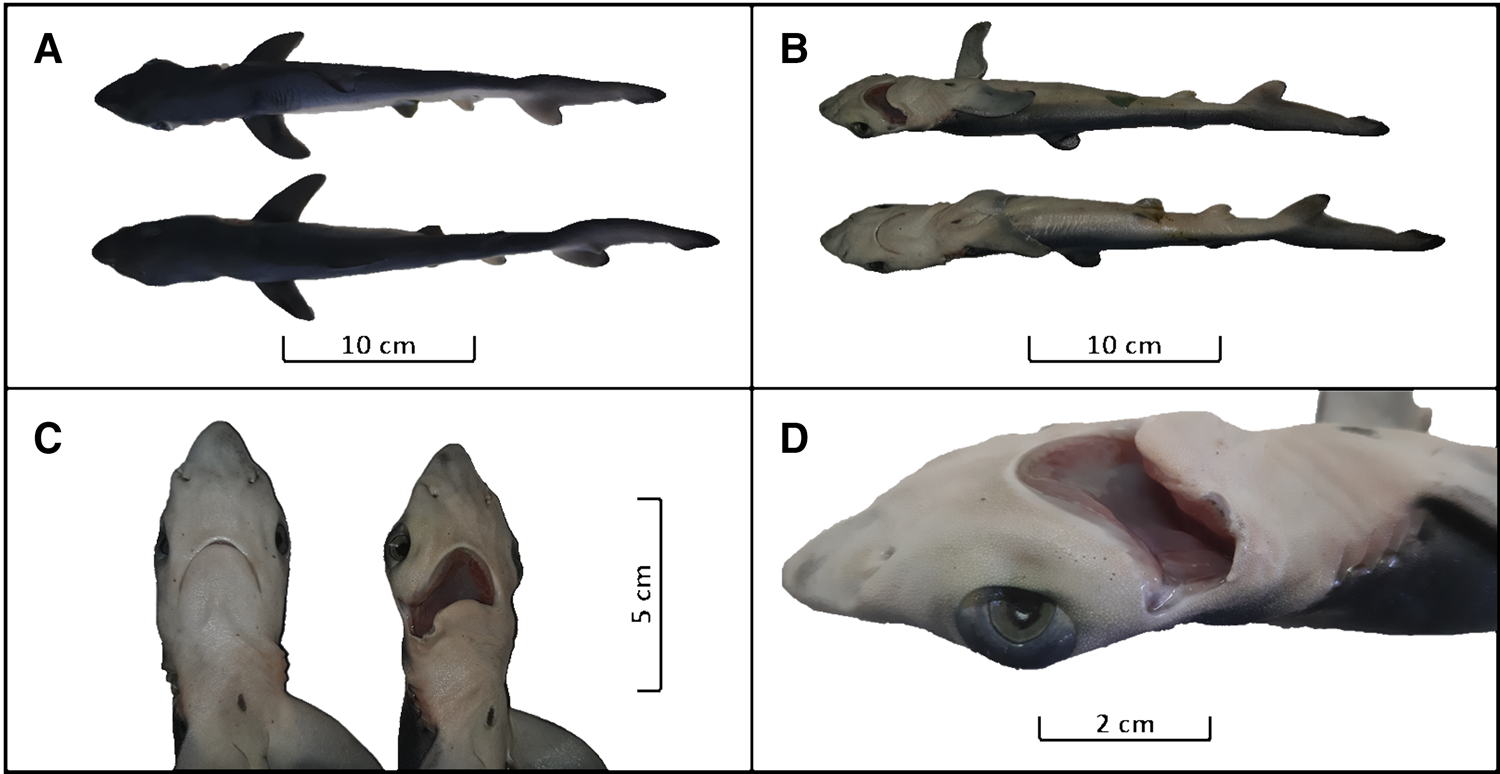
Fig. 2. Prionace glauca embryo B: (A) dorsal view (upper: embryo B; lower: sibling), (B) lateral diagonal view displaying the malformation in the mouth (upper: embryo B; lower: sibling), (C) ventral view comparing with its sibling (left: sibling; right: embryo B), and (D) zoom to the malformed mouth.
Discussion
According to the classification of morphological anomalies by Hennekam et al. (Reference Hennekam, Biesecker, Allanson, Hall, Opitz, Temple and Carey2013), embryo A presents a major morphology anomaly because it has significant consequences on its health and appearance at the time of evaluation, whilst embryo B has a minor morphological anomaly since there is a low impact on appearance with minimal health consequences.
Even though embryonic development was still in progress, the survival chance after birth would likely be small due to swimming disabilities for embryo A and feeding difficulties for both specimens.
Abnormalities related to cyclopia have been reported before for embryos of other elasmobranch species such as Carcharhinus obscurus (Bejarano-Álvarez & Galván Magaña, Reference Bejarano-Álvarez and Galván-Magaña2013), Galeorhinus galeus (Ramírez-Amaro et al., Reference Ramirez-Amaro, Fernández-Peralta, Serna and Puerto2019) and Squatina californica (synophthalmia; Escobar-Sánchez et al., Reference Escobar-Sánchez, Moreno-Sánchez, Aguilar-Cruz and Abitia-Cárdenas2014), while specific mouth malformations have not been reported for elasmobranchs.
As shown in Table 1, malformations in P. glauca embryos have been widely reported in different marine regions. However, similar reports in the South Atlantic Ocean are less frequent in comparison with the North Atlantic Ocean. In both the Atlantic and Pacific Oceans, the most commonly reported malformations for this species at this stage of development are diprosopia (usually two heads) and twisted vertebral columns (Mancini et al., Reference Mancini, Casas and Amorim2006; Bejarano-Álvarez et al., Reference Bejarano-Alvarez, Galván-Magaña and Ochoa-Báez2011; Galván-Magaña et al., Reference Galván-Magaña, Escobar-Sánchez and Carrera-Fernández2011; Hevia-Hormazábal et al., Reference Hevia-Hormazábal, Pastén-Marambio and Vega2011; Ehemann et al., Reference Ehemann, Marín-Sanz and Barany-González2016; Lamarca et al., Reference Lamarca, Ribeiro, Galheigo and Vianna2017; Pastén-Marambio et al., Reference Pastén-Marambio, Hevia-Hormazábal, Acuña and Vega2018; Rodriguez-Romero et al., Reference Rodriguez-Romero, Simeón-de la Cruz, Ochoa-Díaz and Monsalvo-Spencer2019; Ramírez-Amaro et al., Reference Ramirez-Amaro, Fernández-Peralta, Serna and Puerto2019; Kanagusuku et al., Reference Kanagusuku, Romero and Ramírez-Amaro2020). In this study, we report the second embryo with a kind of cyclopia for the South-western Atlantic Ocean (Ferreira et al., Reference Ferreira, Ferreira and Amorim2002).
Table 1. Morphological abnormalities reported in Prionace glauca embryos worldwide

The abnormalities observed in blue sharks and their relatively frequent occurrence could be explained by their high production of embryos, with a maximum litter size of 135 (Smith, Reference Smith1997). The causes for embryonic abnormalities could include the effects of contaminants (Casarini et al., Reference Casarini, Gomes and Tomas1997; Rosa et al., Reference Rosa, Mariano and Sampaio2004), as elasmobranchs are particularly vulnerable to bioaccumulation and biomagnification of pollutants due to their longevity and high trophic level (Gelsleichter & Walker, Reference Gelsleichter, Walker, Carrier, Musick and Heithaus2010). Moreover, abnormalities such as spinal malformations could be caused by arthritis, injuries, parasites, poor nutrition or tumours (Sadowsky, Reference Sadowsky1971; Schwartz, Reference Schwartz1973; Heupel et al., Reference Heupel, Simpfendorfer and Bennet1999). Theoretically, if population declines resulted in higher levels of inbreeding, this might also increase the likelihood of malformations in embryonic development (Dulvy et al., Reference Dulvy, Fowler, Musick, Cavanagh, Kyne, Harrison, Carlson, Davidson, Fordham, Francis, Pollock, Simpfendorfer, Burgess, Carpenter, Compagno, Ebert, Gibson, Heupel, Livingstone, Sanciangco, Stevens, Valenti and White2014; Lamarca et al., Reference Lamarca, Ribeiro, Galheigo and Vianna2017).
The number of developmental abnormalities reported in sharks has increased over time, although it is uncertain as to whether this relates to anthropogenic impacts or simply an increase in sampling and reporting. More standardized sampling and reporting of embryos would be required to inform on this. Despite the difficulty in making assumptions about possible causes for embryonic abnormalities, reporting of morphological abnormalities needs to be encouraged because it will allow us to better understand their causes, if there are species with a greater predisposition to these malformations, or even to understand the juvenile survival rate, which is an essential parameter for stock assessment.
Acknowledgements
We thank the Projeto Tubarão Azul research project for making it possible to collect individuals. We also thank the crew members and skippers of the fishing boats ‘Sambaqui III’ and ‘Aústria’ for their support in the field activities. Finally, we thank Dr Jim Ellis and the anonymous reviewers for improvements in final version of manuscript.
Financial support
We are grateful to the Organization of American States (OAS) and the Coordenação de Aperfeiçoamento de Pessoal de Nível Superior (CAPES), which provided scholarships for MCT. We also thank IdeaWild for providing computer equipment to MCT.


