


Introduction
Aelurostrongylus abstrusus (Railiet, 1898) is a nematode of the superfamily Metastrongyloidea, which is considered a primary respiratory parasite of cats and infects terminal bronchioles, alveolar ducts, and alveoli (Traversa and Di Cesare Reference Traversa and Di Cesare2013). Cats and wild felids are definitive hosts and become infected through the ingestion of intermediate (snails or slugs) or paratenic hosts (rodents, frogs, lizards, snakes, and birds) (Traversa and Di Cesare Reference Traversa and Di Cesare2013). Domestic cockroaches (American periplaneta) and mice also represent a potential source of infection to cats (Falsone et al. Reference Falsone, Colella, Napoli, Brianti and Otranto2017; Colella et al. Reference Colella, Knaus, Lai, Cantile, Abramo, Rehbein and Otranto2019).
It is a cosmopolitan disease and, although most cats remain subclinically infected or show mild disease (Traversa and Di Cesare Reference Traversa and Di Cesare2013, Reference Traversa and Di Cesare2016), intense chronic cough, dyspnea, tachypnea, wheezing, severe respiratory distress, and death may occur in severely parasitized cats (Traversa and Di Cesare Reference Traversa and Di Cesare2013; Vezzosi et al. Reference Vezzosi, Perrucci, Parisi, Morelli, Maestrini, Mennuni, Traversa and Poli2020). A necropsy-based retrospective study conducted in Brazil showed that 22 (1.5%) of 1,489 cats were parasitized by A. abstrusus and presented different disease degrees, and this was the cause of death in 45.5% of the affected cats (Pereira et al. Reference Pereira, Argenta, Rolim, Oliveira, Sonne, Pavarini and Driemeier2017).
Feline lungworm infections may lead to clinical signs similar to those of bronchitis and feline asthma; thus, lungworm infections may be misdiagnosed as allergic respiratory disease (Trzil and Reinero Reference Trzil and Reinero2014). Additionally, patients with lungworm infection frequently show clinical improvement with symptomatic therapy, making clinical diagnosis even more difficult (Traversa et al. Reference Traversa, Di Cesare and Conboy2010). Radiographically, bronchial to bronchointerstitial lung patterns are typically noted in both diseases, as well as eosinophilia (Trzil and Reinero Reference Trzil and Reinero2014). Some techniques used to detect the presence of A. abstrusus larvae are bronchoalveolar lavage fluid (BALF) cytology and fecal Baermann examination (Traversa and Di Cesare Reference Traversa and Di Cesare2013, Reference Traversa and Di Cesare2016; Trzil and Reinero Reference Trzil and Reinero2014). However, the lack of larvae in these tests does not rule out lungworm infection, and empiric treatment with benzimidazoles or macrolactones may be necessary to exclude this parasitism (Traversa and Di Cesare Reference Traversa and Di Cesare2013; Trzil and Reinero Reference Trzil and Reinero2014).
The objective of this study was to (1) prospectively determine the occurrence of A. abstrusus infection in Brazilian cats with cough or radiographic changes of bronchoalveolar disease through fecal Baermann examination, polymerase chain reaction (PCR), and BALF cytology; (2) evaluate the sensitivity and specificity of the fecal Baermann technique and BALF cytology as diagnostic methods, with fecal PCR serving as the gold standard, and (3) assess the potential risk factors associated with the development of A. abstrusus infection in the examined feline population.
Materials and methods
Animals
This present study was approved by the Animal Ethics Committee (CEUA/UFRGS, approval 33344). The owners of each animal signed an informed consent to participate in this study. Animals included in this study were client-owned cats presented between December 2017 and December 2018 to the Feline Medicine Service (MedFel) of the veterinary teaching hospital of the Universidade Federal do Rio Grande do Sul (UFRGS). Inclusion criteria were i) client-owned cats presented with a history of cough and ii) asymptomatic cats with radiographic abnormalities compatible with bronchopulmonary disease.
Patients were classified according to cough frequency as (i) asymptomatic (no coughing episodes); (ii) patients with mild signs, with sporadic cough and no decrease in quality of life; (iii) patients with moderate signs, with intermittent cough more than three times a week, and (iii) patients with severe clinical signs, with daily episodes of cough.
Study design
Data were recorded for each animal at the time of enrollment (e.g., sex, age, breed, data on the living environment, hunting habits). Physical examination was performed on all cats, and cats were submitted to high-quality lateral and ventrodorsal radiographic views of the thorax. Doppler echocardiography was requested for patients over five years of age and for patients with a history of or abnormalities in auscultation compatible with heart disease.
Blood samples were collected from all cats to perform complete blood count (CBC), serum biochemistry (total proteins, serum albumin and creatinine levels and serum alanine aminotransferase (ALT) and alkaline phosphatase (AF) activity in serum), and feline leukemia virus (FeLV)/feline immunodeficiency virus (FIV) infection status.
Three consecutive fecal samples were collected by the owners. After collection, samples were kept refrigerated at 4ºC for up to 24 hours, and then sent to the Helminthology Laboratory at UFRGS to perform the Willis technique (Willis Reference Willis1921). To perform the Baermann method, 5 g of feces were subjected to a sieve (aperture 100 mm) and placed in a Baermann apparatus (Baermann Reference Baermann1917). The funnel was slowly filled with water (20–25ºC) until half of the fecal sample was immersed in water. The apparatus was left at room temperature for at least 12 hours. By carefully opening the clamp, 2–3 drops were collected and placed on a glass slide, covered with a cover slip, and analyzed microscopically (100x magnification).
Faeces samples were also processed for DNA extraction using the commercial kit MagMAXtm CORE Nucleic Acid Purification (Thermo Fisher Scientific, Waltham, Massachusetts, USA) according to the manufacturer’s recommendations. The duplex-PCR protocol for amplifying the ITS-2 region was carried out as per Annoscia et al. (Reference Annoscia, Latrofa, Campbell, Giannelli, Ramos, Nascimento Dantas-Torres, Brianti and Otranto2014) to enable simultaneous detection of two species. The protocol utilizes AeluroF (5′-GCATTTATGCTAGTGATATC-3′) as a forward primer to amplify 220 bp for A. abstrusus, and TrogloF (5′-GCACTTGAAATCTTCGACA-3′) as a forward primer to amplify 370 bp for Troglostrongylus brevior; one single primer was used as reverse MetR (5′-TAAGCATATCATTTAGCGG-3′). In the analysis, two positive gender controls and negative control with UltraPure ™ DNase / RNase-Free Distilled Water (Invitrogen ™, Carlsbad, CA, USA) were used. The PCR products were subjected to 1.5% agarose gel electrophoresis, subsequently visualized on a Kasvi® LED transilluminator (São José dos Pinhais, Paraná, Brazil).
Cats were submitted to a blind BALF protocol if they were suited to anesthesia procedure with owner agreement. Cats were premedicated with a bronchodilator (terbutaline at a dose of 0.2 mg/kg SC), acepromazine (0.03 mg/kg IM), and meperidine (4 mg/kg IM), and light general anesthesia was induced with propofol (2–4 mg/kg EV). Anesthetic maintenance was performed with isoflurane vaporized in 100% oxygen (100 mL/kg) through orotracheal intubation in a semi-open Baraka system. A soft catheter (e.g., 8F red rubber) was passed down the endotracheal tube and into the lower airway until it lodged in a bronchoalveolar unit. With the catheter in place, a volume of sterile saline (5–15 mL) was rapidly infused and then removed from the lower airway by applying suction to the catheter with a syringe. This procedure was repeated up to 3 times to yield enough sample volume for analysis. Following lavage, the cat’s head was lowered to allow passive drainage of fluid from the airway through the endotracheal tube. This additional fluid was collected in a sterile specimen container. Supplemental oxygen was administered until extubating. Fluid samples were sent for cytology analysis, bacterial culture, and sensitivity profile and fungal culture.
Cytology slides were prepared with BALF samples. Direct cytology slides (at least two for each sample) were prepared by placing 100–200μl of uncentrifuged fluid on a slide and spreading it with a spreader slide. After this, the same BALF sample (2–4 ml) was centrifuged at 1500 rpm for 10 min, most of the supernatant liquid was discarded, the formed pellet was resuspended in a small amount of remaining fluid, and this fluid was used to prepare additional cytology slides (at least two for each sample). Uncentrifuged and centrifuged cytology preparations were stained with Diff-Quik (Romanowsky stain) and evaluated under light microscopy for the presence of parasites. The presence of different inflammatory cells was subjectively graded as mild, moderate, and marked.
Statistical analysis
A descriptive analysis was performed by calculating (i) the mean and the standard deviation of the quantitative variables and (ii) the proportions of the qualitative variables. Descriptive statistics of PCR results were used to evaluate the performance of the BM to diagnose the parasitosis when compared to PCR as the gold standard method, through the sensitivity and specificity. Also, a Poisson Regression with Robust Variance model was used to estimate possible association between the variables (clinical signs, lifestyle, and hunting) and the outcome (positive PCR). Statistical analysis was performed using SAS Studio software and a 5% significance level for the statistical tests were used.
Results
A total of 43 cats were included in this study, and among them, 74% (32/43) were positive for A. abstrusus by PCR and 41% (18/43) in the BM. Of the patients with a positive PCR, 78% (25/32) had abnormalities in the radiographic pattern and 25% (8/32) had eosinophilia on the CBC (Table 1). Twenty-one BALF samples were collected, and cytology did not detect the presence of lungworm larvae in any of the cases. Among parasitized patients, 93% (30/32) were mixed breed cats and 53% were males (17/32), with a mean age of 3.7 years (ranging from five months to eight years), in respect to 56% (18/32) having an outdoor lifestyle and 43% (14/32) hunting habits.
Table 1. Results of the evaluations of the 43 cats included in the study, according to the classification of the clinical sign of cough; detection of Aelurostrongylus abstrusus by PCR and by Baermann Method, cells present in the cytology of bronchoalveolar lavage samples, predominant pattern of thoracic radiography, presence of eosinophilia on blood count, FeLV antigen and FIV antibodies detection test

a Polimerase chain reaction
b Baermann method
c Bronchoalveolar lavage
d Eosinophilia
e Feline immunodeficiency virus
f Feline leukemia virus
A total of 41% patients (18/43) were positive for A. abstrusus by the Baermann method of the feces, and 16% of them (3/18) were co-infected with one of the other helminths: Toxocara spp., Ancylostoma spp., and Dipylidium spp. The presence of E. aerophilus or other pulmonary parasites was not observed in the coproparasitological techniques.
Only 21/43 cats were submitted to BALF due to the lack of agreement of the owners, or because they were unstable to undergo anesthesia. In 14% (6/43) of the patients, there was a diagnosis of a comorbidity, such as heart disease, chronic kidney disease, or hypertension. However, 66.6% (14/21) of them were PCR positive. Cytology showed patterns compatible with mild to moderate bronchitis. No larvae w found in the cytological evaluation of the 21 BALF samples. Pneumonia was diagnosed in 19% (4/21) of the patients, of which 14% (3/21) the culture identified Pasteurella sp. and 5% (1/21) Proteus sp. In the fungal culture, there were no positive results in any of the samples evaluated. In 24% (5/21) of the procedures performed, there was the complication of transient hypoxemia, which resolved with oxygen supply.
Descriptive measures of cats with positive PCR and negative PCR according to the EPF, RX, and eosinophilia results are presented in Table 2. Using PCR as the gold standard in the diagnosis of aelurostrongylosis, the BM presented a sensitivity value of 56.25% and a specificity value of 100%. BALF cytology could not be evaluated because it was done in only 21 cats, none of which were positive for lungworm larvae. Also, a Poisson Regression with Robust Variance model was used to estimate possible association between the variables (clinical signs, lifestyle, and hunting) and the outcome (positive PCR) (Table 3). The variable ‘hunting’ had a prevalence ratio of 0.6198 (0.4331–0.8869). The prevalence of PCR-positive A. abstrusus in hunting cats is approximately 38% higher than in non-hunting cats (p=0.0089).
Table 2. Descriptive measures of the variables results of the Baermann method, presence of radiographic abnormalities, and occurrence of eosinophilia in the hemogram in relation to PCR results for Aelurostrongylus abstrusus of 43 cats with clinical signs of cough or radiographic alterations compatible with bronchopulmonary disease

a Polymerase chain reaction
Table 3. Crude associations between the variables of anamnesis factors in parasitized and non-parasitized patients. The p-values for the crude associations were obtained using the Poison Regression with Robust Variance model

a Polymerase chain reaction
Three patients (3/43=6.9%) included in this study died, one of them due to the severity of the parasitosis (Figure 1A, B), another due to primary lung carcinoma, and the third one due to traumatic diaphragmatic hernia.
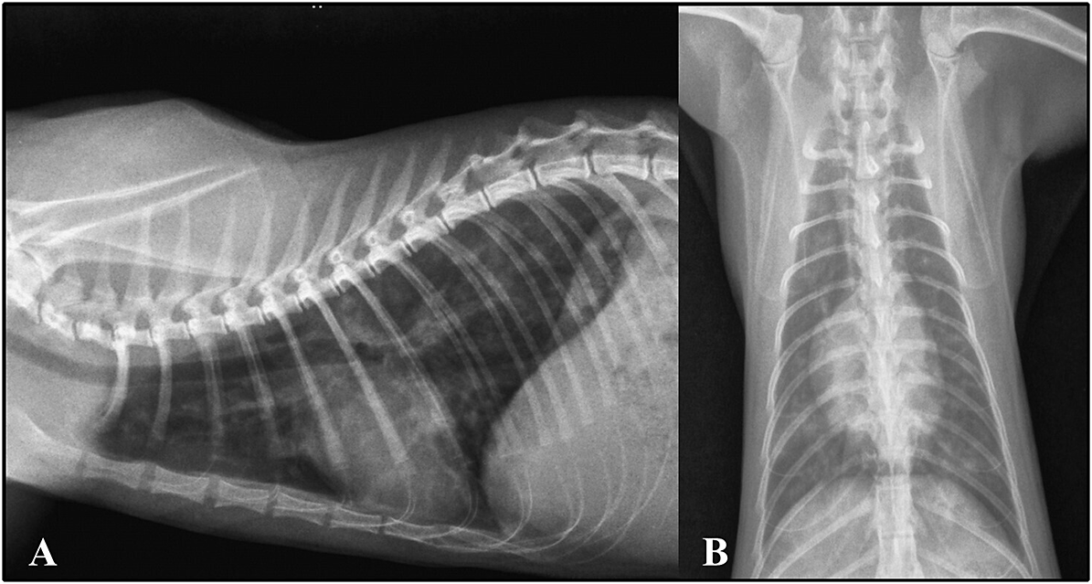
Figure 1. Thoracic radiographs. Left lateral (A) and ventrodorsal (B) positions of a cat infected with Aelurostrongylus abstrusus, showing a bronchial pattern
Discussion
In the current investigation, the prevalence of lungworm infection was found to be higher in cats using both BM (41%) and PCR (74%) compared to the prevalence described in South America by Penagos-Tabares et al. (Reference Penagos-Tabares, Lange, Chaparro-Gutiérrez, Taubert and Hermosilla2018). Prevalence rates reported in the literature vary depending on factors such as lifestyle, geographic origin, and diagnostic methods employed, ranging from 0.21% in Colombia (Echeverry et al. Reference Echeverry, Giraldo and Castaño2012) to 35.3% in Argentina (Cardillo et al. Reference Cardillo, Clemente, Pasqualetti, Borrás, Rosa and Ribicich2014) using the Ritchie and Baermann methods, respectively. The high prevalence observed in the present study could be attributed to the study design, as the enrolled cats exhibited respiratory signs or pulmonary radiographic changes, thus constituting a selected sample of the population. Moreover, it is important to note that the use of fecal diagnosis alone may not reflect the true prevalence among exposed populations (Morelli et al. Reference Morelli, Diakou, Di Cesare, Schnyder, Colombo, Strube, Dimzas, Latino and Traversa2020), and therefore, the use of serological detection methods such as ELISA has been proposed as a useful tool for detecting infection in endemic areas (Vismarra et al. Reference Vismarra, Schnyder, Strube, Kramer, Colombo and Genchi2023).
The prevalence of A. abstrusus in felines varies widely among countries and regions worldwide, with the disease considered endemic in the European continent (Knaus et al. Reference Knaus, Kusi, Rapti, Xhaxhiu, Winter, Visser and Rehbein2011; Echeverry et al. Reference Echeverry, Giraldo and Castaño2012; Barutzki and Schaper Reference Barutzki and Schaper2013; Olsen et al. Reference Olsen, Willesen, Pipper and Mejer2015). In Brazil, several states have reported this parasite, including Minas Gerais (Mundim et al. Reference TCD, Oliveira Júnior, Rodrigues and Cury2004), Mato Grosso (Ramos et al. Reference Ramos, Scheremeta, Oliveira, Sinkoc and Pacheco2013), Goiás (Campos et al. Reference Campos, Garibaldi and Carneiro1974), Rio de Janeiro (Scofield et al. Reference Scofield, Madureira, de Oliveira, Guedes Junior, Soares and da Fonseca2005; Ferreira et al. Reference Ferreira, Souza-Dantas and Labarthe2007), São Paulo (Fenerich et al. Reference Fenerich, Santos and Ribeiro1975; Matsui et al. Reference Matsui, Luzzi, Ferreira, PCD, PRR and André2018), and Rio Grande do Sul (Ehlers et al. Reference Ehlers, Jane de Mattos and Marques SM2013; Rigão et al. Reference Rigão, Franco, Machado and Rosa2019; Ferraz et al. Reference Ferraz, Pires, Santos, Barwaldt, Dallmann, Sapin, Lima, Pinto, Nobre and Nizoli2020). Since this lungworm was confirmed as an important cause of death in cats in our region (Pereira et al. Reference Pereira, Argenta, Rolim, Oliveira, Sonne, Pavarini and Driemeier2017), we were motivated to study its occurrence among our patients presented with cough or evidence of bronchopulmonary disease on radiographic evaluation.
There was a wide age range of parasitized cats included in this study, from kittens to seniors. A study conducted in Brazil in the 1990s indicated that young cats (<4 years old) were more frequently infected with A. abstrusus (Headley Reference Headley2005). However, the nematode can infect cats of any age, regardless of their lifestyle, breed, or sex. Younger cats may be more susceptible to infection due to their less developed immune system, but adult cats also have a cumulative risk of exposure due to hunting (Headley Reference Headley2005; Cavalera et al. Reference Cavalera, Schnyder, Gueldner, Furlanello, Iatta, Brianti, Strube, Colella and Otranto2019; Carruth et al. Reference Carruth, Buch, Braff, Chandrashekar and Bowman2019; Ferraz et al. Reference Ferraz, Pires, Santos, Barwaldt, Dallmann, Sapin, Lima, Pinto, Nobre and Nizoli2020). Hunting increases the chances of cats ingesting intermediate and paratenic hosts (Knaus et al. Reference Knaus, Kusi, Rapti, Xhaxhiu, Winter, Visser and Rehbein2011; Traversa et al. Reference Traversa, Lia, Iorio, Boari, Paradies, Capelli, Avolio and Otranto2008a). Additionally, some suspected paratenic hosts can be present inside houses and apartments, exposing indoor cats to infection (Falsone et al. Reference Falsone, Colella, Napoli, Brianti and Otranto2017; Colella et al. Reference Colella, Knaus, Lai, Cantile, Abramo, Rehbein and Otranto2019). This may partly explain why approximately 44% of indoor cats had positive PCR results in this study.
The Baermann method is the preferred parasitological method for diagnosing lung parasite infections, and it exhibited 100% specificity in our study. However, the Baermann method can only detect parasites during the patency period and may suffer from reduced sensitivity due to intermittent or low excretion of larvae (Iorio and Traversa Reference Iorio and Traversa2008). Alternative diagnostic tools for feline aelurostrongylosis have been proposed, including molecular detection methods using samples of feces or pharyngeal swabs. In our study, we employed a single-step duplex polymerase chain reaction (duplex-PCR) on the ribosomal internal transcribed spacer 2 region (ITS-2) to simultaneously detect and differentiate T. brevior and A. abstrusus. These two parasites share similar biology and ecological niches and can potentially co-infect cats, but their first-stage larvae (L1) are difficult to differentiate due to morphological similarities (Bowman Reference Bowman2021). Our results align with those of Morelli et al. (Reference Morelli, Traversa, Diakou, Colombo, Russi, Mestek, Chandrashekar, Beall, Paoletti, Iorio, Tsokana, De Cristofaro, Barlaam, Simonato and Di Cesare2022), who confirmed that PCR is more sensitive and specific than the Baermann method for diagnosing A. abstrusus infections in domestic cats. Molecular methods, including PCR, have proven to be powerful in a range of studies, including those focused on diagnostic purposes in clinical cases (Traversa and Guglielmini Reference Traversa and Guglielmini2008; Traversa et al. Reference Traversa, Iorio and Otranto2008b; Di Cesare et al. Reference Di Cesare, Frangipane di Regalbono, Tessarin, Seghetti, Iorio, Simonato and Traversa2014; Hawley et al. Reference Hawley, Johnson, Traversa, Bucy, Vernau and Vernau2016), postmortem evaluations (Crisi et al. Reference Crisi, Traversa, Di Cesare, Luciani, Civitella, Santori and Boari2015; Traversa et al. Reference Traversa, Della, Diakou, Sforzato, Romanucci, di Regalbono, Lorio, Colaberardino and Di Cesare2018), and anthelmintic evaluations (Traversa et al. Reference Traversa, Romanucci, Di Cesare, Malatesta, Cassini, Iorio, Seghetti and Della Salda2014).
No larvae were observed in the BALF cytology samples. According to the literature, A. abstrusus larvae are cytologically detected in 20.8% to 36.4% of naturally infected cats (Crisi et al. Reference Crisi, Johnson, Di Cesare, De Santis, Di Tommaso, Morelli, Pantaleo, Luciani, Schaper, Pampurini and Boari2019, Reference Crisi, Di Cesare, Traversa, Vignoli, Morelli, Di Tommaso, De Santis, Pampurini, Schaper and Boari2020). However, even when evaluating experimental infections using high parasite loads (800 larvae per cat), the number of animals with positive cytology for A. abstrusus larvae was low (3 in 14 cats, representing 21.4%) (Lacorcia et al. Reference Lacorcia, Gasser, Anderson and Beveridge2009). The reasons for our results in the 13 PCR-positive cats that underwent BALF testing may include a low parasite load in the population studied and the fact that not all cats were sampled due to external circumstances (Annoscia et al. Reference Annoscia, Latrofa, Campbell, Giannelli, Ramos, Nascimento Dantas-Torres, Brianti and Otranto2014). It is essential to highlight that cytological findings in aelurostrongylosis are not specific and may overlap with those of other feline airway diseases, unless larvae are detected (Crisi et al. Reference Crisi, Di Cesare, Traversa, Vignoli, Morelli, Di Tommaso, De Santis, Pampurini, Schaper and Boari2020). In this study, some owners did not consent to the BALF procedure for their cats due to concerns about potential respiratory complications from the invasive nature of the procedure. However, among the cats that underwent the procedure, only mild and transient hypoxemia was observed, which was treated with the administration of oxygen. Previous studies by Lacorcia et al. (2019) and Crisi et al. (Reference Crisi, Johnson, Di Cesare, De Santis, Di Tommaso, Morelli, Pantaleo, Luciani, Schaper, Pampurini and Boari2019, Reference Crisi, Di Cesare, Traversa, Vignoli, Morelli, Di Tommaso, De Santis, Pampurini, Schaper and Boari2020) also reported no anesthetic or procedure-related complications.
Blood cell count and thoracic radiography are screening tests for pulmonary parasitosis, but they lack specificity. Only 25% of the cats positive for PCR had eosinophilia, which is consistent with other feline bronchopulmonary diseases (Trzil and Reinero Reference Trzil and Reinero2014). Among parasitized cats, 78% had an abnormal radiographic pattern, including bronchial, interstitial, and nodular patterns. Thoracic imaging can reveal a range of patterns, from multifocal distributions of bronchial, nodular, and unstructured interstitial patterns in early stages to generalized alveolar patterns in severe cases (Ribeiro et al. Reference Ribeiro, JMP, Negrão-Correa, Barçante, Klein and Lima2014; Pennisi et al. Reference Pennisi, Hartmann, Addie, Boucraut-Baralon, Egberink, Frymus, Gruffydd-Jones, Horzinek, Hosie, Lloret, Lutz, Marsilio, Radford, Thiry, Truyen and Möstl2015; Morelli et al. Reference Morelli, Diakou, Colombo, Di Cesare, Barlaam, Dimzas and Traversa2021). These findings may resemble and need to be distinguished from chronic bronchial disease, metastatic neoplasms, and bacterial or mycotic conditions (Pennisi et al. Reference Pennisi, Hartmann, Addie, Boucraut-Baralon, Egberink, Frymus, Gruffydd-Jones, Horzinek, Hosie, Lloret, Lutz, Marsilio, Radford, Thiry, Truyen and Möstl2015; Traversa and Di Cesare Reference Traversa and Di Cesare2016; Morelli et al. Reference Morelli, Diakou, Colombo, Di Cesare, Barlaam, Dimzas and Traversa2021). The correlation between the presence and severity of clinical scores and radiographic changes is only partial, as many infected cats show radiographic changes without evident clinical signs, while cats that show clinical signs often have evident radiographic abnormalities (Traversa and Di Cesare Reference Traversa and Di Cesare2016; Febo et al. Reference Febo, Crisi, Traversa, Luciani, Di Tommaso, Pantaleo, Santori, Di Cesare, Boari, Terragni and Vignoli2019).
Differential diagnoses for lungworms include bacterial infections, as well as other parasitic infections such as pulmonary toxoplasmosis, respiratory mycoses, feline bronchial disease/asthma, airway foreign bodies, and pulmonary tumors (Traversa et al. Reference Traversa, Lia, Iorio, Boari, Paradies, Capelli, Avolio and Otranto2008a; Foster and Martin Reference Foster and Martin2011). Bacterial pneumonia was diagnosed in 19% of the samples collected by BAL, and the bacteria identified from the culture, including Pasteurella sp. and Proteus sp., are described in the literature as being associated with pneumonia, bronchopneumonia, and bacterial bronchitis in cats (Reinero Reference Reinero2010). Thus, the BALF method is important to investigate other causes of bronchopulmonary diseases in addition to parasitic diseases and should be performed whenever possible. In a previous study using BALF in cats (Johnson and Vernau, Reference Johnson and Vernau2011), this test was able to differentiate between pneumonia (29%), neoplasms (15%), bronchitis/asthma (48%), and other conditions (8%).
The clinical signs caused by A. abstrusus may mimic those associated with feline bronchial disease and asthma (Foster and Martin Reference Foster and Martin2011). Moreover, treatment with corticosteroids and bronchodilators may result in a clinical improvement in cats with respiratory parasitosis (Foster and Martin Reference Foster and Martin2011; Trzil and Reinero Reference Trzil and Reinero2014). Therefore, treatment without identification of the etiologic agent may lead to unfavorable outcomes for patients. This highlights the importance of including BM and PCR as essential triage tests for all patients presenting with cough or radiographic abnormalities to identify lungworms as a potential differential diagnosis in the clinical setting.
Conclusions
The study demonstrated that A. abstrusus is a significant cause of bronchopulmonary disease in domestic cats in southern Brazil, with a prevalence of 74% among cats presenting with cough or radiographic abnormalities. Although the Baermann method is a quick, non-invasive, and cost-effective technique for detecting first-stage larvae in feces, its sensitivity is low compared to the PCR method, which is considered the gold standard. In addition, the cytological evaluation of the 21 BALF samples did not reveal any larvae. These findings highlight the importance of using the PCR method as a triage test to accurately diagnose A. abstrusus infections in cats with respiratory signs and to ensure proper treatment.
Data availability statement
All data used in this study are available in the records of the Hospital de Clínicas Veterinárias at Universidade Federal do Rio Grande do Sul.
Authors’ contribution
Renata Fagundes-Moreira: conceptualization, methodology, formal analysis, writing – original draft preparation.
Elissandra Silveira: conceptualization, investigation, data analysis, methodology, writing – original draft preparation.
Vinícius Baggio-Souza: conceptualization, formal analysis, writing – original draft preparation.
Sandra Márcia Tietz Marques: conceptualization, investigation, data analysis, methodology, writing – original draft preparation.
Silvana Bellini Vidor: investigation, data analysis, methodology.
Stela Maris de Jezus Castro: investigation, data analysis, methodology.
Andréia Spanamberg: investigation, data analysis, methodology.
Luan Cleber Henker: investigation, data analysis, methodology.
Saulo Petinatti Pavarini: investigation, data analysis, methodology, supervision, writing – review and editing.
João Fabio Soares: conceptualization, formal analysis, supervision, writing – review and editing.
Fernanda Vieira Amorim da Costa: conceptualization, investigation, methodology, data analysis, project administration, supervision, writing – review and editing.
Funding
This study was supported by research grants from the Conselho Nacional de Pesquisa e Desenvolvimento Científico e Tecnológico (CNPq) (grant number 131701/2018-5 and 312576/2021-8), Coordenação de Aperfeiçoamento de Pessoal de Nível Superior – Brasil (CAPES) (Finance code 001), and Fundação de Amparo à Pesquisa do Rio Grande do Sul (FAPERGS) (Finance code:19/2551–0001842–8). The author JFS is funded by CNPq (grant #312576/2021-8).
Competing interest
Declarations of interest: none
Ethical approval
This work involved non-experimental animals (owned or unowned) and procedures that differed from internationally established and recognized high standards (‘best practice’) of veterinary clinical care for the individual patient. The study, therefore, had ethical approval from an established committee as stated in the manuscript.
Authorizations
All authors approve the submitted version and agree to the copyright conditions.


