


Introduction
Intense debate shrouds the practice of infant sacrifice in Phoenician and Punic societies (see D'Andrea Reference D'Andrea2014 for a summary, see also Smith & Kahila Reference Smith and Kahila1992; Lee Reference Lee1994; Bénichou-Safar Reference Benichou-Safar2005; Smith et al. Reference Smith, Avishai, Greene and Stager2011; McCarty Reference McCarty and Xella2013; Xella et al. Reference Xella, Quinn, Melchiorri and Dommelen2013; Mays Reference Mays, Thompson, Alfonso-Durruty, Crandall and Larsen2014; Schwartz et al. Reference Schwartz, Houghton, Bondioli and Macchiarelli2017; Carroll Reference Carroll2018). Some scholars view the existence of tophets—Phoenician or Punic sanctuaries where infants and young children were cremated, placed in urns and ritually interred—as prima facie evidence for sacrifice; yet others urge caution in the interpretation of these sites, arguing that mortuary treatment may have differed depending on age and social identity (see online supplementary material (OSM) for detailed debate on infant sacrifice at some tophets). Classical texts and inscriptions can be highly informative for reconstructing Phoenician-Punic mortuary customs (Bénichou-Safar Reference Benichou-Safar2004). But variations in rituals between communities, which may not have been recorded in historical accounts (Vella Reference Vella1996; Xella et al. Reference Xella, Quinn, Melchiorri and Dommelen2013), and the inherent biases of such written records (e.g. Carroll Reference Carroll2018: 166, see also OSM), permit space for conflicting interpretations of tophets (e.g. Smith et al. Reference Smith, Avishai, Greene and Stager2013; Schwartz et al. Reference Schwartz, Houghton, Bondioli and Macchiarelli2017).
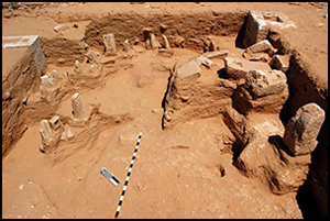
Here, we take a different approach to interpreting the human remains found within these contexts. Inspired by the life-course approach, we explore the lives and deaths of 12 cremated infants and children found at the tophet of Zita (also spelled Zitha), Tunisia (Figure 1)—an urn-field within an urban mound inhabited from the fifth century BC to AD 450 (Ben Tahar et al. Reference Ben Tahar, Drine, Kaufman and Barnard2021; Kaufman et al. Reference Kaufman2021). We go beyond questions of the presence or absence of infanticide in this tophet to examine aspects related to health fragility and remembrance. With this aim in mind, we investigated: 1) the pathological conditions present in the infants and children at Zita; 2) the possible causes of these conditions and what they can tell us about the lives of these young individuals; and 3) how the infants and children were treated after death.

Figure 1. Satellite image of the archaeological site of Zita and photograph showing part of the tophet at Zita (image by Hans Barnard and photograph by Brett Kaufman; measured co-ordinates are projected onto UTM zone 32S (N) of the WGS84 geoid (satellite image courtesy of Google Earth)).
The life-course approach explores “human life as a result of interrelated and cumulative events over the timeframe of individuals, as well as over generations at the community level” (Agarwal Reference Agarwal2016: 130). This approach combines concepts from anthropology, sociology, psychology and history (Inglis & Halcrow Reference Inglis, Halcrow, Beauchesne and Agarwal2018) and connects individuals with the historical and socioeconomic contexts in which they lived (Gilchrist Reference Gilchrist2012). Infants and children are not distinct entities; their social and physiological destinies are subjectively intertwined with their caregivers and social groups (Gowland & Halcrow Reference Gowland and Halcrow2020). The role and significance of infancy and childhood varies among cultural traditions and is usually linked to social and cultural factors, especially morbidity (the state of being unhealthy or symptomatic for a disease or condition) and mortality (the likelihood of death) (Montgomery Reference Montgomery2008). Health can shape the experience of infants and children, how their personhood is perceived and their overall life and mortality. Building on the life-course approach, our research combines biological and archaeological data to contextualise these relationships—between child and caregiver, between health and society—at Zita. Posthumous treatment of the body is reconstructed through the analysis of thermal alterations, skeletal placement within the urns and archaeological context, to explore the practices of burial and remembrance. Such combined analysis and contextualisation allows for a deeper exploration of these individuals and their place in the broader social and economic setting of Zita.
The tophet at Zita
Zita comprises an urban mound of at least 34ha positioned along an ancient trade route from Carthage, in present-day Tunisia, to Lepcis Magna in what is now Libya. The site was occupied from c. 425 BC–AD 450 (Ben Tahar et al. Reference Ben Tahar, Drine, Kaufman and Barnard2021; Kaufman et al. Reference Kaufman2021). The tophet is large and well-preserved and was initially identified from surface finds of more than 600 votive stelae (Drine Reference Drine1991; Drine & Ferjaoui Reference Drine, Ferjaoui, Fantar and Ghaki1991). Targeted excavations of the area yielded 24 undisturbed urns—only 12 of which were analysed due to time and budget constraints—associated with upright stelae facing roughly eastward, plastered basins and pit features containing burned bone, unguentaria (small ceramic or glass bottles) and bowls. Archaeological information generated from the tophet and the overall site allows a glimpse of variation in funeral rituals conducted by the Neo-Punic population at an individual and communal level (‘Neo-Punic’ here denoting the cultural era of Phoenician descendants and neighbouring groups in North Africa following the fall of Carthage; Kaufman et al. Reference Kaufman2021). Chronological determinations from the ceramic assemblage indicate that examined burials were deposited from around 50–30 BC to c. AD 100 (Ben-Tahar et al. Reference Ben Tahar, Drine, Kaufman and Barnard2021), with all the urns dating to after the destruction of the Carthaginian state, akin to a few other Neo-Punic tophets (McCarty Reference McCarty and Xella2013). Ceramic vessels dating to c. AD 200 provide evidence that the tophet continued to be visited and maintained after the cessation of ritual deposition. Excavations, survey and historical surface collections indicate that the area was preserved into the Roman era, with the addition, during this period, of iconography and what may be Latinised inscriptions to ancient Semitic deities (Berger Reference Berger1905; Kaufman et al. Reference Kaufman2021).
Materials and methods
We analysed bone and bone fragments from nine urns and three pit features (Table S1), all of which represent secondary cremation deposits—deposits created after the removal of material from the primary cremation location (Cerezo-Román Reference Cerezo-Román2015). Radiographs of complete urns with intact contents were made to identify the locations of bones (Figure S1) and these urns were subsequently micro-excavated in the laboratory at the Musée de Zarzis in Tunisia (Figures 2 & 3). This allowed for the reconstruction of bone placement inside the urn.

Figure 2. Micro-excavation of urn L. 1025 (photograph by Jessica Cerezo-Román).

Figure 3. Ulna from urn L. 1025 (pictured in Figure 2). The warping of the distal end (towards the right of the image) is caused by fire (figure by Jessica Cerezo-Román).
Following micro-excavation and cleaning, standard protocols for osteological data collection were followed (Buikstra & Ubelaker Reference Buikstra and Ubelaker1994; Cunningham et al. Reference Cunningham, Scheuer and Black2016). Detailed methods are available in the OSM and, in brief, involved the compilation of a full skeletal inventory, bone metrics and morphological observations. Age-at-death was estimated following accepted standards (Buikstra & Ubelaker Reference Buikstra and Ubelaker1994; AlQahtani et al. Reference AlQahtani, Hector and Liversidge2010; Cunningham et al. Reference Cunningham, Scheuer and Black2016; see OSM). When present, evidence of pathological conditions and skeletal trauma were also recorded using accepted protocols (Buikstra & Ubelaker Reference Buikstra and Ubelaker1994; Buikstra Reference Buikstra2019), which involved a detailed account of the location, extent, severity and type of new bone formation (see OSM). Thermal alterations limited some differential diagnoses of pathologies and trauma, but enough skeletal material remained to make assessments based upon lesion distribution and skeletal elements present. Thermal alterations to osseous material and body manipulation were analysed according to the variables of predominant colour, degree of shrinkage, maximum length measurements, degree and pattern of fracture and cremation weights.
Results
A total of 12 individuals was identified: seven infants (less than 2 years at death) in urns, two children (2 to 12 years at death) in urns, two children in pit features and one potentially slightly older individual (12 years ± 2.5 months) in a pit feature (Table 1). All the deposits contained only one individual, based on the skeletal elements that were present, lack of duplication of bone type and ontogenetic ages. No evidence of ante- or perimortem trauma is identifiable in any individual. Nine individuals show changes to bone structure or morphology that could be suggestive of scurvy, a condition caused by a deficiency of vitamin C and characterised by the micro-haemorrhaging of blood vessels that leads to patches of new bone formation throughout the body (Figure 4).
Table 1. Estimated age and pathology of cremated infants and children at Zita. Individuals with an estimated age-at-death of less than two years are considered ‘infants’, while those between 2 and 12 years are considered ‘children’. For scurvy, lesions are classified as suggestive or diagnostic following Snoddy (2018). For more detailed differential diagnoses of pathological lesions, see OSM.


Figure 4. Pathological changes to bone possibly associated with scurvy: a) maxilla from urn L. 1033 showing active abnormal woven bone on the anterior surface; b) sphenoid from urn L. 2064 showing abnormal micro- and macroporosity (figure by Jessica Cerezo-Román).
Two individuals have skeletal pathologies related to respiratory tract infection (Figures 5 & 6) and abnormal bone growth on the internal surface of the cranium (the endocranium) associated with inflammation or haemorrhaging of the meningeal vessels that surround the brain (Figure 7). Nine individuals have pathological lesions suggestive of anaemia, a condition that affects the ability of red blood cells to transport oxygen around the body. A total of four individuals show porosity of the upper orbit (cribra orbitalia) and the bones of the cranial vault (porotic hyperostosis) (Figure 8), while three individuals have just porotic hyperostosis (included here is individual L. 2120). Furthermore, one individual has just cribra orbitalia and one individual has cribra orbitalia and a band of decreased enamel thickness (enamel hypoplasia) on the lower left permanent canine (Tables 1 & S1). Nine individuals also present periostitis of nonspecific origin (identified osteologically as new bone that forms on the outer surface of bones in response to inflammation of the overlying tissue—the periosteum) (Figures 3 & 9, see also Table S1 for more detail).

Figure 5. Maxillary sinuses with active (left) and healed (right) periosteal reactions (figure by Jessica Cerezo-Román).

Figure 6. Active (urns L. 1033 and L. 1025) and healed (urn L. 2068) periosteal reactions on rib shafts (figure by Jessica Cerezo-Román).

Figure 7. Endocranial new bone growth showing grooves associated with the meningeal blood vessels (figure by Jessica Cerezo-Román).

Figure 8. Examples of cribra orbitalia (figure by Jessica Cerezo-Román).

Figure 9. Examples of periosteal reactions: urn L. 1023 tibia shaft with active periosteal reaction, urn L. 1025 humeri shaft with active periosteal reaction and urn L. 1008 unidentified long bone with active periosteal reaction (figure by authors).
The individual deposits contained most of the skeletal elements, suggesting that the skeletal elements were thoroughly collected from the pyres and the deposits contained fairly complete bodies of individuals. In general, all the skeletal areas of the body were represented at some level. The appearance of the bones is consistent with their being burned with flesh present or moisture in the bones: the remains were warped and they exhibited horizontal, vertical and concentric fractures caused by the fire. Depending on the duration of and exposure to the fire, bone colour changes, from unburned ivory colour to brown and black in light and partially burned remains exposed to temperatures over 300±50°C to less than 500±50°C (Ellingham et al. Reference Ellingham, Thompson, Islam and Taylor2015). Initial calcination of the bone causes a wide range of grey colouring that is usually associated with temperatures higher than 500±50°C to less than 700±50°C. Full calcination is associated with white and blue colours and is seen in temperatures over 700±50°C (Ellingham et al. Reference Ellingham, Thompson, Islam and Taylor2015). Eleven of the 12 individuals were cremated at a high temperature and/or for a long time (Figure 10). Bones burned at high temperatures and/or for a long time usually are white in colour, calcined, warped and present concentric, vertical and horizontal fractures caused by fire. One individual (feature 1023) presented a mix of burned and unburned skeletal elements.

Figure 10. Colour of infant and child bones at Zita. Depending on the duration of and exposure to the fire, bone colour changes from unburned to brown, black, grey and finally white when the bones are calcined and have been exposed to the fire for a long time or high temperature (figure by Jessica Cerezo-Román).
Following cremation, the bones and ashes were placed in urns or pits. Infants are represented by fewer bone fragments than children (Table 2). Eight of the nine individuals excavated from the urns were not placed in anatomical order inside the urns after cremation (Figures 1–3, Table S3). The skeletal elements were concentrated in the middle and on the bottom of the urns. In one urn (L. 2064), the long bones were placed first, followed by the axial and cranial bones. In two instances (L. 1025 in Figure S1 and L. 2099), the bottom of the urn appears to have been intentionally lined with sand before the remains were placed inside (Table S2). For detailed osteological and thermal alterations analyses, see the OSM.
Table 2. Bone weights and maximum length.

Discussion
Across various cultures, archaeological, ethnographic and historical claims of infant sacrifice are often associated with contexts showing high infant mortality (Smith & Kahila Reference Smith and Kahila1992; Lee Reference Lee1994; Mays Reference Mays, Thompson, Alfonso-Durruty, Crandall and Larsen2014) and focus on emotional detachment by the parents (Cannon & Cook Reference Cannon and Cook2015) and detachment from emotional distress in the wider community (Carroll Reference Carroll2011). Historical records detail many ways in which infants were killed or disposed of, such as exposure, neglect, drowning, smothering or trauma (Scott Reference Scott1999). Few of these would leave any archaeological traces or direct evidence on the bones. We did not find direct evidence of perimortem trauma or violence on the remains of the Zita infants or children. We did, however, identify evidence of poor health at a systemic level, which likely reduced their quality of life and may have contributed to their deaths.
At Zita, the relatively good state of preservation of the cremated bones allowed assessment of pathological conditions. While the life-course approach often centres on how early stages of life affect later stages—both across an individual life and throughout generations—we build on this framework and use a longitudinal perspective of the individuals to explore some key aspects of their life, principally their health and how these likely had an impact on their treatment in death. The individuals that died young had experienced health problems that were of sufficient duration and intensity to leave marks on their skeleton, allowing us to reconstruct some of their impact through time (Wood et al. Reference Wood, Milner, Harpending and Weiss1992). Most of the infants and children at Zita suffered from poor health or disease during their life and around the time of death. Of the 12 individuals analysed, seven infants and three children demonstrate pathological alterations to bones consistent with scurvy, anaemia and physiological stress or infection and many of these lesions were active when they died.
A total of nine individuals present evidence that is suggestive of scurvy, a condition caused by a deficiency of vitamin C that may reflect the individual's and their mother's nutritional deficiencies. Skeletal lesions appear only in advanced cases of scurvy (Brickley et al. Reference Brickley, Ives and Mays2020: 43–74). From a life-course perspective, it is very likely that these children and their breast-feeding mothers were undernourished. The age of weaning could also have influenced and possibly exacerbated any nutritional deficiencies. Moses and colleagues (Reference Moses, Kaufman, Drine, Barnard, Ben Tahar, Jerray and Daniels2019) examined zooarchaeological material from Zita contemporaneous with the tophet and none of the animals identified are good sources of vitamin C. Macrobotanical remains from Zita identified olives, colocynth, grapes, wheat and barley (Kaufman et al. Reference Kaufman2021). Of these, only grapes and olives are a source of vitamin C, but it is possible that both were processed for export (as wine and oil, respectively) as Zita was enfolded within the Roman industrialised export economy. Under these circumstances, local access to these staples, and the key micronutrients they contain, may have been limited.
Infants and children with scurvy are highly susceptible to secondary infections and usually have comorbidities, such as anaemia and vitamin D deficiency (Lewis Reference Lewis2018). Six individuals had cribra orbitalia and two individuals (L. 1025 and L. 1008) had ‘hair-on-end’ lesions in the bones of the cranial vault typical of anaemia (Tables 1 & S1). These last two individuals also have abnormal endocranial bone growth consistent with inflammation or haemorrhaging of the meningeal vessels (Schultz Reference Schultz2001; see Table 1 and OSM). This type of pathological lesion could be associated with meningitis (see OSM) but may also form as a primary or secondary response to bacterial infections (including otitis media, measles, typhoid fever, tuberculosis and pneumonia; Kim-Farley Reference Kim-Farley and Kiple1993) and haemorrhaging of the dura mater (the outermost layer of connective tissue that surrounds and protects the brain) arising from vitamin C deficiencies or minor trauma can also cause similar lesions (Cecconi et al. Reference Cecconi, Mallegni and D'Anastasio2007; Lewis Reference Lewis2018). Individuals L. 1025 and L. 1008, therefore, likely had comorbidity of anaemia and scurvy, associated with vitamin C deficiency.
Several individuals show healed and active subperiosteal reactions likely related to periostitis of nonspecific origins (see OSM) in addition to the previously mentioned manifestations of scurvy. Subperiosteal new bone formation in the bones of individuals younger than four years is challenging to interpret because normal appositional growth—whereby bone diameter is increased through the surface deposition of new bone tissue—produces a similar appearance on the bone surface as active periosteal reaction deposits associated with pathological conditions (Kwong et al. Reference Kwong, Friello and Semba2004; Lewis Reference Lewis2018). Consideration of the location, severity (thicker than 2mm) and expression of subperiosteal new bone formation does, however, suggest that many of the active deposits are pathological in origin (De Silva et al. Reference De Silva, Evans-Jones, Wright and Henderson2003; Kwong et al. Reference Kwong, Friello and Semba2004; Weston Reference Weston2008; Rittemard et al. Reference Rittemard, Colombo, Desbarats, Dutailly, Dutour and Coqueugniot2019). At Zita, the majority of infants and children with subperiosteal reactions also displayed skeletal manifestations associated with scurvy, suggesting that these lesions could be associated.
Anaemia could be inferred from skeletal changes in nine individuals, possibly caused by thalassemia, dietary iron deficiency or malaria. Due to the high degree of fragmentation of the bones it was not possible to narrow down the cause(s) of the anaemia. Zooarchaeological and macrobotanical analyses at Zita suggest that sources of protein and iron were locally available, including livestock, olives, wheat, grapes and the Atlas pistachio (Pistacia atlantica) (Kaufman et al. Reference Kaufman2021). At the Roman site of Leptiminus, stable isotope analysis of human skeletal remains found that breastfeeding children, and potentially also mothers and wet nurses, consumed a different diet from the rest of the population (Keenleyside et al. Reference Keenleyside, Schwarcz, Stirling and Lazreg2009). At Zita, we suggest that either proteins and iron were not regularly consumed by infants and children, and maybe also their mothers or wet nurses, or that other causes of iron deficiencies existed, such as parasitic infections, thalassemia or malaria.
One child (L. 2099) had cribra orbitalia and enamel hypoplasia. Based on the location of the hypoplastic lesion on the crown of the left mandibular canine, this individual likely experienced a period of stress in early childhood—2.5 to 4.5 years ± 3 months (AlQahtani et al. Reference AlQahtani, Hector and Liversidge2010)—possibly related to problems during weaning. However, this child possibly also suffered from scurvy, anaemia and infections of the respiratory tract (the latter diagnosis being based on small areas of micro- and macroporosity on the left side of the nasal cavity; Tables 1 & S1), any of which could have contributed to physiological stress in early childhood.
Further evidence of respiratory tract infection was found in infant L. 2120, who had active and healed woven bone on one rib (Kroegel & Antony Reference Kroegel and Antony1997; Davies-Barrett et al. Reference Davies-Barrett, Owens and Eeckhout2021). Poor air quality or bacterial and viral infections of the sinuses and respiratory tract are factors that contribute to osteological changes in the thorax and the frontal and maxillary sinuses (Boocock et al. Reference Boocock, Roberts and Manchester1995; Davies-Barrett et al. Reference Davies-Barrett, Owens and Eeckhout2021). Although unlikely, other diseases that manifest similar pathological changes—such as leprosy, nasal polyps, trauma, asthma and fungal allergies (Hamilos Reference Hamilos2000)—cannot be ruled out. Kaufman and colleagues (Reference Kaufman2021) found evidence for the extensive production of metals, ceramics (including amphorae for wine and olive oil export) and charcoal at Zita, representing an overall increase in the pace of industrialisation. These activities probably produced excessive smoke that would have been routinely inhaled by inhabitants of the site. Based on the broader archaeological context and pathological changes, an overall degradation in air quality and an increase in pollution were possible factors in the development of respiratory tract infections in infants and children at Zita.
Several studies have argued that infant and childhood mortality patterns are better understood within a specific social and environmental context (e.g. Hodson Reference Hodson2017). At Zita, it is evident that many individuals suffered from poor nutrition and poor health at the time of death. The exact cause(s) of many of the lesions is unclear, as are the specific infections from which they suffered and, ultimately, what contributed to their deaths. It is clear, however, that the infants and children buried at this site had severe health problems at the time of death.
Publications that include osteological analysis of human remains from the Mediterranean region in the first millennia BC and AD are limited but some serve as useful comparisons for our data. At the tophet of Dougga in northern Tunisia, for example, no evidence of trauma was found in the cremated remains of infants (Aounallah et al. Reference Aounallah2020). Adults from the nearby Roman site of Pupput had cribra orbitalia and joint disease was also observed in their vertebrae (Aïcha & Griesheimer Reference Aïcha and Griesheimer2004), while low frequencies of enamel hypoplasia, cribra orbitalia and possibly rickets were also found in adults at Leptiminus (Stirling et al. Reference Stirling, Mattingly and Ben Lazreg2001), suggesting that health problems experienced at some Roman sites were of a sufficient degree to leave marks in the skeletons of some individuals, albeit in low frequencies.
On the other hand, high levels of pathological conditions have been found in uncremated infants and children from the suburbs of Roman sites in Britain (e.g. Fairgrieve & Molto Reference Fairgrieve and Molto2000; Catalano et al. Reference Catalano, Minozzi, Pantano, Quilici and Quilici Gigli2001; Carroll Reference Carroll2018; Killgrove Reference Killgrove and Smith2020). Poor sanitary conditions and osteological indicators of infectious diseases have been observed in other contemporaneous sites, such as Pompeii and Ostia in Italy (Scobie Reference Scobie1986; Schwartz et al. Reference Schwartz, Houghton, Bondioli and Macchiarelli2017; Carroll Reference Carroll2018), and similar conditions at Zita could have contributed to a high rate of infant mortality. Cholera, dysentery, gastroenteritis, malaria and smallpox, among many others, could have been present in the broader region and had an influence on infant mortality (Schwartz et al. Reference Schwartz, Houghton, Bondioli and Macchiarelli2017; Killgrove Reference Killgrove and Smith2020). The manner of death of the infants and children at Zita cannot be established; there is no evidence of perimortem trauma, or violence, but it is likely that poor health was a major contributor to their demise.
If child sacrifice was practiced at Zita, then it is possible that sick children could have been sacrificed preferentially over healthy children, thereby introducing bias into our data, but conclusive evidence for sacrifice has not yet been uncovered. Whether the infants and children were sacrificed or not, the urns filled with cremated bone represent only one stage of the funeral process. Cremation rituals include many events and practices involving a series of decisions made before, during and after the cremation. In tophet remains, only the last stage of this process—the burial—is preserved, usually with a votive stela (some with signs of fire) on top or nearby that may or may not have been used as a burial marker (Figures S1 & S2).
The selection of cremation as a funeral treatment and the placement of the burned remains in vessels seems to be predominantly associated with infants and children in Punic contexts. Cremation became more common from the first to the mid-third centuries AD in North Africa (Moore & Stirling Reference Moore and Stirling2021) and some authors suggest that the tophet can be viewed as a sanctuary and cemetery for infants that died of natural causes (Bénichou-Safar Reference Benichou-Safar2005; Schwartz et al. Reference Schwartz, Houghton, Bondioli and Macchiarelli2017). At Zita, we cannot definitively state if the tophet was a cemetery or a sacrificial area (or both), as no adult necropolis has yet been located and thus comparisons between adult and child burials cannot be made. There is, however, evidence of significant attention, care and investment in the treatment of the individual infants and children buried at Zita by mourners or those in charge of the funerary rituals.
Differential mortuary treatment between infants and children and older individuals has been attributed to notions of emerging personhood and the gradual integration of children into society in a variety of cross-cultural studies (e.g. Scott Reference Scott1999; Norman Reference Norman2002; Cannon & Cook Reference Cannon and Cook2015; Hodson Reference Hodson2017; Carroll Reference Carroll2018). Variation within tophet deposits and sanctuaries could highlight the agency of mourners and enable us to view these mourners as active agents and not just as passive individuals following rituals without resistance or alteration. Ceramic typology of the urns from Zita suggests that the tophet was used for at least 150 years (Ben-Tahar et al. Reference Ben Tahar, Drine, Kaufman and Barnard2021). The tophet was maintained as a sacred space well after deposits ceased, and it remains undisturbed until the present day. Following cremation, the bones were collected with almost complete recovery, placed in an urn (in nine of 12 cases) and buried with great care (Tables 2 & S2). In some instances, a bed of sand was first placed on the bottom of the vessel (L. 1025 and L. 2099), or the burial pit was carefully lined with pot sherds (Figures S2 & S3, Table S2); in others, the care with which the remains were placed inside the vessels is shown by the completeness of the fragile cremated long bones (Figure 3, Table S1). The care taken in handling the remains meant that bone preservation was sufficient to allow detailed osteological analysis.
Conclusions
Inspired by the life-course approach, we examined the life, health and treatment in death of 12 infants and children found in the tophet at Zita. By exploring these burials in a more contextualised manner and not limiting the focus of our research to the presence or absence of infanticide, we were able to reconstruct a complex picture of some aspects of the life and death at Zita.
We found indications of chronic systemic health problems that potentially contributed to the deaths of these 12 individuals, health problems that were likely also present in mothers of some of the infants. The infants, children and their caregivers experienced a wide array of environmental, social and economic hardships at Zita. Following their death, these infants and children were cremated and buried with care and respect. Their burial place was maintained and left undisturbed, reflecting the social significance of these individuals, remembrance of place and individuals and the importance of the tophet to the society. Movement away from the Punic rural economy to an industrialised Roman surplus economy, starting in the first century AD, potentially had a substantial impact on the population of Zita. The presence of scurvy may indicate that some infants and children (and likely some adults) did not eat or did not have access to the vitamin C-rich agricultural products, such as grapes (exported as wine) and olives (exported as oil) grown nearby, a common byproduct of a colonial occupation. Increasing industrial activities would also have contributed to environmental pollution, poor air quality and sanitation problems. The overexploitation of resources to amass a surplus for export can impact the health of a population at many levels and particularly in the most vulnerable sectors of society: infants, children and nursing mothers.
Acknowledgements
We are grateful to the Institut National du Patrimoine Tunisie for their support. All the data are available from the authors upon request.
Funding statement
Research was facilitated by the Institut National du Patrimoine Tunisie, with sponsorship provided by the National Geographic Society Committee for Research and Exploration (grant number 9696-15), the Cotsen Institute of Archaeology at UCLA, the Institute for Field Research, the Joukowsky Institute for Archaeology of the Ancient World at Brown University, the Institute for Cultural Heritage and History of Science & Technology at the University of Science and Technology Beijing, the National Natural Science Foundation of China (grant number 51850410507), the Rust Family Foundation and the University of Oklahoma.
Supplementary material
To view supplementary material for this article, please visit https://doi.org/10.15184/aqy.2024.85.


 Open access