


Introduction
Mortuary practices, both modern and ancient, vary substantially around the world and a growing awareness of multistage practices in prehistory is enhancing our understanding of mortuary treatment globally (Robb et al. Reference Robb, Elster, Isetti, Knüsel, Tafuri and Traverso2015; Crozier Reference Crozier2018; Kerner Reference Kerner2018; Daubaras Reference Daubaras2020). One factor to consider when discussing corpse manipulation and multistage burial is whether such practices are the result of funerary processes or of non-funerary activities such as perimortem violence or post-funerary exhumation and reburial. The funeral process or ‘funerary cycle’ (Weiss-Krejci Reference Weiss-Krejci, Agarwal and Glencross2011) is a transitional period between the biological death and the social death of an individual, during which the living rearrange their lives and mourn the dead. The cycle may consist of several stages, including preparation or treatment of the body, a temporary or final deposition and extended funerary rites that take place after burial (Weiss-Krejci Reference Weiss-Krejci, Agarwal and Glencross2011). Each of these stages can leave its marks on the body and may thus be archaeologically traceable. As the treatment of the body is an important aspect of the funerary cycle, it provides a lens for understanding the funerary practices and rituals of the archaeological culture in question (Duday et al. Reference Duday, Cipriani and Pearce2009; Crozier Reference Crozier2018; Kerner Reference Kerner2018).
Another factor that must be considered is intentionality, as the body can also be altered accidentally (Knüsel Reference Knüsel2014). When intentional, modifications to the body provide effective and convincing evidence for the reconstruction of post-mortem treatment (Lorkiewicz Reference Lorkiewicz2011; Mazza et al. Reference Mazza, Acosta, Guarido, Buc and Loponte2018; Mariotti et al. Reference Mariotti, Muntoni and Belcastro2020). These anthropogenic taphonomic signatures can take a variety of forms and reflect combinations of changes to the corpse, such as post-mortem decapitation, evisceration (removal of the organs of the abdomen and thorax), dismemberment and the artificial restoration of body shape (Weiss-Krejci Reference Weiss-Krejci, Agarwal and Glencross2011). Evidence for deliberate body treatment can also be derived from reconstructions of butchery techniques based on the distribution and direction of cutmarks (Pickering & Hensley-Marschand Reference Pickering and Hensley-Marschand2008).
Archaeological evidence for deliberate modification of corpses and multistage burial processes has been found in many regions of the world, particularly in Mesolithic and Neolithic contexts (Aufderheide Reference Aufderheide2003; Boz & Hager Reference Boz, Hager, Osterholtz, Baustian and Martin2014). Some of the earliest evidence comes from Taforalt in Morocco, where bones dating to 15 000–12 000 BP bear physical traces of dismemberment and defleshing (Mariotti et al. Reference Mariotti, Belcastro and Condemi2016). Post-mortem decapitation is apparent in the Pre-Pottery Neolithic of the Near East, such as at the site of Tell Qaramel in Syria between 11 700 and 10 700 BP (Kanjou et al. Reference Kanjou, Kuijt, Erdal and Kondo2015). In Neolithic Britain, excarnation and dismemberment following initial interment of human remains have been recorded in Orkney (Reilly Reference Reilly2003), as has decapitation at West Tump Long Barrow in Gloucestershire (Smith & Brickley Reference Smith and Brickley2004). Post-mortem modification of human remains has also been demonstrated at numerous Neolithic sites in continental Europe, including Lepinski Vir and Vlasac in Serbia (Wallduck & Bello Reference Wallduck and Bello2016) and Scaloria Cave in Italy (Robb et al. Reference Robb, Elster, Isetti, Knüsel, Tafuri and Traverso2015), and at the Mesolithic site of Margaux Cave in Belgium (Toussaint Reference Toussaint2011).
In southern China, the reconstruction and interpretation of corpse treatment is not often attempted. In the past 25 years, a total of 84 graves containing individuals with deliberate disarticulation of some joints have been recorded at the Neolithic site of Dingsishan. The burials show disarticulation of the cranium or limb bones while articulation of the smaller labile joints was maintained (Guangxi Team et al. 1998; Kerner Reference Kerner2018). Scholars have argued that these bodies were actively disarticulated prior to the advance of decomposition and to interment (Fu Reference Fu2002; Li et al. Reference Li, Wang, Fu, Dobney, Li, Chen and Yu2013). Further analysis of the pre-deposition handling of bodies at this site will allow new insights into the intentional funerary practices of this period and region.
Archaeological context
The Dingsishan site (22°43’48”N, 108°28’6”E) is a well-preserved cemetery within shell mounds and is currently the oldest known Holocene open-air site in southern China (Figure 1). Occupation appears to span four phases, of which phases II and III are identified as Dingsishan Culture (for more detailed information, see Fu Reference Fu2002; Li et al. Reference Li, Wang, Fu, Dobney, Li, Chen and Yu2013). Despite the calibrated date of 11 041–11 965 BP on a sample of shell for phase II at the site (Guangxi Team et al. 1998), there remains debate about the chronology of phase IV (dated to 6000 BP by Guangxi Team et al. (1998) and 4500 BP after Zhang & Hung (Reference Zhang and Hung2012); also see Table 1). Here, we follow the typological chronology outlined in Table 1 (see Zhu et al. Reference Zhu, Li, Chen, Fu and Hu2021). Phases I to III represent hunter-gatherers (Guangxi team et al. 1998; Zhu et al. Reference Zhu, Li, Chen, Fu and Hu2021) while the presence of cultivated rice phytoliths in phase IV suggests the possibility of rice-farming (Zhao et al. Reference Zhao, Lu and Fu2005).

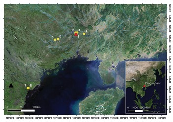
Figure 1. The location of sites in China and Vietnam with evidence for corpse manipulation: 1) Dingsishan; 2) Huiyaotian; 3) Qiujiang; 4) Lingwu; 5) Chongtang; 6) Hecun; 7) Đa Bút; 8) Con Co Ngua (maps created by Z.Q. Ye using QGIS v. 3.4.7).
Table 1. Typological, radiocarbon and calibrated dates for the Dingsishan and Huiyaotian sites.

Note: a) Guangxi Team et al. (1998); b) Fu (Reference Fu2002); c) Zhu et al. (Reference Zhu, Li, Chen, Fu and Hu2021); d) Chen (Reference Chen2016); e) Li et al. (Reference Li, Wang, Fu, Dobney, Li, Chen and Yu2013); f) dates modelled in OxCal v.4.4, using IntCal20 calibration curve (Bronk Ramsey Reference Bronk Ramsey2009; Reimer et al. Reference Reimer2020); g) Zhang & Hung (Reference Zhang and Hung2012); h) for comparison, radiocarbon and calibrated dates are also given for the Huiyaotian site, which is generally considered as a settlement strongly related to Dingsishan Phase III in the same period; i) Matsumura et al. (Reference Matsumura, Hung, Zhen and Shinoda2017).
To date, 336 burials containing 355 individuals interred in various burial positions have been identified. Applying Knüsel's (Reference Knüsel2014) terminology to field records from the Dingsishan site, individuals are categorised in five burial positions: extended (n = 2); limb-flexed (n = 113); contracted (lower limb flexed closely to the torso and vertebral column vertically oriented, n = 50), hyper-flexed (beyond the normal range of movement and anatomical articulation, n = 88) and unidentified (fragmentary assemblages, n = 102). Archaeologists have suggested that limb-flexed and contracted burials are the main funerary rituals in the Early and Middle Neolithic of southern China (Zhang & Hung Reference Zhang and Hung2012; Li et al. Reference Li, Wang, Fu, Dobney, Li, Chen and Yu2013), so the presence of 88 hyper-flexed individuals in 84 burials could be interpreted as resulting from a special, intentional form of interment. Among these burials, 87 individuals in 83 burials belong to phase III and only one belongs to phase IV. According to previous burial and anatomical analyses, decapitation, evisceration and disarticulation of joints are features of corpse manipulation at Dingsishan, and are not restricted to hyper-flexed burials (Fu Reference Fu2002). Hyper-flexed burials have been observed at five sites in Neolithic southern China (Hecun, Qiujiang, Dingsishan, Lingwu and Huiyaotian, see Figure 1), suggesting a constrained geographic distribution for this burial tradition (He et al. Reference He, Yang and Ning2009; Li et al. Reference Li, Wang, Fu, Dobney, Li, Chen and Yu2013; Qin Reference Qin and Wang2015; Matsumura et al. Reference Matsumura, Hung, Zhen and Shinoda2017). The cemeteries of the Đa Bút Culture in northern Vietnam also feature a similar pattern of depositional bodily modification to the Dingsishan site (Viet Reference Viet2005; Trinh & Huffer Reference Trinh and Huffer2015).
Several archaeological and ethnological hypotheses have been proposed to account for the abnormal skeletal representations at this site, though no thorough osteological examination has previously been reported. Qin (Reference Qin2010) suggests that the hyper-flexed burials found at Dingsishan are variants of limb-flexed burials: when group members died away from the site, the corpses were disarticulated, packed for convenience of transportation or release of rigor mortis, and later buried in a limb-flexed-like position. Using the historical record and ethnography of the later period of southern China as an analogy, Pan (Reference Pan2004) argues that disarticulation served to prevent the spirit's revenge. The arrangement of the bodies may also suggest that soft, perishable containers or quadrangular boxes were used in the burial process (Kerner Reference Kerner2018, Figure 2).

Figure 2. Examples of hyper-flexed, contracted and limb-flexed burials at Dingsishan. M65 (deposit code), M107 and M191 are hyper-flexed burials; M59 and M34 are limb-flexed burials; M24 is a contracted burial cutting through the burial M34 (photographs by X.G. Fu; figure by Z.Q. Ye & F.J. Li).
Such conjecture is enlightening, but a discussion of burial diversity is not the focus of this article. Instead, we analyse cutmarks on human bone from Dingsishan and how these may relate to methods of preparing bodies for interment. This study applies an integrated approach in which both archaeological and osteological information are considered in order to reconstruct funerary processes. Field records, including descriptions, images and hand drawings of burial deposits, are reviewed as background information for the analysis of osteological modifications. Within this context, we address the following questions: Are cutmarks present on all hyper-flexed individuals? What are the details of corpse manipulation at Dingsishan (e.g. the sex or age difference)? Is there any specific selection of location within the body for manipulation? By analysing the anthropogenic modification of the skeletons, we aim to reconstruct processes of individual manipulation, including initial preparation of the body, further corpse treatment and final placement, at Dingsishan.
Materials and methods
The complete assemblage of human bone from Dingsishan was examined. A team from Sun Yat-sen University and the Institute of Archaeology at the Chinese Academy of Social Sciences completed sex and age-at-death estimation following the standards of Lovejoy (Reference Lovejoy1985) and Bruzek (Reference Bruzek2002). Following the morphological criteria of Potts and Shipman (Reference Potts and Shipman1981), we undertook initial observations with a hand lens to distinguish different types of cutmarks and to differentiate them from excavation damage, antemortem injury, animal trampling, root etching and blood vessel impressions (Fernandez-Jalvo & Andrews Reference Fernandez-Jalvo and Andrews2016; McKinley & Smith Reference McKinley, Smith, Mitchell and Brickley2017). To confirm the exact position of each cutmark, microscratch observation and measurement were conducted with a handheld microscope (Dino-Lite Pro AM413ZT) under a magnification of 20–50×. In conjunction with DinoXcope for MacOS, we collected high-magnification images and orientation data for the cutmarks.
The position of each cutmark was recorded relative to standard human anatomical posture (extended supine). Referring to Pickering and Hensley-Marschand's (Reference Pickering and Hensley-Marschand2008) criteria of measurement, cutmark angles equate to the degree of excursion from parallel relative to the long axis of the specimen, regardless of the distal or proximal end of the bones (0–180°, whereby 0° denotes a mark which is parallel to the bone long axis and pointing to the distal end, and 180° denotes a mark parallel to the long axis but pointing to the proximal end). Under DinoXcope software, measurements were made twice on each mark and the average was taken as the final result following the criteria above (see online supplementary material (OSM) Table S1).
In this study, corpse manipulation was determined by representation of burial position and osteological observation. To evaluate any age- or sex-based preferential manipulation of individuals, percentage prevalence of cutmarks in different age-at-death categories were calculated, and Chi-square tests were performed to examine the relationship between estimated sex and evidence of manipulation using the SPSS Statistics software (v. 19.0). Various human behaviours may ‘mark’ bones, including slicing, chopping, gnawing and puncturing (Shipman Reference Shipman1981). To avoid a priori assumptions of specific intent, the term ‘cutmark’ is used here as a general term for marks made by artefacts, as defined by Potts and Shipman (Reference Potts and Shipman1981).
Results
Palaeodemographic information on manipulated corpses
The 88 individuals with hyper-flexed burial positions are classed as manipulated corpses, as the degree of joint flexion in the skeletons exceeds that achievable during normal motion. Microscratch observation identified cutmarks on 54 of the hyper-flexed individuals. Two individuals buried in a contracted position (M131 & M213) and one individual with an unknown burial type (M134-3) also bore cutmarks. A total of 91 skeletons from phases III and IV may therefore be classed as manipulated remains, including 54 males, 22 females and 15 unidentified individuals. The Chi-square test indicates no significant difference in the sex of skeletons that were manipulated (χ2 = 1.866, p = 0.172) compared with the overall population of phases III and IV (males = 169, females = 92). Table 2 shows that manipulation was most prevalent among individuals between the ages of 24 and 55 (80.77% of the known-age individuals). This indicates that manipulation was not constrained by the sex of an individual but was primarily reserved for adults.
Table 2. Comprehensive information on manipulated individuals at Dingsishan.
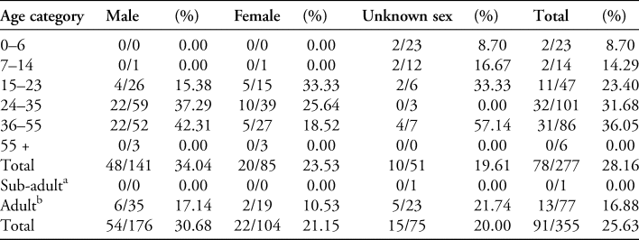
Note: number of manipulated individuals/ total individuals in each category; unknown age category: a sub-adult <18 years old; b adult ≥18 years old.
Types of modification marks
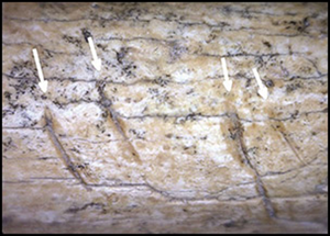
Microscratch observation found 212 cutmarks on 57 individuals at the Dingsishan site. The cutmarks can be categorised into three types. Scraping marks were identified in 13 cases (6.13%) on 11 individuals (Figure 3a & b). They were found on the exterior of the ribs, as well as on the diaphyses of femora, humeri and tibiae, where muscles are tightly attached. Chopping marks were identified in 20 cases (9.43%) on 11 individuals (Figure 3c) and were typically located on the posterior wall of the thorax, the neck and head of the femora, and around the elbow, in areas usually covered by thick layers of ligaments and tendons. The remaining 179 examples (84.43%) are slicing marks (Figure 3d–g), exhibiting relatively simple structures; all are fine, short lines with V-shaped cross-sections (Shipman Reference Shipman1981). In 96 instances, multiple slicing marks were found around muscle attachment sites, particularly on the patellae, vertebrae, clavicles and scapulae. Similarities in the morphology of slicing marks between skeletons suggest that modifications were made by the same kind of tool, possibly the sharp-edged tektite or obsidian tools that abound at the Dingsishan site (Li et al. Reference Li, Wang, Fu, Dobney, Li, Chen and Yu2013; Kerner Reference Kerner2018).

Figure 3. Types of cutmarks at Dingsishan: a & b) scraping mark on the shaft of humerus of M41-2 and femur of M108; c) chopping marks on the right femur of M273; d) slicing mark on the right tibial shaft of M270; e) parallel-line slicing mark on the left clavicle of M55; f) parallel-line slicing mark on the right patella of M55; g) clustered-line slicing mark on the left radius of M213 (figure by Z.Q. Ye & F.J. Li).
Distribution of the cutmarks
The distribution of cutmarks is summarised below by body segment (n denotes the number of cutmarks). No cutmarks were observed on the bones of the cranium or mandible (Figure 4, Tables 3 & S1).
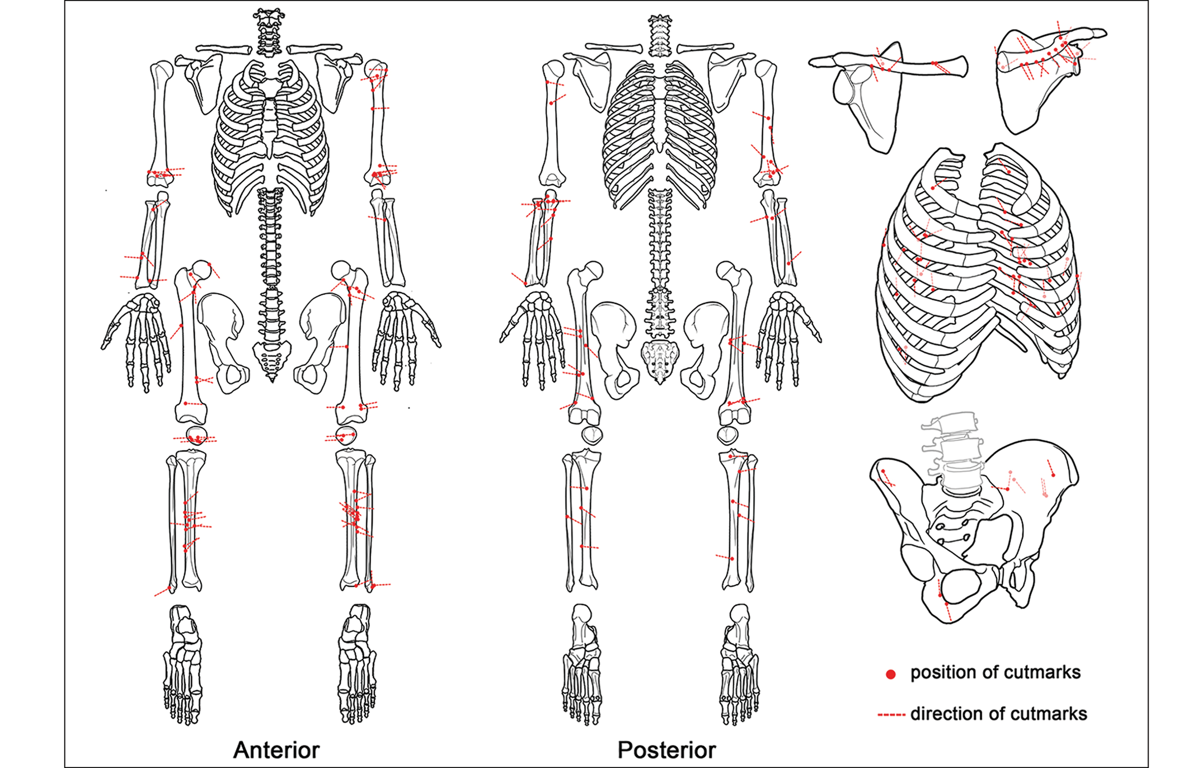
Figure 4. The distribution and directionality of cutmarks on the Dingsishan human skeletons (figure by Z.Q. Ye & F.J. Li).
Table 3. The cutmark distribution of different skeletal regions.
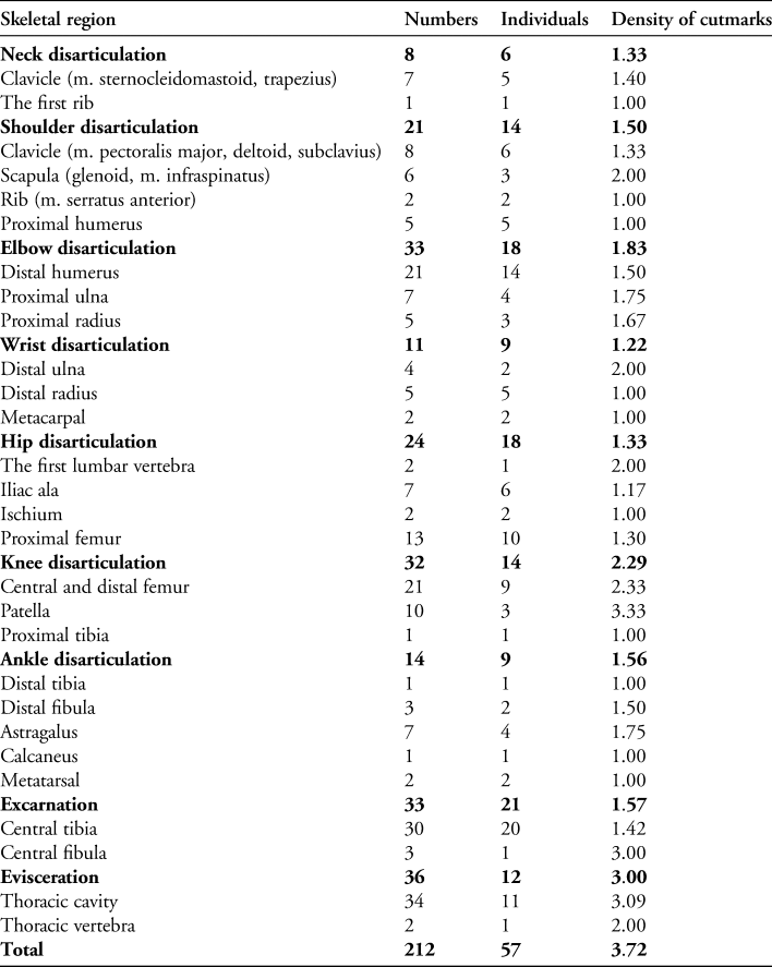
Note: the density of cutmarks is equal to the number of cutmarks divided by the number of manipulated individuals.
Axial skeleton:
Ribs (n = 37). The greatest density of cutmarks is found around the thorax. Three scraping marks (one each on M38, M126-1 and M193, see Table S1), located on the exterior of the thorax, may be the byproduct of neck and shoulder disarticulations. Other cutmarks are scattered on the inferior aspects of the fourth to twelfth ribs, demarking the posterolateral thoracic cavity (n = 34). Excavation reports demonstrate that at least 15 individuals were found with skulls interred within the thoracic cavity; together with the cutmarks this suggests the act of evisceration. Five chopping marks on the left, right and interior of the ribs of M38 (Figure 5) indicate the force used to cut and separate this individual's viscera.

Figure 5. Cutmarks on the internal surface of the ribs of M38 (figure by Z.Q. Ye & F.J. Li).
Vertebrae (n = 4). Though severance of the body through the waist may be observed from the depositional arrangement of some skeletons (e.g. M107 & M191 in Figure 2), only four slicing marks are identified on the vertebrae of two individuals (M38 & M264).
Upper limbs:
Clavicles (n = 15). In total, 11 clavicles show slicing marks (Figure 6). Generally, these occur as parallel or clustered lines in two areas: around the origin of the sternocleidomastoid muscle or the insertion of the trapezius muscle—both of which are involved in the rotation and flexion of the head—and at the attachment of the pectoralis major, deltoid and subclavius muscles, which facilitate movement of the upper arm.

Figure 6. Parallel and clustered cutmarks on the clavicles of M63 and M135 (figure by Z.Q. Ye & F.J. Li).
Scapulae (n = 6). Cutmarks are located around the glenoid cavity and at the attachment of the infraspinatus muscle (one on M41-1, two on M92 and three on M235, Table S1).
Humeri (n = 26). Five cutmarks are identified on the proximal part of the humerus of 15 individuals, located around the attachment site of the shoulder capsule ligament and the coracobrachialis, subscapular, deltoid and infraspinatus muscles. Twenty-one marks on 15 individuals are distributed over the humeral shaft and around the elbow.
Ulnae and radii (n = 21). Towards the elbow, cutmarks are identified at the attachment of muscles linking the upper and lower arm. Towards the wrist, slicing marks are found at the attachments of muscles linking the forearm and hand.
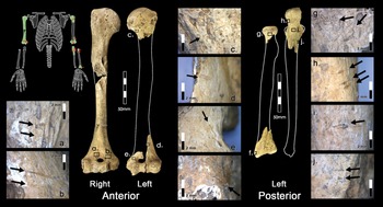
Ten cutmarks were found on the bones of the right and left arms of M213, their distribution suggesting an intention of arm disarticulation (Figure 7). All the cutmarks are directionally orientated towards the left side of the corpse. As experimental research illustrates that cutting motions generally move toward the operators for the convenience of hand movement (Pickering & Hensley-Marschand Reference Pickering and Hensley-Marschand2008), the cutmarks on M213 suggest that the operator occupied a relatively fixed position beside the body during corpse disarticulation. An understanding of the process of disarticulation is demonstrated in the distribution of cutmarks, particularly in the focus on areas of muscular or tendonous attachment and in the more varied orientation of marks around the elbow that accommodate muscle flow and bone structure.

Figure 7. Cutmarks found on the upper limbs of M213 (figure by Z.Q. Ye & F.J. Li).
Lower limbs:
Pelvis (n = 9). Four cutmarks are located at the site of attachment for the iliacus muscle on the interior of the ilium. Two cutmarks to the exterior ilium may have severed the gluteus maximus, medius and minimus muscles. Three cutmarks are found on the rims of the greater sciatic notch of two individuals (Table S1).
Femora (n = 34). The femora are the second most intensive area of modification, with cutmarks concentrating around the hip and knee joints. Cutmarks on the femoral head, neck and intertrochanteric line, as well as along the medial linea aspera of the femoral shaft, suggest that soft tissue around the hip, including the iliofemoral ligament and the iliacus and adductor muscles, was severed to disarticulate the joint. Cutmarks on the distal portions of femora also indicate disarticulation of the knee by severing the muscles that crossed the joint, such as gastrocnemius, biceps femoris and vastus medialis and lateralis.
Patellae (n = 10). Concentrated on the anterior surfaces of patellae, slicing marks are oriented to suggest the removal of the quadriceps tendons and the medial and lateral retinacula.
Tibiae and fibulae (n = 38). Cutmarks are concentrated around the attachment sites of the tibialis anterior muscle and the tibiofibular ligaments (n = 24), but are also observed on the posterior (n = 7) and medial (n = 2) aspects of the tibiae and around the knee and ankle joints (n = 5). Most of the tibiofibular cutmarks do not appear to be related to disarticulation, but instead may indicate the excarnation of the muscle that controls upward and inward movement of the foot—the tibialis anterior. These cutmarks may have been made for the relaxation and adjustment of foot posture following rigor mortis. Cutmarks to the posterior tibia can only be made in the absence of intact muscles, thus natural decomposition or anthropogenic excarnation probably preceded further treatment in this area of the body.
Discussion
Deliberate and skilful manipulation
Cutmarks on skeletal remains can be considered an unintentional ‘by-product’ of mortuary preparation practices, including excarnation and disarticulation (McKinley & Smith Reference McKinley, Smith, Mitchell and Brickley2017; Mazza et al. Reference Mazza, Acosta, Guarido, Buc and Loponte2018). Analysis of the spatial distribution, frequency and directionality of cutmarks can therefore allow us to reconstruct corpse manipulation and the Neolithic body treatment process at Dingsishan. Consideration of cutmark location and the functionality of the corresponding muscular attachments (Figure 4 and Table S1) suggests that corpse treatment at Dingsishan included disarticulation (143 cutmarks at 94 joints in 47 individuals), evisceration (36 cutmarks in 11 individuals) and excarnation (33 cutmarks in 21 individuals). Disarticulation and defleshing marks were found on bones in both hyper-flexed and contracted burials, indicating deliberate limb manipulation through disarticulation, rigor mortis relaxation and posturing.
Cutmarks are evident on just under two-thirds of the hyper-flexed individuals (61.36%) and occur at low frequency within each of these individuals. The location and frequency of cutmarks are consistent with manipulation during the early and late stages of decomposition, as demonstrated by previous micro-morphometric analyses of cut-marks on disarticulated human bones (Wallduck & Bello Reference Wallduck and Bello2016). Given the presence of numerous slicing and chopping marks inside thoracic and abdominal cavities, it is probable that mortuary treatment was undertaken on fresh corpses at Dingsishan; similar percussive treatment in a later stage of decomposition would have resulted in distinctive fracturing of the ribs.
The orientation of cutmarks varies by functional aim and by body part (Figure 8). Defleshing (excarnation) marks show the greatest consistency in orientation; they scratch obliquely to the long axis of bones with angles around 50–75°, indicating an intention to sever the muscle fibres. Cutmarks associated with evisceration concentrate at around 80–110° and demonstrate a consistent strategy of vertical dissection along the ribs. In contrast, cutmarks associated with disarticulation show a wider range of orientations, indicating the complexity of the process and the variety of approaches.

Figure 8. Rose diagrams showing orientations of cutmarks associated with different intentions and body parts: a) disarticulation; b) evisceration; c) excarnation; d & e) upper and lower limbs. The cutmarks are grouped every 5° and demonstrate a range in cutting angle from 0–180°. The number of the cutmarks is indicated by the length of the bin (diagrams created by Z.Q. Ye using ggplot2 v. 3.4.0 in R v. 4.2.2).
A variant of local Neolithic burial customs
Treatment of the corpse in the peri-mortem or post-mortem period in different cultures stimulates substantial discussion (Craig et al. Reference Craig, Knüsel, Carr, Pearson and Thorpe2005; Duday et al. Reference Duday, Cipriani and Pearce2009; Crozier Reference Crozier2018; Kerner Reference Kerner2018), but there is no diagnostic method for linking certain modifications (e.g. cutmarks) to specific practices (Weiss-Krejci Reference Weiss-Krejci, Rakita, Buikstra, Beck and Williams2005, Reference Weiss-Krejci, Agarwal and Glencross2011). In most cases, interpretations of behaviour are developed from certain historic and cultural contexts. The microscratch data presented here provide new evidence for the reconstruction of the function of corpse manipulation at Dingsishan.
No specific evidence for cannibalism, such as gnawing marks or thermal signatures (Jamieson Reference Jamieson1983; Kuckelman et al. Reference Kuckelman, Lightfoot and Martin2002; Cáceres et al. Reference Cáceres, Lozano and Saladié2007), is found in this collection and only 15.57 per cent of identified cutmarks can be defined as the result of defleshing. All of the burials were located in similar subterranean pits with limited accompanying furniture (e.g. natural or shaped fragments of stone, ceramics and shellfish) (Kerner Reference Kerner2018) and, aside from differences in the placement of the bodies within the graves, there is nothing to indicate that manipulation was used as a mark of honour or degradation, which might lead to interpretations of human sacrifice, necrophobia or other value judgements (Aufderheide Reference Aufderheide2003; Tsaliki Reference Tsaliki and Murphy2008). Moreover, the observation of cutmarks on bones from both hyper-flexed and contracted burials suggests that manipulated individuals were subsequently arranged for interment.
Variation in the positioning of cutmarks on bones and of disarticulated remains within burials means that no two individuals shared a pattern of post-mortem modification, although ultimately all manipulated individuals had flexed joints and were compressed into a limited space (Kerner Reference Kerner2018). We interpret this as evidence that, beyond the maintenance of a limb-flexed position, each individual was processed in a different way depending on multiple factors, including body size, age at death, level of rigor mortis and manner of death. This suggests that, although there are common aspects of interment—mature adults being, for example, more frequently in receipt of overt manipulation—the processing of corpses was variable for each body. In the ‘distant transport’ hypothesis, disarticulation, excarnation of extremities and evisceration with thoracic interment of skulls may be interpreted as aspects of funerary treatment associated with packaging the corpse for transport (Qin Reference Qin2010), possibly by mobile hunter-gatherers. Considering the cramped distribution of burials (Li et al. Reference Li, Wang, Fu, Dobney, Li, Chen and Yu2013) and the possibility of grave space organisation (Kerner Reference Kerner2018), these processes could also be seen as accommodations within the funerary practice to fit individuals into pre-designated burial spaces within the whole cemetery.
Conclusion
Identification of anthropogenic modifications to bodies and burial in postures beyond the normal anatomical range denote a funerary process specific to Neolithic phases III and IV at Dingsishan. No differences in burial treatment between the sexes are apparent but mature adults dominate the manipulated assemblage. The compressed form of burial and large variation in the placement and orientation of cutmarks within each skeleton points to two likely interpretations of the underlying aim of the manipulation: the ‘distant transport’ hypothesis (Qin Reference Qin2010) and the ‘space organisation’ hypothesis (Kerner Reference Kerner2018). Lack of consistency in the positioning of disarticulated remains and in burial postures fits the profile of unstandardised and non-ritualistic behaviour.
Yet the deliberate and accurate cutmarks on skeletons imply several sequential and highly organised mortuary processes. Considerable effort was placed on the preparation, management and implementation of corpse treatment, and these goal-oriented and strategic manipulations would require a measure of co-operation, experience and even technical knowledge. Thus, this process could be interpreted as a respectful mourning and/or an attempt at consistency with the wider limb-flexed burial custom.
This study provides new information on post-mortem processing of human remains in Neolithic southern China and opens the possibility of further comparisons with similar deposits from other geographic areas. Furthermore, given the intimate interaction between Neolithic cultures in Southeast Asia, application of the combined microscratch and archaeothanatological method employed here at other sites within the region may shed light on the origins and influences of Neolithic funerary practices more generally.
Acknowledgements
We are grateful to the following scholars for their valuable advice: Professor Qiang Lin, Professor Zhen Li, Professor Fang Qin and Professor Anyi He in Guangxi Institute of Cultural Heritage and Archaeology of China, Professor Nguyen Lan Cuong in the Archaeological Society of Vietnam, Dr Jenny Kerner in Muséum National d'Histoire Naturelle and du Musée de l'Homme of France, Dr Da Di in Université de Genève of Switzerland, Yue Zhang in the Australian National University of Australia, Ying Yu in Sun Yat-sen University of China, Boyu Chen in Guangdong Institute of Cultural Heritage and Archaeology of China. We are also grateful to the editor and two reviewers whose professional advice greatly improved the article.
Funding statement
This study is supported by the National Social Science Foundation of China (13CKG002) and the co-operative project of Sun Yat-sen University and Guangzhou Municipal Institute of Cultural Heritage and Archaeology (23000-71020804).
Supplementary material
To view supplementary material for this article, please visit https://doi.org/10.15184/aqy.2024.57.