


Introduction
Conodonts are an extinct group of jawless, boneless fishes that first appeared in the Cambrian and went extinct at the end of the Triassic. Their fossil record primarily consists of mineralized oral and oropharyngeal denticle-bearing elements (referred to as “conodont elements”) analogous to teeth (Murdock et al. Reference Murdock, Dong, Repetski, Marone, Stampanoni and Donoghue2013), and these elements are frequently preserved. As a result, conodont fossils are found in marine rocks on all continents and can be very common in some localities. While some workers dispute the vertebrate affinities of conodonts (Blieck et al. Reference Blieck, Turner, Burrow, Schultz, Rexroad, Bultynck and Nowlan2010; Turner et al. Reference Turner, Burrow, Schultze, Blieck, Reif, Rexroad, Bultynck and Nowlan2010), most work in the last 30 years has suggested a vertebrate affinity based on studies of preserved soft tissues, composition of tooth-like elements, and phylogenetic analyses (Aldridge Reference Aldridge, Briggs, Smith, Clarkson and Clark1993; Donoghue et al. Reference Donoghue, Forey and Aldridge2000; Donoghue Reference Donoghue2001; Terrill et al. Reference Terrill, Henderson and Anderson2018). Although the exact relationships between conodonts and other vertebrates are still uncertain, recent work has suggested conodonts are closely affiliated with cyclostomes, a group that consists of hagfish and lampreys (Donoghue and Rücklin Reference Donoghue and Rücklin2016; Terrill et al. Reference Terrill, Henderson and Anderson2018; Miyashita et al. Reference Miyashita, Coates, Farrar, Larson, Manning, Wogelius, Edwards, Anné, Bergmann, Palmer and Currie2019).
The conodont fossil record is unusual among vertebrates. The lack of bones has led to a dearth of body fossils, similar to other boneless vertebrates such as lampreys and hagfish (Miyashita et al. Reference Miyashita, Coates, Farrar, Larson, Manning, Wogelius, Edwards, Anné, Bergmann, Palmer and Currie2019). The record of their mineralized elements is, however, among the best of any group of organisms (Foote and Sepkoski Reference Foote and Sepkoski1999) from the upper Cambrian through the Upper Triassic. Their relative abundance and high evolutionary rates have made conodonts ideal index fossils for biostratigraphy, where they are used to define many important geologic boundaries such as the Permian/Triassic (Yin et al. Reference Yin, Zhang, Tong, Yang and Wu2001) and the Frasnian/Famennian boundaries (House et al. Reference House, Menner, Becker, Klapper, Ovnatanova and Kuz'min2000). Conodonts have been well documented and analyzed since they were first described by Pander (Reference Pander1856) and have been the subject of numerous paleoecological studies over the last half century (Clark Reference Clark1974, Reference Clark1984; Barnes Reference Barnes1976; Ji and Barnes Reference Ji and Barnes1994; Donoghue and Purnell Reference Donoghue and Purnell1999; Armstrong and Smith Reference Armstrong and Smith2001; Jarochowska et al. Reference Jarochowska, Viira, Einasto, Nawrot, Bremer, Männik and Munnecke2017), yet their role in ecosystems, such as their dietary niches, remains elusive.
The lack of knowledge surrounding conodont trophic paleoecology is problematic for the understanding of marine ecology in the Paleozoic and Triassic, as highlighted by a recent study reexamining the Devonian nekton revolution (Whalen and Briggs Reference Whalen and Briggs2018). The Devonian nekton revolution refers to the apparent rapid occupation of the water column during the Devonian Period, with nektic biodiversity rising from 15% of non-benthic taxa at the end of the Silurian to approximately 85% by the end of the Devonian (Klug et al. Reference Klug, Kröger, Kiessling, Mullins, Servais, Frýda, Korn and Turner2010). By simply including conodonts as probable nektic swimmers, Whalen and Briggs (Reference Whalen and Briggs2018) showed that nektonism had not risen suddenly in the Devonian, but rather gradually since the first appearance of conodonts in the Cambrian. Given the importance of conodonts in the shift toward nektic environmental occupation, understanding the dietary niches of conodonts is vital to our understanding of how and why animals began to increasingly occupy the water column.
While the question of what conodonts ate remains uncertain, there have been a number of studies directed at understanding how conodont elements may have been used to capture and process food. Microwear analyses have shown that conodont elements frequently possess wear facets and damage consistent with use in food processing (Purnell Reference Purnell1995; Purnell and Jones Reference Purnell and Jones2012), with wear patterns indicating occlusion similar in complexity to mammals in platform elements (Donoghue and Purnell Reference Donoghue and Purnell1999). Dental function is further supported by microstructural analysis of conodont crown tissues (tissues thought to be partially or completely exposed in the oral cavity; Donoghue Reference Donoghue2001), while occlusion between platform elements is further supported by structural modeling of stress distribution during function (Martínez-Pérez et al. Reference Martínez-Pérez, Rayfield, Purnell and Donoghue2014). Mechanical models created based on clusters of conodont elements preserved in positions close to their relative orientation in the apparatus in vivo have been used to formulate hypotheses for how the oral apparatus may have functioned to capture food (Goudemand et al. Reference Goudemand, Orchard, Urdy, Bucher and Tafforeau2011). Due to these efforts, the current hypothesis is that the preserved conodont elements did indeed function as teeth, without any direct evidence of dietary preferences being known. It has been suggested that size of conodont elements could be used as a proxy for trophic position (Balter et al. Reference Balter, Martin, Tacail, Suan, Renaud and Girard2019; Ginot and Goudemand Reference Ginot and Goudemand2019), with larger elements possibly being indicative of a more carnivorous diet (Ginot and Goudemand Reference Ginot and Goudemand2019). Using size as a proxy for trophic position has been observed to be somewhat inconsistent in modern cyclostomes, however, with a positive correlation among lampreys and no statistical relationship among hagfish (Romanuk et al. Reference Romanuk, Hayward and Hutchings2011). Conodont body size also may not be directly related to conodont element size, so inferring conodont trophic positioning based on element size is heavily reliant on assumptions that may not be well founded.
One possible path to explore conodont dietary niches is to examine the chemistry preserved in conodont elements. The crown of a conodont element is comparable in composition to vertebrate tooth enamel; it is hypermineralized and composed of hydroxyapatite. As such, conodont elements are highly resistant to diagenetic processes and often record chemical signatures that reflect original biological composition (Barham et al. Reference Barham, Joachimski, Murray and Williams2012). Thus far there have only been a few studies that applied chemical analysis to the question of conodont trophic structure using calcium isotopes and organic carbon isotopes (Shirley et al. Reference Shirley, Grohganz, Bestmann and Jarochowska2018; Balter et al. Reference Balter, Martin, Tacail, Suan, Renaud and Girard2019; Zhuravlev Reference Zhuravlev2020), which concluded that several Devonian conodont taxa were low-level consumers that most likely fed on plankton. While compelling, data reported in these studies still show a significant variation in isotopic values within each taxon and do not address the very large diversity of morphologies that exist throughout the conodont fossil record.
In this study, we analyzed the Sr/Ca and Ba/Ca content of several co-occurring Silurian conodont species from the Gotland succession in Sweden. Sr and Ba are common trace elements in vertebrate hard tissues, substituting for Ca in bones and teeth in vivo (Reynard and Balter Reference Reynard and Balter2014; Shirley et al. Reference Shirley, Grohganz, Bestmann and Jarochowska2018). The relative abundances of these trace elements can be affected by several ecological factors. In terrestrial vertebrates, the ratio is dominated by diet, with carnivores typically having lower Sr/Ca and Ba/Ca ratios than herbivores (Gilbert et al. Reference Gilbert, Sealy and Sillen1994; Burton et al. Reference Burton, Price and Middleton1999; Blum et al. Reference Blum, Taliaferro, Weisse and Holmes2000; Peek and Clementz Reference Peek and Clementz2012; Kohn et al. Reference Kohn, Morris and Olin2013). The proposed mechanism for this trophic proxy is the biopurification of Ca, a biological process in which Ca preferentially accumulates in the skeleton over trace elements such as Sr and Ba, which results in an organism having lower Sr/Ca and Ba/Ca ratios relative to their dietary source (Burton et al. Reference Burton, Price and Middleton1999; Peek and Clementz Reference Peek and Clementz2012). The relationship between trophic positioning and Sr/Ca and Ba/Ca ratios has also been observed in marine vertebrates (Peek and Clementz Reference Peek and Clementz2012); however, additional factors may impact these ratios. It has been observed that Sr and Ba can bioaccumulate near the base of marine food webs (Peek and Clementz Reference Peek and Clementz2012), while water temperature has been shown to also impact Sr/Ca and Ba/Ca ratios in the bones of captive seabreams (Balter and Lécuyer Reference Balter and Lécuyer2010; Balter et al. Reference Balter, Lécuyer and Barrat2011). To date, there have been very few examinations of Sr/Ca and Ba/Ca ratios in modern fish apatite beyond the studies mentioned, while examinations of Sr/Ca and Ba/Ca ratios preserved in modern fish otoliths (which are composed of biogenic aragonite) are much more numerous. Studies on otoliths have shown Sr/Ca and Ba/Ca ratios are significantly impacted by water temperature (Martin and Thorrold Reference Martin and Thorrold2005; Nelson et al. Reference Nelson, DeVries and Wright2018) and salinity (Diouf et al. Reference Diouf, Panfili, Labonne, Aliaume, Tomás and Chi2006; Martin and Thorrold Reference Martin and Thorrold2005; Zimmerman Reference Zimmerman2005; MacDonald and Crook Reference Macdonald and Crook2010; Nelson et al. Reference Nelson, DeVries and Wright2018). While otoliths do not consist of apatite, these results still reinforce the observations of Balter and Lécuyer (Reference Balter and Lécuyer2010) that temperature is an important variable to consider when making interpretations, while indicating that salinity should be considered as well. Finally, it is worth noting that, apart from the study by Peek and Clementz (Reference Peek and Clementz2012), there have been no comprehensive studies of marine trophic relationships between vertebrates using Sr/Ca and Ba/Ca ratios preserved in apatite, so the application of these methods to a fossil conodont community is not only novel from a paleontological perspective but represents one of the very few attempts to characterize the trophic structure of any marine community using these methods.
As conodont elements are not completely homogenous in terms of tissue composition, we investigate here whether histological differences between taxa might confound our interpretations of the Sr/Ca proxy. Conodont elements are composed of lamellar (or hyaline) tissue, finely laminated apatite that is considered structurally and chemically analogous to vertebrate enamel (Donoghue Reference Donoghue1998; Terrill et al. Reference Terrill, Jarochowska, Henderson, Shirley and Bremer2022: SI fig. 2). A second tissue is known as white (or albid) matter due to its white appearance in reflected light microscopy. White matter is often found in the denticles of conodont elements, but the distribution and volume vary greatly between taxa. It has different crystallographic properties (Trotter et al. Reference Trotter, Fitz Gerald, Kokkonen and Barnes2007; Atakul-Özdemir et al. Reference Atakul-Özdemir, Warren, Martin, Guizar-Sicairos, Holler, Marone, Martínez-Pérez and Donoghue2021) and elemental composition than the lamellar tissue (Trotter and Eggins Reference Trotter and Eggins2006). The compositional variation is therefore very important to determine; if white matter contains varying amounts of Sr, Ba, or Ca in comparison to the lamellar tissue, it could have a significant impact on the measured Sr/Ca and Ba/Ca ratios measured in taxa with systematically different proportions of these tissues.
In addition to Sr/Ca and Ba/Ca ratios, we also analyzed Sr isotopes preserved in different ontogenetic stages of Ozarkodina confluens. This was done to test the hypothesis that some conodonts may have displayed anadromous behavior, possibly migrating to freshwater environments to spawn. Sr isotope ratios in conodont elements are believed to directly reflect the water in which they lived (Bertram et al. Reference Bertam, Elderfield, Aldridge and Conway-Morris1992; Armstrong et al. Reference Armstrong, Pearson and Griselin2001). Sr isotope ratios are consistent throughout the world's oceans at a given moment in geologic time (Veizer Reference Veizer1989), as a result of mixing from fluvial sources of highly variable composition. Sr isotopic ratios should therefore be relatively consistent between individuals that lived exclusively in the marine realm. In contrast, ontogenetically younger individuals that partially inhabited fresh or brackish waters would have been exposed to a wider range of environmental Sr isotope values, resulting in higher variability in the preserved Sr isotope ratios. To determine whether conodonts may have spent time in fresh or brackish water environments (possibly related to spawning activities), comparisons were made between young and adult specimens.
We evaluate here the potential for using Sr/Ca and Ba/Ca ratios to study the trophic structure of conodont communities. This was done by analyzing the Sr/Ca and Ba/Ca ratios preserved in nine different conodont taxa from Gotland, Sweden (see Fig. 1 for examples of each taxon). By combining these data with Sr isotope ratios, in situ analysis of elemental composition through use of a microprobe, and paleoenvironmental information provided by facies analysis, we were able to account for several environmental and biological factors that may have influenced the preserved Sr/Ca and Ba/Ca ratios. This enabled us to test the hypothesis that morphological differences between conodont taxa reflect trophic diversification (Ginot and Goudemand Reference Ginot and Goudemand2019; Petryshen et al. Reference Petryshen, Henderson, De Baets and Jarochowska2020; Kelz et al. Reference Kelz, Guenser, Mazza, Rigo and Jarochowska2021), while also examining whether proxies for the trophic position proposed here are influenced by the histological composition of conodont elements.
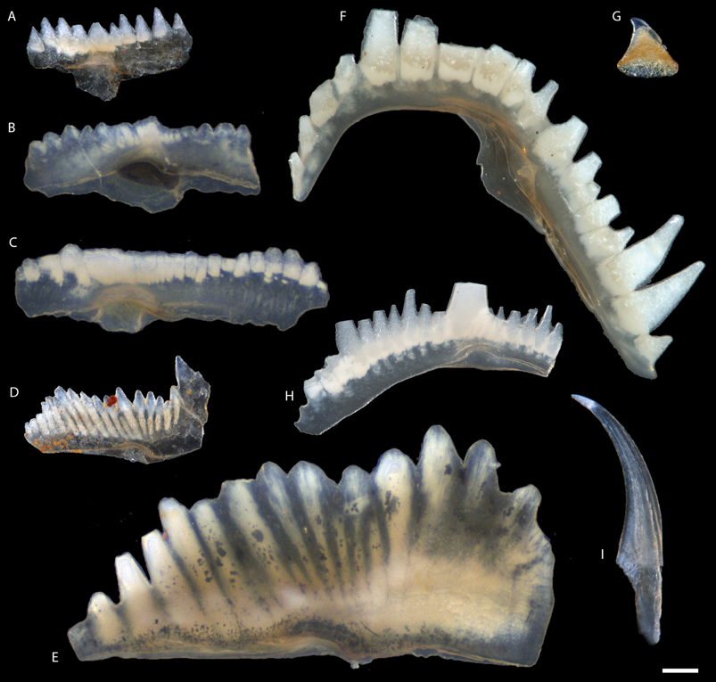
Figure 1. Typical morphologies of P1 elements of taxa discussed in the study. A, Zieglerodina remscheidensis, NRM-PZ Co99 from Ängvards 7, Sundre Fm. (sample G00-26 LJ). B, C, Ozarkodina bohemica from Gothemshammar 9, Halla Fm. (sample EJ-14-407): B, EJ-14-407-0001; C, EJ-14-407-0002. D, Ozarkodina confluens from Storms 2, Sundre Fm., NRM-PZ Co155 (sample G94-42 LJ). E, Ctenognathodus murchisoni from Gothemshammar 9, Klinteberg Fm., EJ-14-410-0001 (sample EJ-14-410). F, Oulodus ?excavatus from Rivviken 2, Hamra or Sundre Fm. NRM-PZ Co153 (sample G04-739 LJ). G, Pseudooneotodus beckmanni from Närshamn 1, Burgsvik Fm., NRM-PZ Co79 (sample G83-11LJ). H, Wurmiella excavata from Barshageudd 2, Sundre Fm., G14-18-OB-0001 (sample G14-18OB). I, Panderodus equicostatus from Närshamn 2, Burgsvik Fm., NRM-PZ Co72 (sample G83-12LJ). Scale bar, 100 μm. Collection identifications starting with NRM-PZ refer to Swedish Museum of Natural History, Stockholm; all other specimens are hosted at Friedrich-Alexander-Universität, Erlangen.
Methods
Conodonts
Conodont assemblages were extracted from 10 rock samples collected in the Swedish island of Gotland. The strata exposed in Gotland represent carbonate deposits formed on a tropical carbonate platform, which during the Silurian Period formed in an epeiric sea on the shelf of Baltica (Baarli et al. Reference Baarli, Johnson and Antoshkina2003). The cratonic position and lack of tectonic deformation led to excellent conodont preservation (Conodont Alteration Index [CAI] = 1) in the absence of substantial burial. They also allowed the development of a detailed stratigraphic interpretation across coeval paleoenvironments represented in the outcrops in Gotland (Calner et al. Reference Calner, Jeppsson and Munnecke2004; Jeppsson et al. Reference Jeppsson, Eriksson and Calner2006). Reviews of depositional environments in the Silurian of Gotland have been compiled by Samtleben et al. (Reference Samtleben, Munnecke and Bickert2000) and Calner et al. (Reference Calner, Jeppsson and Munnecke2004). Rock samples used in this study are summarized in Table 1. Except for the two youngest samples (G14-18OB and G14-19OB), they have been processed at GeoZentrum Nordbayern following the technique of Jeppsson et al. (Reference Jeppsson, Anehus and Fredholm1999), that is, digested in buffered 7% acetic acid and wet sieved. The fraction between 63 μm and 1 mm was retained, separated from lighter residue in sodium polytungstate with the density of 2.83 g × cm−3, and hand-picked. Samples G14-18OB and G14-19OB were processed at Department of Geology at Lund University as described in Bremer et al. (Reference Bremer, Jarochowska and Märss2020).
Table 1. Summary of biostratigraphic ages (according to Jeppsson et al. Reference Jeppsson, Eriksson and Calner2006) and environments represented by each sample in this study, as well as taxa found in those samples. Taxa abbreviations are as follows: Ou., Oulodus; Oz., Ozarkodina; Pa., Panderodus; Ps., Pseudooneotodus; W., Wurmiella; Z., Zieglerodina.

Elemental Abundances
Identifiable elements were hand-picked from digested rock samples and separated into 256 monospecific analytical samples. Thus, conodonts extracted from one rock sample yielded multiple analytical samples. Analytical samples were first cleaned through immersion in a 7% unbuffered acetic acid solution for a minimum of 1 hr. This was done to remove any remaining carbonate material and other contaminating particles attached to the fossil's surface. After removal from acid, each fossil was transferred to a deionized water bath for another hour to rinse off and dilute remaining acid before being placed in clean microfossil slides for transport. Conodont fossils were then weighed using a scale to create analytical samples for measurement. While some individual conodont fossils were massive enough to constitute a complete analytical sample, other analytical samples were amalgamations of multiple specimens belonging to the same taxon. Minimum sample mass was 20 μg, with most analytical samples having a mass exceeding 50 μg (see Terrill et al. Reference Terrill, Jarochowska, Henderson, Shirley and Bremer2022: SI table 1).
Analytical samples were then digested in 30 μl of 3 M nitric acid. Digestion was typically complete after just 1 hr; however, all samples were allowed to digest for at least 2 hr. One milliliter of deionized water was then added to dilute the analytical sample, which was mixed using a vortex machine. Samples were further diluted by a factor of 5 to 50, depending on the initial mass of the sample, using deionized water. An internal calibration standard was then added before each analytical sample was again mixed using the vortex machine. Samples were then loaded into a tray and sampled using a Thermo Scientific iCAP Qc quadrupole inductively coupled plasma mass spectrometry (ICP-MS) mass spectrometer in the Department of Geology at Friedrich-Alexander-Universität Erlangen-Nürnberg and measured in triplicate. Results were compared with the Bone Ash NIST SRM 1400 standard. Full results are presented in Terrill et al. (Reference Terrill, Jarochowska, Henderson, Shirley and Bremer2022: SI table 1).
Electron Microprobe Analysis
In addition to analyzing the bulk Sr/Ca and Ba/Ca content of conodont elements, we employed an electron microprobe to examine the potential variation in composition between tissue types. Conodont elements can generally be divided into two major tissue types, the crown tissue and the basal tissue, based on differences visible under the light microscope, crystallographic properties, and composition. The basal tissue is less mineralized and more organic than the crown tissue (Müller and Nogami Reference Müller and Nogami1971; Donoghue Reference Donoghue1998). As a result, basal tissue is more likely to experience diagenetic alteration and is not ideal for chemical analyses (Holmden et al. Reference Holmden, Creaser, Muehlenbachs, Bergstrom and Leslie1996; Wenzel et al. Reference Wenzel, Lécuyer and Joachimski2000). In this study, we only analyzed conodont crowns to eliminate the possible impacts of basal tissues on our results.
Using an electron microprobe, we measured the variation in Sr concentrations between white matter and lamellar tissue in three cross-sectioned conodont crowns. The measurement was set up with the following elements to be detected: Na, F, Ba, and P (as P2O5). The sections were prepared following the methods described by Shirley et al. (Reference Shirley, Bestmann and Jarochowska2020). The measurements were made at University of Calgary electron microprobe facility using a JEOL JXA-8200 equipped with five wavelength-dispersive spectrometers using a spot size of 5 μm to minimize sample damage.
All conodont specimens for microprobe analysis were P1 elements of Ozarkodina confluens (for anatomical notation, see Purnell et al. Reference Purnell, Donoghue and Aldridge2000). Specimens EJ-14-111-3 and EJ-14–407-016 were collected from rock samples using the same methods as described in “Conodonts.” They derive from samples EJ-14-111 and EJ-14-407, collected in the uppermost part of the “Gothemshammar member” (informal unit) of the Halla Formation (Homerian, Wenlock) at Gothemshammar 9 (57°36′33.46″N, 18°47′46.32″E; Jarochowska et al. Reference Jarochowska, Bremer, Heidlas, Pröpster, Vandenbroucke and Munnecke2016). EJ-14-111-3 was a subadult specimen, visibly smaller and with fewer denticles than the others. EJ-14-407-016 was characterized in terms of histology and chemical composition by Shirley et al. (Reference Shirley, Grohganz, Bestmann and Jarochowska2018). Specimen Co0000203 comes from rock sample G89-797 in the collection of Lennart Jeppsson housed at Swedish Museum of Natural History. It was collected at Linde 3 (Gorstian, Ludlow; 57°16′30.22590″N, 18°22′52.29829″E) and processed at the Department of Geology at Lund University. The elements were embedded individually in an epoxy resin (Körapox 398, Kömmerling) and allowed to harden for at least 24 hr. They were then ground using a progression of 800 and 1200 grit silicon carbide until the optimal section plane was reached. The sections were polished mechanically using a 1 μm diamond spray for 20 min and chemically in OP-S Nondry colloidal silica suspension for 2 min using a Logitech WG2 polishing head. Polished specimens were coated with 3.8 nm of carbon to prevent charging.
Statistical Evaluation of Electron Microprobe Results
The differences in chemical composition between the lamellar tissue and the white matter were evaluated by fitting a mixed-effects general linearized model using the lme4 package (Bates et al. Reference Bates, Maechler, Bolker and Walker2015) for R Software v. 3.5.1 (R Core Team 2018). A model with tissue as a fixed effect and specimen as a random effect was compared against the null model accounting only for the variability between specimens. This comparison was carried out individually for all elements that could be detected using the microprobe, that is, values above the detection limit (Table 2). Instead of using p-values, likelihood ratios were used to evaluate the strength of the fixed effect; to that end, the estimates of the effects were chosen to optimize the log-likelihood criterion.
Table 2. Comparison of the chemical composition of the lamellar tissue (N = 30; 18 for BaO) and the white matter (N = 10; 6 for BaO) obtained with electron microprobe using mixed-effects generalized linear models. The values have been rounded to reflect the precision of the microprobe; for Ba, the difference between estimated effects of the tissues lies below the detection limit. AIC, Akaike information criterion.

Previous research by Trotter and Eggins (Reference Trotter and Eggins2006) and Shirley et al. (Reference Shirley, Grohganz, Bestmann and Jarochowska2018) indicated that changes in Sr composition can also be detected within the lamellar tissue. To evaluate this, measurements from the lamellar tissue have been divided into two classes: (1) center, close to the growth axis; and (2) side, close to the specimen edge. The role of the position within the lamellar tissue was evaluated using a mixed-effects model with position as a fixed effect and specimen as a random effect, analogous to the model comparing the tissues.
The total number of measurements depended on the detection limits of the microprobe. For CaO, SrO, Na2O, F, and P2O5, all 40 measured points yielded values above the detection limit (10 in the white matter, 30 in the lamellar tissue). Ba measurements yielded results above the detection limit at 24 points only, the remainder was coded for the needs of the analysis as zero. The complete dataset of measurements is provided in Terrill et al. (Reference Terrill, Jarochowska, Henderson, Shirley and Bremer2022: SI table 2).
Sr Isotope Ratios
For this study, we analyzed the 87Sr/86Sr ratio preserved in 13 elements belonging to Oz. confluens. Three ontogenetic stages were distinguished qualitatively under the light microscope based on the size of the element and the number of denticles. Conodonts with a low CAI (as determined through visual inspection and comparison with defined standards) and no visual evidence of recrystallization have been demonstrated to be good sources of original Sr isotope data and have been used as a proxy for determining global marine values through geologic time (Kurschner et al. Reference Kurschner, Becker, Buhl and Veizer1992; Diener et al. Reference Diener, Ebneth, Veizer and Buhl1996; McArthur et al. Reference McArthur, Howarth, Shields, Zhou, Gradstein, Ogg, Schmitz and Ogg2020). Marine Sr isotope ratios are considered to be globally consistent due to the Sr residence time being much longer than the global ocean mixing rate (Veizer Reference Veizer1989). Marine values are only expected to vary under specific circumstances, such as in a restricted basin, where connection between an inland sea and the larger ocean is minimal or absent. Freshwater or brackish water ecosystems can, however, show significant variation, as the Sr content will be mostly controlled by local inputs such as bedrock weathering (Veizer Reference Veizer1989). As conodont elements grew through external apposition (Dzik Reference Dzik2008), they potentially retain a record of the elemental composition of waters they inhabited through their ontogeny. If a conodont species were to spend time in a coastal river or estuary (to spawn, for example), the Sr isotope ratios preserved in the oral/oropharyngeal elements would be expected to show variations depending on ontogeny and would also vary with respect to species that lived entirely in marine settings.
All Sr isotope ratios were measured using thermal ionization mass spectrometry (TIMS) at the University of Calgary. Measurement error ranged between 0.000004 and 0.000042 and averaged 0.000008 across all analyses. Before analysis, elements were first exposed to diluted acetic acid to remove any diagenetic material that might be present on the outer surface of the fossils. They were then digested in 0.5 ml of 3.0 M nitric acid and 30 μl of peroxide. An ion-exchange procedure was then performed to concentrate the Sr ions before analysis. The results are reported in Terrill et al. (Reference Terrill, Jarochowska, Henderson, Shirley and Bremer2022: SI table 3).
Results
Elemental and Isotopic Composition of Conodont Crowns
Sr/Ca and Ba/Ca ratios are summarized in Figure 2 and Terrill et al. (Reference Terrill, Jarochowska, Henderson, Shirley and Bremer2022: SI table 1). Of the nine taxa analyzed in this study, five (Oulodus excavatus, Ozarkodina bohemica, Panderodus equicostatus, Wurmiella excavata, and Zieglerodina remscheidensis) produced measurements with distinct and well-constrained ranges of Sr/Ca and Ba/Ca values. Panderodus equicostatus contained generally high Ba/Ca and Sr/Ca ratios, while W. excavata and Ou. excavatus both contained low Ba/Ca and Sr/Ca ratios. Oz. bohemica also recorded low Sr/Ca and Ba/Ca ratios; however, there was more variability in Ba/Ca ratios than seen in other taxa. Zieglerodina remscheidensis recorded moderate ratios, with lower Sr/Ca and Ba/Ca ratios than Pa. equicostatus, but higher values than Ou. excavatus and W. excavata. Two taxa (Ctenogathodus spp. and Ozarkodina confluens) recorded much more variable Sr/Ca ratios, with some specimens recording the highest Sr/Ca ratios in this study, while Ba/Ca ratios of these taxa were consistently low in most measurements. The two remaining species (Panderodus unicostatus and Pseudooneotodus beckmanni) were only found in a single assemblage. Panderodus unicostatus recorded Sr/Ca and Ba/Ca ratios in line with Pa. equicostatus, while Ps. beckmanni recorded low Sr/Ca ratios and very high Ba/Ca ratios.

Figure 2. Ba/Ca vs. Sr/Ca values in crown tissues of nine conodont taxa from 10 rock samples in this study. Polygons encompass the complete range of values in each taxon. Samples are arranged according to the interpreted paleoenvironment represented in each rock sample.
In addition to Sr/Ca and Ba/Ca ratios, Sr isotope data were also collected from Oz. confluens. Dental elements representing different ontogenetic stages and different horizons were selected and analyzed for 87Sr/86Sr, with the results summarized in Figure 3. The 87Sr/86Sr ratios clearly show very little variation within a rock sample regardless of ontogeny, with almost all elements within a horizon reporting values within the margin of error (see Terrill et al. Reference Terrill, Jarochowska, Henderson, Shirley and Bremer2022: SI table 2).

Figure 3. Strontium isotope ratios preserved in Ozarkodina confluens from different rock samples and stratigraphic ages (see Terrill et al. Reference Terrill, Jarochowska, Henderson, Shirley and Bremer2022: SI table 3). Data points have been spread horizontally to increase readability, as the overlap is substantial.
Chemical Differences between the Lamellar Tissue and the White Matter
Sr, Ba, P, and Na content differed substantially between the three specimens of Oz. confluens (Fig. 4 A–C,F). After correcting for this interspecimen variation using a mixed-effects model, the effect of tissue type on the concentration of Sr and P, as well as on the Sr/Ca ratio, had more support (Table 2) than the null model. Sr and the Sr/Ca ratio were higher in the lamellar tissue (effect estimate for the Sr/Ca ratio = 0.010; Table 2) than in the white matter (effect estimate for the Sr/Ca ratio = 0.003). White matter, in turn, was enriched in P (effect estimate = 40.669 ppm) compared with the lamellar tissue (effect estimate = 39.479 ppm).

Figure 4. Microprobe analysis of white matter and lamellar tissue from three specimens of Ozarkodina confluens (A–F) and an elemental contrast image of the specimen EJ-14-407-016 (G).
Ba content in all three specimens was slightly higher in the white matter compared with the lamellar tissue (Fig. 4D), but this difference disappeared after accounting for intraspecimen variability (effect estimate 0.015 ppm for both tissues). The null model explaining the overall variation in Ba content by interspecimen differences had more support (AIC = −196.18) than the full model explaining it also by differences between the tissues (AIC = −194.18). Therefore, no systematic differences in Ba content between both tissues could be demonstrated.
Variations in F and Na content could be compared between tissues only in two specimens: Co0000203 and EJ-14-407-016, as no measurements of these elements were obtained in the white matter of EJ-14-111-3 (Fig. 4E,F). In the two specimens, F content was minimally higher in the white matter (effect estimate 3.241 ppm) than in the lamellar tissue (effect estimate 3.359 ppm). The absolute content was very consistent between measurements in either tissue and varied little between the specimens. Na content was similar across the lamellar tissue of all three specimens (effect estimate 0.934 ppm) and twice as high as in the white matter in the two specimens where it was measured (effect estimate 0.456 ppm). For both elements, F and Na, the model accounting for differences between the tissues had more support (Table 2).
Variation of the Sr/Ca Ratio in the Lamellar Tissue during Ontogeny
It has been previously shown that the Sr content can also vary within the lamellar tissue in relation to its position within the element, that is, between inner (older) and outer (younger) growth layers (Shirley et al. Reference Shirley, Grohganz, Bestmann and Jarochowska2018). Therefore, we tested whether this variation is manifested in the Sr/Ca ratio between growth stages. We assigned each of the 34 microprobe measurements taken within the lamellar tissue to either growth center (older, N = 9) or the side of the element (younger, N = 25) and compared it against a model accounting only for the differences between specimens (Fig. 5). In one of the specimens, EJ-14-111-3, measurements could only be made in the outer part, but not in the center. In spite of strong variation between the specimens, the model accounting for the position within the element as the main driver of Sr/Ca had more support (AIC = −288.07) than the null model accounting for this variation only (AIC = −284.59, log ratio = 5.5). The effects of the position as estimated in the full model were 0.0079 for the central (ontogenetically older) position and 0.0116 for the marginal (ontogenetically younger) position, indicating a significantly higher Sr/Ca ratio at younger ontogenetic stages.

Figure 5. Comparison of the Sr/Ca ratios between ontogenetic stages in three specimens: green, young (central part of the element); white, adult (outer part of the element). Specimen no. EJ-14-111-3 was ontogenetically younger than the remaining two; measurements in its center were not successful.
Discussion
When interpreting the ecological significance of Sr/Ca and Ba/Ca ratios preserved in vertebrate tissues of marine organisms, several variables need to be considered. These include the trophic position of the organism (diet), water chemistry (concentrations of dissolved ions), water temperature, salinity, and taxonomic effects. As this study was primarily interested in the trophic structure of the Gotland Silurian conodont community presented here, the other variables identified were considered first, with the relative importance of these variables being detailed in Figure 6. We focused on the variability of Sr/Ca and Ba/Ca between individuals and taxa within individual assemblages, as opposed to cross-site or cross-time differences, which could be driven by changes in seawater temperature or composition. We hypothesized that if individual taxa occupied trophic levels that were different but stable between communities, they would show a consistent order along the Sr/Ca and Ba/Ca axes. Even if absolute values within communities were shifted owing to different background values of temperature or Sr concentration in the seawater, the trophic order shown by the proxy is expected to be preserved. Based on this hypothesis, we first discuss whether observed Sr/Ca and Ba/Ca variability is consistent with trophic diversity, and we then address histological and environmental factors.

Figure 6. Summary of factors affecting Sr/Ca and Ba/Ca ratios compiled from the literature discussed in this study. The arrow direction indicates an increase in the corresponding variable, while the weight represents the relative importance of that variable in our interpretations. The most important variables are trophic level followed by white matter composition, with salinity followed by dissolved ion concentration (Bath et al. Reference Bath, Thorrold, Jones, Campana, McLaren and Lam2000; Kraus and Secor Reference Kraus and Secor2003) and temperature providing a limited influence on our results.
Effect of Diet
The final variable considered here is that of trophic positioning. Ba and Sr can both substitute for Ca in vertebrate tissue due to similarities in their charge and ionic radius (Balter Reference Balter2004). Both trace elements are, however, selected against during metabolic processes, as described in Balter (Reference Balter2004). As diet is a primary source for Ba and Sr, this results in a decrease in the concentration of both elements at higher trophic levels (Gilbert et al. Reference Gilbert, Sealy and Sillen1994; Blum et al. Reference Blum, Taliaferro, Weisse and Holmes2000; Balter Reference Balter2004; Peek and Clementz Reference Peek and Clementz2012). This produces a noticeable trend in Ba/Ca and Sr/Ca ratios, with vertebrates inhabiting higher trophic positions preserving lower Ba/Ca and Sr/Ca ratios than those in lower trophic positions (Blum Reference Blum, Taliaferro, Weisse and Holmes2000; Balter Reference Balter2004; Peek and Clementz Reference Peek and Clementz2012). We see such a trend in our data; the two Panderodus species analyzed here have relatively high Ba/Ca and Sr/Ca, suggesting a lower trophic position, while Wurmiella excavata and Oulodus excavatus have relatively low ratios, suggesting a higher trophic position. Zieglerodina remscheidensis and Ozarkodina bohemica preserve more intermediate Ba/Ca and Sr/Ca ratios, suggesting a trophic position between Panderodus and both W. excavata and Ou. excavatus. The three remaining taxa in this study fall outside the expected Ba/Ca to Sr/Ca relationship based on trophic positioning. Pseudooneotodus beckmanni preserved very high Ba/Ca and low Sr/Ca ratios, a unique result in this study, suggesting a specialized dietary niche. The final two taxa, Ozarkodina confluens and Ctenognathodus spp., both have low to moderate Ba/Ca ratios and moderate to high Sr/Ca ratios, with significant variability in Sr/Ca ratios. The variability in these two taxa makes interpreting their trophic position challenging.
To evaluate whether environmental variables, taxon effects (abundance of white matter), or trophic positioning are responsible for the taxonomic signals in Ba/Ca and Sr/Ca ratios obtained here, it is important to consider what is already known about these taxa, including composition, morphology, environmental distribution (Fig. 7), and occlusion patterns. This will allow a general evaluation of the estimated trophic structure, as detailed earlier.

Figure 7. Environmental distribution of conodont taxa in this study. Distributions are based on data obtained from the Paleobiology Database (methods are described in Terrill et al. Reference Terrill, Jarochowska, Henderson, Shirley and Bremer2022). Due to a lack of entries, Ctenognathodus was examined as a genus for the purposes of this figure. The distribution of Zieglerodina remscheidensis is incomplete due to a lack of sufficient data. This is the result of taxonomic classification issues that are still ongoing.
Conodont Distribution
Basin-scale models of lateral and vertical distribution of conodont taxa can be broadly divided into two groups. The depth-stratification model group assumes that conodonts had a pelagic mode of life and different taxa occupied different levels in the water column (Seddon and Sweet Reference Seddon and Sweet1971). The lateral segregation models assume a nekto-benthic mode of life, in which different taxa occupied environmental niches tied to the conditions on the seafloor (Barnes and Fåhræus Reference Barnes and Fåhræus1975). Lateral biofacies supporting the latter model have been identified in multiple time slices (e.g., Clark Reference Clark1984), but consistent findings of certain taxa in black shales indicate they were constrained to the upper part of the water column (e.g., Heckel Reference Heckel1991; Dzik Reference Dzik1999). Comparisons with extant biological models (Klapper and Barrick Reference Klapper and Barrick1978), quantitative analyses of distribution across facies (Zhang and Barnes Reference Zhang and Barnes2004; Jarochowska et al. Reference Jarochowska, Viira, Einasto, Nawrot, Bremer, Männik and Munnecke2017), and geochemical comparisons between conodont taxa and between conodont elements and the surrounding strata all highlight that individual taxa followed one type of distribution or the other, resulting in death assemblages that are a mixture of pelagic and nekto-benthic species (Herrmann et al. Reference Herrmann, Barrick and Algeo2015; Wheeley et al. Reference Wheeley, Jardine, Raine, Boomer and Smith2018).
Ozarkodinins
The majority of the taxa in this study belong to ozarkodinids, the most diverse group among “complex” (i.e., non-coniform) conodonts (Donoghue et al. Reference Donoghue, Purnell, Aldridge and Zhang2008). The dental function of their elements has been widely reconstructed from patterns of microwear on their surfaces (Purnell Reference Purnell1995; Purnell and Jones Reference Purnell and Jones2012) and in histological sections (Donoghue and Purnell Reference Donoghue and Purnell1999; Shirley et al. Reference Shirley, Grohganz, Bestmann and Jarochowska2018), from mechanical properties (Jones et al. Reference Jones, Evans, Siu, Rayfield and Donoghue2012b), and from clusters preserving occlusion (Martínez-Pérez et al. Reference Martínez-Pérez, Rayfield, Purnell and Donoghue2014). For ozarkodinins (represented here by Ozarkodina, Wurmiella, and Zieglerodina), articulated clusters of relatively primitive forms are known (Pollock Reference Pollock1969; Nicoll and Rexroad Reference Nicoll, Rexroad and Aldridge1987). These clusters indicate rotational occlusion of the P1 elements, which has been reconstructed for W. excavata (Jones et al. Reference Jones, Evans, Rayfield, Siu and Donoghue2012a). This species is distinguished by outstandingly sharp, bladed denticles that bear evidence of in vivo wear. Combined with results of structural modeling of stress distribution during function, these characteristics are interpreted as evidence for processing tough, viscoelastic food (Jones et al. Reference Jones, Evans, Rayfield, Siu and Donoghue2012a,Reference Jones, Evans, Siu, Rayfield and Donoghueb).
Ozarkodina confluens (Fig. 1D) differs from W. excavata (Fig. 1H) by having a straight, higher, blunter blade and comparably shorter troughs between denticles. Based on Baltic sections, Viira (Reference Viira1999) suggested a shoal and open-shelf environment for Oz. confluens and a more basinward distribution of W. excavata, but a quantitative analysis by Barrick (Reference Barrick1983) indicated uniform frequencies of W. excavata across lithofacies, consistent with a pelagic type of distribution. Both taxa are found up to hypersaline, periodically emerged environments (Viira and Einasto Reference Viira and Einasto2003; Jarochowska et al. Reference Jarochowska, Viira, Einasto, Nawrot, Bremer, Männik and Munnecke2017).
The relatively high amounts of white matter in Oz. confluens and W. excavata should be expected to result in low Sr/Ca ratios in both taxa. We see this in W. excavata, which also has low Ba/Ca ratios. In the case of Oz. confluens, we see substantially more intraspecific variability in Sr/Ca ratios and much higher values despite the high concentrations of white matter. This may be the result of more variation in white matter concentrations between individuals. These two species likely lived in the same environments, but differed in their trophic niches, with functional morphology and chemical composition reported here suggesting W. excavata feeding on a higher trophic level.
Other ozarkodinins examined here, Oz. bohemica (Fig. 1B,C) and Z. remscheidensis (Fig. 1A), are zonal taxa with much shorter stratigraphic ranges and could not be investigated in as many assemblages as the previous two. The synonymy of Z. remscheidensis has been revised multiple times (Jarochowska et al. Reference Jarochowska, Bremer, Yiu, Märss, Blom, Mörs and Vajda2021), making it very difficult to analyze reports on its distribution. A quantitative analysis by Jarochowska et al. (Reference Jarochowska, Viira, Einasto, Nawrot, Bremer, Männik and Munnecke2017) indicated a pelagic type of distribution for Oz. bohemica. Zieglerodina remscheidensis has shorter, more compact denticles than W. excavata and less white matter than Oz. bohemica or Oz. confluens. These two taxa hold similar positions relative to other taxa in Ba/Ca and Sr/Ca ratios, but do not co-occur in any assemblages in this study. It is possible they may have filled similar ecological niches and intermediate trophic positions.
Prioniodinins
Functional morphology and ecology of prioniodinin species studied here, Ctenognathodus sp. (Fig. 1E) and Ou. excavatus (Fig. 1F), is less understood. Ctenognathodus is considered a shallow-water specialist (Strömberg Reference Strömberg1997; Viira and Einasto Reference Viira and Einasto2003; Jarochowska et al. Reference Jarochowska, Viira, Einasto, Nawrot, Bremer, Männik and Munnecke2017). This preference for shallow-water habitats may also include hypersaline environments. This may be partly responsible for the high variability in the Sr/Ca ratios, corresponding with relatively low Ba/Ca ratios. Not enough reports of this large and relatively rare genus are available to afford a quantitative analysis, but it shares many traits with Oz. confluens: thick P1 elements with long, compact denticles, a very high proportion of white matter, and a miniature basal cavity (for anatomical notation, see Supplementary Fig. 2 and Purnell et al. [Reference Purnell, Donoghue and Aldridge2000]). As with Oz. confluens, variability in the high concentrations of white matter may also serve to increase the variability seen in the Sr/Ca ratios, further obscuring the trophic signal. Typical S elements and, in certain species, P elements have peg-like denticles, rounded in cross-section (for anatomical notation, see Purnell et al. Reference Purnell, Donoghue and Aldridge2000). Such morphology indicates a very distinct type of occlusion from that performed by the sharp blades of W. excavata, which may be consistent with a lower (less predatory) trophic position.
Peg-like, thick, long, and clearly separated denticles formed entirely by white matter are characteristic of all elements in the large species Ou. excavatus, for which no clusters and no reconstructions of occlusion currently exist. In Upper Ordovician and lower Silurian strata, various species of Oulodus are characteristic of shallow-water biofacies (Nowlan and Barnes Reference Nowlan and Barnes1981; Watkins and Kuglitsch Reference Watkins and Kuglitsch1997; Jing et al. Reference Jing, Stouge, Ding, Wang and Zhou2017). Oulodus excavatus retains very low Ba/Ca and Sr/Ca ratios, overlapping with W. excavata, suggesting a similar combination of trophic position and white matter concentrations are responsible for the observed chemical data. Given the relatively small environmental overlap between W. excavata and Ou. excavatus, it is possible the two taxa filled similar trophic positions without directly competing with each other.
Panderodus
Among taxa examined here, the coniform genus Panderodus (Fig. 1I) consistently had the highest Sr/Ca ratios, which would correspond to the lowest position in the trophic chain. This genus has been regarded as pelagic, even in the nekto-benthic model (Barnes and Fåhræus Reference Barnes and Fåhræus1975). Although its apparatus is nearly homodont compared with “complex” conodonts, mechanical properties compatible with dental tools that were used to restrain prey and then cut it have been identified by Murdock et al. (Reference Murdock, Dong, Repetski, Marone, Stampanoni and Donoghue2013) and elaborated upon in the occlusal model by Murdock and Smith (Reference Murdock and Smith2021). The model included grasping, piercing, and cutting elements. Panderodus is also proposed to have been venomous based on the presence of a lateral groove on its elements (Szaniawski Reference Szaniawski2009). In contrast to these interpretations based on morphology, histological sections have not revealed wear or damage (Leonhard et al. Reference Leonhard, Shirley, Murdock, Repetski and Jarochowska2021), characteristic for “complex” conodonts and indicative of processing hard food (Donoghue and Purnell Reference Donoghue and Purnell1999; Martínez-Pérez et al. Reference Martínez-Pérez, Rayfield, Purnell and Donoghue2014; Shirley et al. Reference Shirley, Grohganz, Bestmann and Jarochowska2018). These characteristics suggest that Panderodus had a distinct trophic niche and did not consume food requiring shearing, crushing, or grinding, so it might have been limited to soft-bodied or very small prey.
Pseudooneotodus
Pseudooneotodus (Fig. 1G) is possibly the least understood conodont genus, with squat conical elements and trimembrate apparatuses proposed for two of its species (Barrick Reference Barrick1977; Armstrong Reference Armstrong1990). It has been assigned to conodonts based on histology (Sansom Reference Sansom1996). Pseudooneotodus beckmanni is a long-ranging species (Sheinwoodian to Emsian; Corradini Reference Corradini2007). Armstrong and Owen (Reference Armstrong and Owen2002) placed the genus in Upper Ordovician shelf (as opposed to oceanic) facies, whereas Barrick et al. (Reference Barrick, Kleffner, Gibson, Peavey and Karlsson2010) reported dominance of this genus in offshore, argillaceous strata; therefore, the environmental niche remains unknown. As only a small number of Ps. beckmanni elements were analyzed, it is difficult to interpret the trophic position of this taxon. However, given the very high Ba concentrations observed, we suggest it may have preyed upon Ba-rich food sources, as Ba transfer from the diet to enamel is well documented (e.g., Healy and Ludwig Reference Healy and Ludwig1968; Austin et al. Reference Austin, Smith, Bradman, Hinde, Joannes-Boyau, Bishop, Hare, Doble, Eskenazi and Arora2013). There is some evidence that diet affects Ba/Ca ratios in modern fish bone, although few studies have examined this question (Balter and Lécuyer Reference Balter and Lécuyer2010). It has previously been shown that Sr and Ba can bioaccumulate near the base of marine food webs, which could then be reflected at higher trophic levels (Peek and Clementz Reference Peek and Clementz2012). Some modern planktic foraminifera such as Globorotalia have been observed to significantly concentrate Ba in their tests, with Ba/Ca ratios nearly 10 times those of other foraminiferal taxa (Lea and Boyle Reference Lea and Boyle1991) while maintaining Sr/Ca ratios that are largely comparable to those of other taxa (Anand and Elderfield Reference Anand and Elderfield2005). It should be noted, however, that planktic foraminifera did not exist in the Paleozoic, so another Ba-rich food source would need to be identified.
Histology
We compare our observations with vertebrate enamel, structurally similar to conodont lamellar tissue. Vertebrate bone and dentine, although mineralized with apatite, are structurally and developmentally very distinct from conodont crown tissues and, thus, not discussed here. Variability in Sr and Ba concentrations, as well as in the Sr/Ca ratio, along crown enamel is known from terrestrial vertebrates. It has been attributed to two factors: dust intake driven by seasonality or other climatic variability (e.g., Kohn et al. Reference Kohn, Morris and Olin2013; De Winter et al. Reference de Winter, Snoeck and Claeys2016; Tacail et al. Reference Tacail, Kovačiková, Brůžek and Balter2017) or changes in elemental composition along the tooth related to enamel maturation (e.g., Montgomery et al. Reference Montgomery, Evans and Horstwood2010) and weaning in mammals (Tacail et al. Reference Tacail, Kovačiková, Brůžek and Balter2017). The former factor is difficult to account for without knowing the life span of a conodont individual (see Dzik [Reference Dzik2008] and Leonhard et al. [Reference Leonhard, Shirley, Murdock, Repetski and Jarochowska2021] for speculations on this topic), but marine organisms in tropical environments are less likely to record strong seasonal variations than those from terrestrial, temperate settings. Nothing is known of a process akin to enamel maturation in conodonts, but multiple studies point to them maintaining their teeth throughout the entire life span (e.g., Müller and Nogami Reference Müller and Nogami1971; Donoghue and Purnell Reference Donoghue and Purnell1999; Shirley et al. Reference Shirley, Grohganz, Bestmann and Jarochowska2018).
The most important aspect distinguishing the growth and histology of examined conodont taxa is the abundance of white matter. Compositional differences between white matter and lamellar tissue could therefore have a very significant impact on the preserved Sr/Ca and Ba/Ca ratios. From our microprobe analysis of Oz. confluens, we observed substantially lower Sr concentrations and slightly higher Ba concentrations in white matter than in lamellar tissue. As previously discussed, Ctenognathodus and Oz. confluens both contain a very high amount of white matter in the denticles of their oral elements; however, the proportion of white matter is somewhat variable. It is likely that this is responsible for the high variability seen in these two taxa, particularly in the preserved Sr/Ca ratios.
Effects of Seawater Composition
Within a single assemblage, it is possible that individual conodonts may have traversed different environments during life. However, geographic variations in the availability of dissolved ions are unlikely to be a significant factor in this study, given the proximity of all studied environments within a single carbonate platform. Past studies have shown Sr/Ca ratios to vary by up to 3% in modern oceanic waters (De Villiers Reference De Villiers1999), while a more recent work suggests this variability could be greater than 30% (Lebrato et al. Reference Lebrato, Garbe-Schönberg, Müller, Blanco-Ameijeiras, Feely, Lorenzoni, Molinero, Bremer, Jones, Iglesias-Rodriguez, Greeley, Lamare, Paulmier, Graco, Cartes, Barcelos e Ramos, de Lara, Sanchez-Leal, Jimenez, Paparazzo, Hartman, Westernströer, Küter, Benavides, da Silva, Bell, Payne, Olafsdottir, Robinson, Jantunen, Korablev, Webster, Jones, Gilg, du Bois, Beldowski, Ashjian, Yahia, Twining, Chen, Tseng, Hwang, Dahms and Oschiles2020). However, this variability is global and is minimized in this study through the analysis of local communities. Dissolved Ba does not significantly change in concentration in near-surface waters (water depths less than 100 m) that lack a significant freshwater input (Cao et al. Reference Cao, Siebert, Hathorne, Dai and Frank2016; Bates et al. Reference Bates, Hendry, Pryer, Kinsley, Pyle, Woodward and Horner2017), which suggests a minimal impact on our data, considering the relatively shallow-water environments in this study. One possible exception to this would be if a conodont species were to exhibit anadromous behavior, splitting the life cycle between freshwater and marine environments. Recent work has suggested that modern cyclostomes (lampreys and hagfish) are the closest living relatives to conodonts (Terrill. et al Reference Terrill, Henderson and Anderson2018; Miyashita et al. Reference Miyashita, Coates, Farrar, Larson, Manning, Wogelius, Edwards, Anné, Bergmann, Palmer and Currie2019, Reference Miyashita, Gess, Tietjen and Coates2021). As modern lampreys are largely anadromous (Kelly and King Reference Kelly and King2001), we explored this possibility using Sr isotope ratios in Oz. confluens, one of the taxa with the greatest observed variability in Sr/Ca ratios in this study. If Oz. confluens was spawning in freshwater, this would be reflected in the 87Sr/86Sr isotope ratio, with smaller (ontogenetically younger) individuals having higher (freshwater) values than large adult individuals. The observed variability in the 87Sr/86Sr isotope ratio is very small with no discernible pattern with regard to individual size (Fig. 3). This observation coupled with a close adherence to expected marine values from the Silurian of Gotland (Terrill et al. Reference Terrill, Jarochowska, Henderson, Shirley and Bremer2022: SI fig. 1) strongly supports a purely marine habitat for Oz. confluens, effectively ruling out the possibility of anadromous behavior and the associated geographic variation in dissolved ions being responsible for the observed variations in Sr/Ca and Ba/Ca in this taxon. As other taxa in this study do not show the same variability in Sr/Ca as Oz. confluens, it is unlikely any of them had an anadromous life cycle.
Effects of Ambient Temperature
Water temperature was also a variable to consider, as experimental studies have shown significant impacts on Sr/Ca and Ba/Ca ratios in fish otoliths (Eldson and Gillanders Reference Eldson and Gillanders2002; Martin et al. Reference Martin, Thorrold and Jones2004; Nelson et al. Reference Nelson, DeVries and Wright2018), as well as bone and enamel apatite (Balter and Lécuyer Reference Balter and Lécuyer2010; Balter et al. Reference Balter, Lécuyer and Barrat2011), with a positive correlation between temperature and both ratios in water warmer than 15°C. During the Silurian, Gotland was located in the tropics, with oxygen isotopes suggesting surface-water temperatures comparable to (or slightly warmer than) modern tropical seas (24°C–40°C; Wenzel et al. Reference Wenzel, Lécuyer and Joachimski2000; Cummins et al. Reference Cummins, Finnegan, Fike, Eiler and Fischer2014). Water temperature also does not typically vary more than 2°C–4°C with depths less than about 100 m in modern tropical environments (Rebert et al. Reference Rebert, Donguy and Eldin1985; Venancio et al. Reference Venancio, Mulitza, Govin, Santos, Lessa, Albuquerque, Chiessi, Tiedemann, Vahlenkamp, Bickert and Schulz2018), and it is probable this relationship would have been similar in the Silurian. As none of the assemblages in this study come from deep-water facies, water temperature is unlikely to have varied significantly between different taxa preserved in a single rock sample. Furthermore, the epeiric nature of the Silurian Gotland sea (Baarli et al. Reference Baarli, Johnson and Antoshkina2003) would limit ocean depths and minimize the likelihood of a significant thermocline. As we do not directly compare absolute values between geologic samples, temperature changes over geologic time are not a factor in our analysis.
Effect of Salinity
The final environmental variable to consider is salinity. Salinity has been shown to correlate positively with Sr/Ca ratios while correlating negatively with Ba/Ca ratios in fish otoliths (Eldson and Gillanders Reference Eldson and Gillanders2002; Martin et al. Reference Martin, Thorrold and Jones2004; Macdonald and Crook Reference Macdonald and Crook2010; Nelson et al. Reference Nelson, DeVries and Wright2018). This effect is particularly pronounced at lower salinities under 10 psu (practical salinity unit) (Macdonald and Crook Reference Macdonald and Crook2010; Nelson et al. Reference Nelson, DeVries and Wright2018), which is considerably lower than typical modern marine environments, which average approximately 35 psu. Salinity therefore has a significant impact on Sr/Ca and Ba/Ca ratios in vertebrates inhabiting estuaries and coastal marshes (Martin and Thorrold Reference Martin and Thorrold2005; Macdonald and Crook Reference Macdonald and Crook2010; Nelson et al. Reference Nelson, DeVries and Wright2018) but is significantly less impactful at the higher salinities found in open-marine environments. Unfortunately no studies on the effect of salinity on Sr/Ca and Ba/Ca ratios in vertebrate skeletal issues are available to date.
Based on the environmental distribution of taxa in this study and Sr isotope ratios in Oz. confluens, all conodonts examined here lived exclusively in marine environments. It is unlikely that changes in salinity would explain differences in Sr/Ca and Ba/Ca ratios between taxa. The only possible exception is Ctenognathodus, a taxon that is often associated with hypersaline environments (Strömberg Reference Strömberg1997; Jarochowska et al. Reference Jarochowska, Viira, Einasto, Nawrot, Bremer, Männik and Munnecke2017). While it is possible that the preference of Ctenognathodus for hypersaline environments may contribute somewhat to its high Sr/Ca and low Ba/Ca ratios, it is unlikely to be a dominant factor, due to the decreasing impact of salinity above 10 psu (Macdonald and Crook Reference Macdonald and Crook2010; Nelson et al. Reference Nelson, DeVries and Wright2018).
Diagenesis
Conodonts are famously resistant to diagenesis in conditions presented by marine sediment and shallow burial, represented by conodonts studied here (Trotter and Eggins Reference Trotter and Eggins2006). This stability applies particularly to trace elements incorporated in the hydroxyapatite crystal lattice, whereas rare earth elements, Th and U—not of interest here—are subject to postmortem uptake. Among known bioapatites in which elemental systematics have been investigated in marine diagenetic environments, conodont crown tissues are the least prone to exchange of Sr and Ca (Holmden et al. Reference Holmden, Creaser, Muehlenbachs, Bergstrom and Leslie1996). Sr isotope values can be used to screen for diagenetic Sr uptake, and in this study, the values (Fig. 3, Supplementary Fig. 1) indicated no detectable deviation from ambient seawater. Furthermore, we employed cleaning, which has been shown to remove almost all diagenetically incorporated Sr (Hoppe et al. Reference Hoppe, Koch and Furutani2003). We did observe an increase of Sr/Ca between internal layers of the lamellar tissue and the outermost ones; this was noted previously by Trotter and Eggins (Reference Trotter and Eggins2006) and Shirley et al. (Reference Shirley, Grohganz, Bestmann and Jarochowska2018), as well as by Holmden et al. (Reference Holmden, Creaser, Muehlenbachs, Bergstrom and Leslie1996), who attributed it to diagenesis. In contrast, higher-resolution studies in coniform conodonts by Leonhard et al. (Reference Leonhard, Shirley, Murdock, Repetski and Jarochowska2021) showed no detectable change throughout the crown tissue, interpreted to reflect homogenous diet and environmental conditions. It is not known whether Sr/Ca variability within conodont crowns is primarily due to diagenesis or to in vivo factors such as diet or migration. The Ca/P proxy used to assess the preservation state of bioapatite marks conodonts as the least altered (Holmden et al. Reference Holmden, Creaser, Muehlenbachs, Bergstrom and Leslie1996: Hoppe et al. Reference Hoppe, Koch and Furutani2003). Even if some diagenetically enriched Sr contributed to our results, the burial conditions were the same for all elements within any given assemblage, making the relative Sr/Ca ratios comparable between elements, even if absolute values were shifted. This interpretation could be questioned in the future if major differences in the diagenetic susceptibility between taxa can be detected, for example, owing to their different surface-to-volume ratios.
Comparison with Other Studies
In recent years, there have been a few attempts to study the trophic position of conodonts using geochemical methods such as carbon isotopes (Zhuravlev and Smoleva Reference Zhuravlev and Smoleva2018; Zhuravlev Reference Zhuravlev2020; Zhuravlev et al. Reference Zhuravlev, Plotitsyn, Gruzdev, Frank-Kamenetskaya, Vlasov, Panova and Lessovaia2020) and Ca isotopes (Balter et al. Reference Balter, Martin, Tacail, Suan, Renaud and Girard2019), all of which focused on Devonian or Mississippian conodont genera such as Polygnathus, Hindeodus, Icriodus, and Palmatolepis. These studies have concluded that conodonts were likely low-level or first-order consumers, feeding largely on zooplankton, but were unable to meaningfully distinguish trophic differences between different conodont taxa. The results of this study strongly suggest different trophic positions for different taxa, which accounts for the wide variety of morphologies within a sampled conodont community. These results are particularly compelling, considering the epeiric nature of the Silurian Gotland sea greatly limits environmental variations that otherwise may have impacted observed Sr/Ca and Ba/Ca ratios, as different conodont taxa have clearly been shown to occupy the water column at depths above and below the thermocline in other regions based on variations in oxygen isotope ratios in conodont elements (Herrmann et al. Reference Herrmann, Barrick and Algeo2015; Wheeley et al. Reference Wheeley, Jardine, Raine, Boomer and Smith2018).
There remain many unanswered questions regarding conodont trophic positions and ecological function, but this study provides a model that can be expanded upon to further study these questions. More conodont communities in different geologic settings could be analyzed to assess the validity of studying conodont trophic structure beyond coastal environments in an epeiric sea using Sr/Ca and Ba/Ca ratios. Conodont elements also become more complex later in the Paleozoic, with many developing platforms that have been compared to mammalian molars in their morphological complexity (Donoghue and Purnell Reference Donoghue and Purnell1999). Determining the impact these more complex elements had on conodont trophic structure may help to elucidate how conodonts managed to remain a significant taxonomic group in marine environments throughout most of the Paleozoic and Triassic. More study should also be given to the compositional variations between different tissue types within conodont elements, given the wide use of conodonts in geochemical studies.
This study represents one of only a few to study the trophic structure of a marine community of vertebrates using Sr/Ca and Ba/Ca ratios preserved in vertebrate tissues in either a paleontological or modern context. Further examination of the impact of trophic positioning on the Sr/Ca and Ba/Ca ratios preserved in the bioapatite of modern marine vertebrates is needed to further confirm the results of this study.
Acknowledgments
We thank C. Hanke and R. Van Geldern for the ICP-MS analyses and R. Marr for assistance with the microprobe. D.F.T. was supported by DAAD. E.J. and B.S. were supported by Deutsche Forschungsgemeinschaft (project JA 2718/3-1). Some research costs were supported by an NSERC Discovery Grant to C.M.H. We are grateful to the anonymous reviewer and the handling editor J. Huntley for their suggestions, which helped improve the article.
Data Availability Statement
Results of ICP-MS and microprobe measurements as well as the R code used for analyses are available as an electronic archive under https://doi.org/10.17605/OSF.IO/NCZB6.


 Open access