


Introduction
The Association of Biomolecular Resource Facilities (ABRF) is a unique association comprised of over 1500 members working directly in shared biotechnology cores or in support of biotechnology research laboratories (Figure 1). The ABRF promotes education and career advancement for scientists, core administrators, and staff in many ways. Some of these include interactions at conferences, a quarterly journal, publication of research group studies, and conference scholarships. The society also sponsors multi-center research studies designed to help members incorporate new biotechnologies into their laboratories.

Figure 1: Banner logo for the ABRF.
ABRF Mission
The overall mission of the ABRF is to define excellence for shared scientific resources worldwide. This shared vision includes efforts to accelerate breakthroughs in scientific discovery; to provide a commitment to advance the integration of technologies, education, communication, and reproducible research in the operations of shared scientific resources worldwide; to provide support and training in best practices, research excellence, and reproducibility in scientific discovery; and to promote a collaborative community that cultivates professional development and technological innovation. As a member organization of the Federation of American Societies for Experimental Biology (FASEB), ABRF members previously participated in a white paper on Maximizing Shared Resources (https://www.faseb.org/resources/shared-research-resources). This document details the critical role of cores and shared resources in research, and outlines opportunities to improve upon and further leverage the capabilities of shared resources for the benefit of the research community worldwide.
ABRF Research Groups
Often referred to as the heart and soul of the ABRF, research groups (RGs) are organized by ABRF members to advance specific biotechnologies and analytical techniques for the benefit of cores and research laboratories. This is done primarily by developing research studies whereby participating laboratories can gauge their ability to perform given analytical techniques and, importantly, gauge the effectiveness of those techniques or methodologies in real laboratory situations. RG members typically participate for up to three years to design, conduct, and report results of a specific study project.
Current ABRF RGs address numerous areas of biotechnology. The Genomics Group (GRG, https://old.abrf.org/research-group/genomics-research-group-grg) includes core facilities working in various microarray and next-gen sequencing platforms, and conducts studies utilizing genomics bioinformatics, genome editing, and metagenomics and the microbiome. The DNA Sequencing RG (DSRG, https://www.abrf.org/dna-sequencing-dsrg-) conducts studies to assess the capabilities of DNA sequencing laboratories. The Proteomics Research Group (PRG; Empowering Team Science, https://www.abrf.org/assets/docs/proteomics_research_groups_2020.pdf) includes facilities with expertise in metabolomics, proteome bioinformatics, and proteomic standards. The Glycoprotein Research Group (gPRG, https://old.abrf.org/research-group/glycoprotein-research-group-gprg) addresses the best techniques and strategies to study glycoproteins. The related fields of flow cytometry and imaging are divided into the Flow Cytometry Research Group (FCRG, https://www.abrf.org/flow-cytometry-fcrg-), which addresses the exploration of the various effects that cell sorting may have on sorted material, and thus the effects on downstream analysis by other shared facilities, and the Light Microscopy Research Group (LMRG, https://www.abrf.org/light-microscopy-lmrg-) discussed below. The Workflow Interest Network (WIN, https://www.abrf.org/workflow-interest-network-win-) collaborates with the other ABRF research groups to identify key factors that contribute to poor reproducibility and inter-laboratory variability.
Light Microscopy Research Group
The LMRG, founded in 2008, is an international group of imaging and image analysis scientists who conduct experiments to characterize variability in light microscopy systems and develop performance standards for light microscopy experiments. The strategy for developing standards is to ask a diverse range of core facility directors and microscopy laboratories to perform identical tests following a standard protocol and to then report their results. The data are then analyzed to identify key areas of variability and/or error, which inform the development of quality assurance protocols designed to address these areas. The LMRG has conducted three surveys that assess various aspects of widefield and confocal microscope performance.
The first study, conducted in 23 laboratories from 8 different countries, assessed short- and long-term uniformity of illumination, co-registration of signals across various wavelengths of light, and field illumination uniformity (Figure 2) [Reference Stack1]. This study established the “then” state-of-the-art standards for microscope performance through a series of simple but efficient tests, and it established performance standards to assist cores in maintaining instrument performance. The second study [Reference Cole2] reported results from 55 laboratories in 18 countries and examined objective lens quality, resolution, and the accuracy of wavelength information from spectral detectors and spectral separation algorithms (Figure 3). As expected, the largest sources of errors in these studies were failure to follow protocols and improper use of the microscope. These findings emphasized the importance of core facilities in properly educating users of imaging technology and analysis.

Figure 2: LASER stability (intensity) versus time (3 h) demonstrating both an acceptable (solid line, 488 nm) and two unacceptable confocal systems including improper warm-up (dashed line, 633 nm) and a poorly operating 543 nm laser due to several possible causes (dotted line). The acceptance criteria for illumination stability could not exceed 10% (long-term). This test is designed to simulate stability during the capture of multiple specimens. Used with permission [Reference Stack1].

Figure 3: Example data (spectral accuracy materials and data output) collected during Study 2 of the LMRG. A. Photo of a typical mirror slide. B. Lambda stack of images collected on a ZEISS 710 confocal with 3 nm resolution in reflection mode using a mirror slide. Although a striped interference pattern appears at various wavelengths, this does not affect the quality control measurements. C. Intensity versus wavelength plot showing discrete laser reflections with peaks corresponding to laser wavelengths. Used with permission [Reference Cole2].
The analysis of the point spread functions (PSF) of a microscope is often used to determine the resolution and to help identify any problems within the optical path. While this is an important test to perform, many view it as too difficult. To help the imaging community, the LMRG published a methods paper with a detailed procedure for collecting and analyzing PSF. This paper describes how to prepare fluorescent microsphere samples, configure a confocal microscope to properly collect 3D confocal image data of the microspheres, and perform PSF measurements [Reference Cole3].
In an ongoing third study, LMRG members are assessing laser scanning confocal microscope performance for imaging 3D specimens using a simple fluorescent bead-based 3D sample to test the effects of imaging depth on object intensity, signal-to-noise, and PSF size and distortion. Over 60 facilities/laboratories have collected data using these 3D samples on 12 different confocal systems to analyze data using detailed protocols for producing the 3D samples and performing quality assurance tests. Data analyses for this study are currently ongoing and a manuscript is being prepared.
In an additional ongoing study, members of the LMRG are assessing reproducibility in quantitative image analysis. Making analysis code, pipelines, and custom software openly accessible improves the ability of one person to repeat another person's analysis. However, in image analysis there are often differing, but equally valid, strategies for doing the same task (as well as erroneous strategies). In these cases, if multiple individuals analyze the same dataset using their own valid independent strategies, will they reach the same result? Conversely, is a result robust if it can only be produced using one specific analysis pipeline, but not using other valid strategies? This is the “next level” of reproducibility and the focus of the fourth study.
The LMRG has developed synthetic image sets with different biologically relevant objects (nuclei; fluorescence in situ hybridization (FISH) staining) and are asking volunteers to segment the objects and report both their results and their analysis strategy (Figure 4). The goal is not to identify a superior segmentation strategy, but rather to use the data to measure variation between users and identify probable sources of the variation. The LMRG study will characterize variability in quantitative image analysis and provide guidelines for best practices in minimizing that variability. To participate in this study please visit: https://sites.google.com/view/lmrg-image-analysis-study/home.

Figure 4: 3D reconstructions of example image data sets used by the LMRG to assess reproducibility in quantitative image analysis and to measure variation between users to identify probable sources of variation. A and B show example images of nuclei; C and D show example FISH staining in C. elegans. A and C show images with high signal-to-noise and relatively low clustering (high spacing between objects). B and D show low signal-to-noise and relatively high clustering (objects are closely spaced).
International Outreach and Collaborations
The 2022 ABRF annual meeting (March 27–30, 2022, Palm Springs, CA, https://www.abrf.org/abrf-annual-meeting) will feature two interactive sessions on enabling science in Africa. The African scientific community is a critical part of combating threats to global health, food security, and environmental conservation. In spite of this, researchers throughout the continent are commonly overlooked by well-intentioned technology dissemination programs. This marginalizes a large, important, and talented part of the global scientific community and denies the world an opportunity to learn from them.
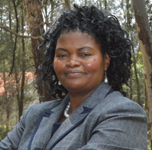
Professor Jane Catherine Ngila (Figure 5) is currently the Acting Executive Director of the African Academy of Sciences (AAS). The AAS is a non-aligned, non-political, not-for-profit pan-African organization with the vision to transform lives on the African continent through science. During the ABRF annual meeting, Dr. Ngila will provide details of the framework on the Equipment for Africa project. The project's aim is to develop programs and funding sources to relocate quality laboratory equipment no longer in use in core labs and to place them into prequalified laboratories in the five regions of Africa. A crucial part of the project includes research exchange visits for skills and capacity building in scientific instrument techniques. A major goal of the venture is to help facilitate the development of shared facilities as Centers of Excellence in Africa and to ensure sustainability of education and research in the continent.

Figure 5: Professor Jane Catherine Ngila, Acting Executive Director of the African Academy of Sciences (AAS).
In addition to Professor Ngila's presentation outlining the perspective of the AAS, representatives from Great Britain and Germany will attend the session and highlight their current efforts to relocate mass spectrometers to laboratories in Africa and Bangladesh. These successful efforts set precedents for relocating instruments and provide a framework to build on within the Equipment for Africa project.
Another topic to be discussed is the Africa Microscopy Initiative (AMI). AMI is a multi-pronged, continent-wide undertaking aimed at directly addressing the inequitable access to advanced imaging techniques throughout Africa. The initiative includes (i) a unique Partners in Teaching (PiTCH) program that pairs international imaging scientists with their African counterparts in offering microscopy workshops, (ii) the Imaging Africa annual microscopy bootcamp course, (iii) the Program for Equipment Exchange and Reutilization (PEER), (iv) the Microscopy Matters webinar series, and (v) a fully equipped imaging center that is accessible at no cost to African scientists through peer-reviewed proposals. AMI also seeks to synergize with other regional communities and capacity-building efforts, such as the African BioImaging Consortium, and TReND in Africa.
We hope this article raises awareness of these ABRF programs. For further information about the ABRF annual meeting and/or participation in the ABRF efforts to improve opportunities for the African Academy of Sciences, please contact Richard Cole.


