


Overview
Cells are the fundamental units of tissues in multicellular organisms. Animal cells are sealed sacs constructed of extremely thin (≈5 nm) lipid bilayer plasma membranes, spanning across which are various membrane proteins. Crucially, the membrane separates an intracellular biochemical compartment, the cytoplasm, from the extracellular environment. This separation enables gradients, or differences in concentration, of ions and small molecules to be maintained across the membrane, and acts to contain the cytoplasmic proteins and enzymes involved in metabolism, as well as organelles, or intracellular membrane compartments. Particularly importantly for the nervous system, the membrane is also an excellent electrical insulator: it is energetically very unfavourable for charged entities like free ions and electrons to jettison their interactions with polar water molecules in order to cross through the uncharged, non-polar hydrocarbon interior of the lipid bilayer membrane, and so transporting them across the membrane is normally very difficult. This high resistance allows an electrical potential difference to be maintained across the membrane – the membrane potential. An electrical potential difference is equivalent to a difference in the ‘concentration’ of unbalanced charges between the two sides of the membrane.
Cells come in a myriad of different types and shapes, with specialised adaptations and purposes. In the brain, neurons are the cells which carry out the rapid electrical signalling underlying sensation, reflexes, decisions and motor action. Neurons are supported by another large population of cells, called glial cells, or glia (which are roughly as numerous as neurons, ≈1010 in the human brain). Glial cells associate intimately with neurons, wrapping around them and providing energy support, transporting and recycling the signalling molecules released by neurons (transmitters) at neuron-to-neuron connections, or synapses. Glia are less studied than neurons, but much new research is shedding light on diverse and important roles of these cells in brain physiology and pathology.
Neurons are highly diverse in shape, but generally have (1) a cell body – a relatively large-diameter compartment of the cell containing the cell nucleus; (2) many dendrites, fine branching elongations, which are covered with synaptic connections from other neurons; and (3) an axon, a tubular elongation of the membrane, which conducts electrical signals to the output synaptic connections of the neuron and may be very long and branched. The axon usually arises from the cell body (in some cases, though, from a dendrite). The structure of the neuron enables the collection of large numbers of synaptic inputs from many different presynaptic neurons, processing of this information, and then dispatching an output, expressing some kind of computation or decision based on the input, to many other neurons. For example, a typical brain neuron might receive on the order of 10,000 input synaptic connections from other cells, and send its output through axonal branches to a similar number of downstream neurons. This extraordinarily high connectivity of individual cells to others in a network, and the ability to modify the strength of these connections in response to patterns of electrical activity, explains the complexity of input–output relations of the brain, and its ability to learn associations between inputs and to store vast amounts of information.
The human brain (Figure 1.1.1) is distinctive in having a particularly large outer mantle or cerebrum, with the ‘neocortex’, a surface layer some 2–3 mm in thickness, overlying phylogenetically older ‘allocortex’. The cerebral cortex is most elaborately folded in humans, giving a total area of something like a quarter of a square metre. The neocortex, which is crucially important for many of the higher cognitive functions of the brain, has a stereotypical structure throughout, albeit with some variation from area to area, with six layers distinguished according to the type and density of neuron cell bodies present, and the arrangements of axons.
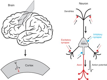
Figure 1.1.1 The cortex of the brain contains billions of neurons. A typical neuron has a soma, or cell body, fine branching membrane extensions called dendrites, and an axon. Information flows in the form of electrical activity arriving from other neurons in the network, to synapses on the dendrites, then electrical current flow to the soma and axon, where an action potential is generated, carrying information down the axon, which forms synapses on other cells. The mapping of the spatiotemporal pattern of synaptic inputs to the timing of the output axonal action potentials defines the information processing function of the neuron.
There are many different types of neuron in the neocortex. These are distinguished, for instance, by:
whether they make only nearby, local axonal connections or long-range connections
their inhibitory or excitatory function
morphology
expression of particular peptides (somatostatin, cholecystokinin) or calcium-binding proteins (parvalbumin, calbindin)
secreted neurotransmitters (commonly glutamate in excitatory cells and gamma-aminobutyric acid (GABA) in inhibitory cells, but also including others such as acetylcholine, dopamine, serotonin, glycine)
other features.
Estimates of the number of different types of cortical neuron vary, and even definitions of ‘type’, but up to 50 or more neuronal cell types have been identified from single-cell gene expression patterns.
In the next section, we consider the universal features of how neurons operate as biophysical machines, to carry out their function of electrical and chemical processing of information.

