Skip to main content Accessibility help
×
Hostname: page-component-77c89778f8-7drxs Total loading time: 0 Render date: 2024-07-19T17:36:37.019Z Has data issue: false hasContentIssue false

Chapter J3 - Structure and dynamics studies

from Part J - Nuclear magnetic resonance

Published online by Cambridge University Press:  05 November 2012

Igor N. Serdyuk
Affiliation:
Institute of Protein Research, Moscow
Nathan R. Zaccai
Affiliation:
University of Bristol
Joseph Zaccai
Affiliation:
Institut de Biologie Structurale, Grenoble
Get access

Summary

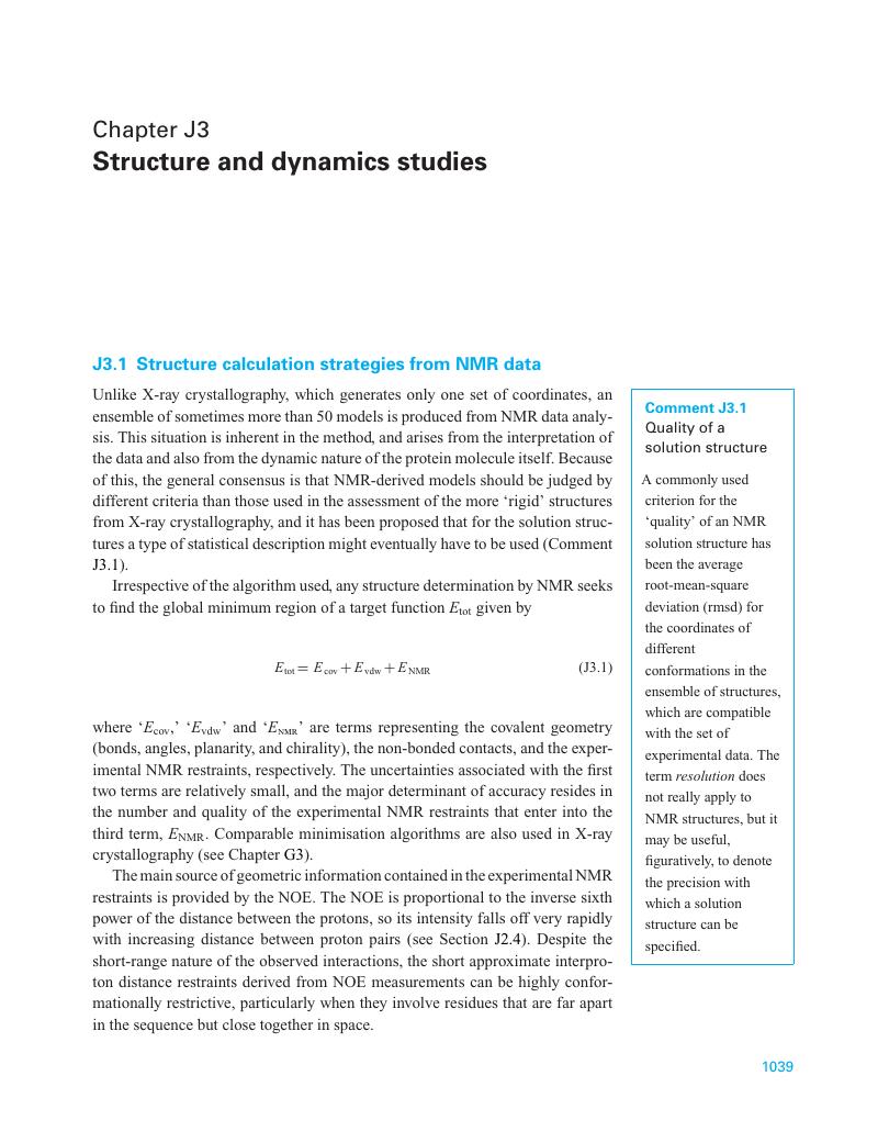
Image of the first page of this content. For PDF version, please use the ‘Save PDF’ preceeding this image.'
Type
Chapter
Information
Methods in Molecular Biophysics
Structure, Dynamics, Function
, pp. 1039 - 1075
Publisher: Cambridge University Press
Print publication year: 2007

Access options

Get access to the full version of this content by using one of the access options below. (Log in options will check for institutional or personal access. Content may require purchase if you do not have access.)

References

Wuthrich, K., Wider, G., Wagner, G., and Braun, W. (1982). Sequential resonance assignments as a basis for determination of spatial protein structures by high resolution proton nuclear magnetic resonance. J. Mol. Biol., 155, 311–319.CrossRefGoogle ScholarPubMed
Spronk, C. A. E. M., Linge, J. P., Hilbers, C. W., and Vuister, G. W. (2002). Improving the quality of protein structures derived by NMR spectroscopy. J. Biomolecular NMR, 22, 281–289.CrossRefGoogle ScholarPubMed
Tjandra, N., Garrett, D. S., Gronenborn, A. M., Bax, A., and Clore, G. M. (1997). Defining long range order in NMR structure determination from the dependence of heteronuclear relaxation times on rotational diffusion anisotropy. Nat. Struct. Biol., 4, 443–449.CrossRefGoogle ScholarPubMed
Tjandra, N., Omichinski, J. G., Gronenborn, A. M., Clore, G. M., and Bax, A. (1997). Use of dipolar 1H–15N and 1H–13C couplings in the structure determination of magnetically oriented macromolecules in solution. Nat. Struct. Biol., 4, 732–738.CrossRefGoogle ScholarPubMed
Wuthrich, K. (1995). NMR – This other method for protein and nucleic acid structure determination. Acta Cryst., D51, 249–270.Google Scholar
Wider, G., and Wuthrich, K. (1999). NMR spectroscopy of large molecules and multimolecular assemblies in solution. Curr. Opin. Struct. Biol., 9, 594–601.CrossRefGoogle ScholarPubMed
Garret, D. S., Seok, Y.-J., et al. (1997). Solution structure of the 30 kDa N-terminal domain of enzyme I of the Escherichia coli phosphoenolpuruvate: sugar phosphotransferase system by multidimensional NMR. Biochemistry, 36, 2517–2530.CrossRefGoogle Scholar
Flaux, J., Bertelsen, E. B., Horwich, A. L., and Wuthrich, K. (2002). NMR analysis of a 900K GroEL–GroES complex. Nature, 418, 207–211.Google Scholar
Wuthrich, K. (2000). Protein recognition by NMR. Nat. Struct. Biol. 7, 188–189.CrossRefGoogle Scholar
Takahashi, H., Nakanishi, T., Kami, K., Arata, Y., and Shimada, I. (2000). A novel NMR method for determining the interfaces of large protein–protein complexes. Nat. Struct. Biol. 7, 220–223.Google ScholarPubMed
Zidek, L., Stefl, R., and Sklenar, V. (2001). NMR methodology for the study of nucleic acids. Curr. Opin. Struct. Biol., 11, 275–281.CrossRefGoogle Scholar
Tjandra, N., Tate, S., Ono, A., Kainosho, M., and Bax, A. (2000). The NMR structure of a DNA dodecamer in an aqueous dilute liquid crystalline phase. JACS, 122, 6190–6200.CrossRefGoogle Scholar
Mollova, E. T., Hansen, M. R., and Pardi, A. (2000). Global structure of RNA determined with residual dipolar coupling. JACS, 122, 11561–11562.CrossRefGoogle Scholar
Tian, F., Al-Hashimi, H. M., Craighead, J. L., and Prestegard, J. H. (2001). Conformational analysis of a flexible oligosaccharide using residual dipolar coupling. JACS, 123, 485–492.CrossRefGoogle Scholar
Ishima, R., and Torchia, D. (2000). Protein dynamics from NMR, Nat. Str. Biol., 7, 740–743.CrossRefGoogle ScholarPubMed
Stejskal, E. O., and Tanner, J. E. (1965). Spin diffusion measurements: spin echoes in the presence of a time dependent field gradient. J. Chem. Phys., 42, 288–292.CrossRefGoogle Scholar
Jones, J. A., Wilkins, D. K., Smith, L. J., and Dobson, C. M. (1997). Characterization of protein unfolding by NMR diffusion measurements. J. Biomolecular NMR, 10, 199–203.CrossRefGoogle Scholar
Smith, S. O., and Peersen, O. B. (1992). Solid-state NMR approaches for studying membrane protein structure. Ann. Rev. Biophys. Biomol. Str., 21, 25–47.CrossRefGoogle ScholarPubMed
Opella, S. J., and Stewart, P. L. (1989). Solid-state nuclear magnetic resonance structural studies of proteins. Meth. Enzymol., 176, 242–275.CrossRefGoogle ScholarPubMed
Marassi, F. M., Ma, C., et al. (1999). Correlation of the structural and functional domains in the membrane protein Vpu from HIV-1. PANS, 96, 14336–14341.CrossRefGoogle ScholarPubMed
Smith, L. J., Redfield, C., et al. (1994). Comparison of four independently determined structures of human recombinant interleikin-4. Struct. Biol., 1, 301–310.CrossRefGoogle Scholar
Schwabe, J. W. R., Chapman, L., Finch, J. T., Rhodes, D., and Neuhaus, D. (1993). DNA recognition by the oestrogen receptor: From solution to the crystal. Structure, 1, 187–204.CrossRefGoogle ScholarPubMed
Prestegard, J. H., Valafar, H., Glushka, J., and Tian, F. (2001). Nuclear magnetic resonance in the era of structural Genomics. Biochemistry, 40, 8677–8685.CrossRefGoogle ScholarPubMed
Jacobs, R. E., Ahrens, E. T., Meade, T. J., and Fraser, S. E. (1999). Looking deeper into vertebrate development. TIBS, 9, 73–76.Google ScholarPubMed

Save book to Kindle

To save this book to your Kindle, first ensure coreplatform@cambridge.org is added to your Approved Personal Document E-mail List under your Personal Document Settings on the Manage Your Content and Devices page of your Amazon account. Then enter the ‘name’ part of your Kindle email address below. Find out more about saving to your Kindle.

Note you can select to save to either the @free.kindle.com or @kindle.com variations. ‘@free.kindle.com’ emails are free but can only be saved to your device when it is connected to wi-fi. ‘@kindle.com’ emails can be delivered even when you are not connected to wi-fi, but note that service fees apply.

Find out more about the Kindle Personal Document Service.

Available formats
×

Save book to Dropbox

To save content items to your account, please confirm that you agree to abide by our usage policies. If this is the first time you use this feature, you will be asked to authorise Cambridge Core to connect with your account. Find out more about saving content to Dropbox.

Available formats
×

Save book to Google Drive

To save content items to your account, please confirm that you agree to abide by our usage policies. If this is the first time you use this feature, you will be asked to authorise Cambridge Core to connect with your account. Find out more about saving content to Google Drive.

Available formats
×