Skip to main content Accessibility help
×
Hostname: page-component-78c5997874-dh8gc Total loading time: 0 Render date: 2024-11-17T22:07:20.688Z Has data issue: false hasContentIssue false

Chapter 4 - Intermediate Malignant and Malignant Tumors of Soft Tissue Featuring a Spindle Cell Morphology

Published online by Cambridge University Press:  10 May 2019

Angelo Paolo Dei Tos
Affiliation:
Università degli Studi di Padova, Italy
Get access

Summary

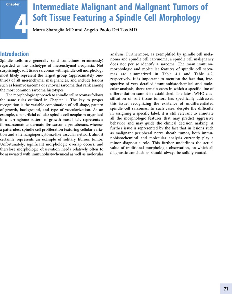
Image of the first page of this content. For PDF version, please use the ‘Save PDF’ preceeding this image.'
Type
Chapter
Information
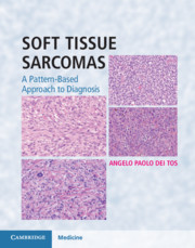
Soft Tissue Sarcomas
A Pattern-Based Approach to Diagnosis
, pp. 71 - 218
Publisher: Cambridge University Press
Print publication year: 2018

Access options

Get access to the full version of this content by using one of the access options below. (Log in options will check for institutional or personal access. Content may require purchase if you do not have access.)

References

Primary Sources

Abbott, JJ, Erickson-Johnson, M, Wang, X, Nascimento, AG, Oliveira, AM. Gains of COL1A1-PDGFB genomic copies occur in fibrosarcomatous transformation of dermatofibrosarcoma protuberans. Mod Pathol. 2006;19:1512–18.Google Scholar
Abbott, JJ, Oliveira, AM, Nascimento, AG. The prognostic significance of fibrosarcomatous transformation in dermatofibrosarcoma protuberans. Am J Surg Pathol. 2006;30:436–43.Google Scholar
Calonje, E, Fletcher, CD. Aneurysmal benign fibrous histiocytoma: clinicopathological analysis of 40 cases of a tumour frequently misdiagnosed as a vascular neoplasm. Histopathology. 1995;26:323–31.Google Scholar
Calonje, E, Fletcher, CD. Myoid differentiation in dermatofibrosarcoma protuberans and its fibrosarcomatous variant: clinicopathologic analysis of 5 cases. J Cutan Pathol. 1996;23:30–6.Google Scholar
Calonje, E, Mentzel, T, Fletcher, CD. Cellular benign fibrous histiocytoma. Clinicopathologic analysis of 74 cases of a distinctive variant of cutaneous fibrous histiocytoma with frequent recurrence. Am J Surg Pathol. 1994;18:668–76.CrossRefGoogle ScholarPubMed
Doyle, LA, Fletcher, CD. Metastasizing “benign” cutaneous fibrous histiocytoma: a clinicopathologic analysis of 16 cases. Am J Surg Pathol. 2013;37:484–95.CrossRefGoogle Scholar
Doyle, LA, Mariño-Enriquez, A, Fletcher, CD, Hornick, JL. ALK rearrangement and overexpression in epithelioid fibrous histiocytoma. Mod Pathol. 2015;28:904–12.Google Scholar
Fiore, M, Miceli, R, Mussi, C, et al. Dermatofibrosarcoma protuberans treated at a single institution: a surgical disease with a high cure rate. J Clin Oncol. 2005;23:7669–75.Google Scholar
Kaddu, S, McMenamin, ME, Fletcher, CD. Atypical fibrous histiocytoma of the skin: clinicopathologic analysis of 59 cases with evidence of infrequent metastasis. Am J Surg Pathol. 2002;26:3546.CrossRefGoogle ScholarPubMed
McArthur, GA, Demetri, GD, van Oosterom, A, et al. Molecular and clinical analysis of locally advanced dermatofibrosarcoma protuberans treated with imatinib: Imatinib Target Exploration Consortium Study B2225. J Clin Oncol. 2005;23:866–73.CrossRefGoogle ScholarPubMed
Mentzel, T, Beham, A, Katenkamp, D, Dei Tos, AP, Fletcher, CD. Fibrosarcomatous (“high-grade”) dermatofibrosarcoma protuberans: clinicopathologic and immunohistochemical study of a series of 41 cases with emphasis on prognostic significance. Am J Surg Pathol. 1998;22:576–87.CrossRefGoogle ScholarPubMed
Mentzel, T, Schärer, L, Kazakov, DV, Michal, M. Myxoid dermatofibrosarcoma protuberans: clinicopathologic, immunohistochemical, and molecular analysis of eight cases. Am J Dermatopathol. 2007;29:443–8.CrossRefGoogle ScholarPubMed
Reimann, JD, Fletcher, CD. Myxoid dermatofibrosarcoma protuberans: a rare variant analyzed in a series of 23 cases. Am J Surg Pathol. 2007;31:1371–7.CrossRefGoogle Scholar
Singh Gomez, C, Calonje, E, Fletcher, CD. Epithelioid benign fibrous histiocytoma of skin: clinico-pathological analysis of 20 cases of a poorly known variant. Histopathology. 1994;24:123–9.Google ScholarPubMed
Stacchiotti, S, Astolfi, A, Gronchi, A, et al. Evolution of dermatofibrosarcoma protuberans to dfsp-derived fibrosarcoma: an event marked by epithelial-mesenchymal transition-like process and 22q loss. Mol Cancer Res. 2016;14:820–9.CrossRefGoogle ScholarPubMed
Stacchiotti, S, Pantaleo, MA, Negri, T, et al. Efficacy and biological activity of imatinib in metastatic dermatofibrosarcoma protuberans (DFSP). Clin Cancer Res. 2016;22:837–46CrossRefGoogle ScholarPubMed
Takahira, T, Oda, Y, Tamiya, S, et al. Detection of COL1A1-PDGFB fusion transcripts and PDGFB/PDGFRB mRNA expression in dermatofibrosarcoma protuberans. Mod Pathol. 2007;20:668–75.Google Scholar
Terrier-Lacombe, MJ, Guillou, L, Maire, G, et al. Dermatofibrosarcoma protuberans, giant cell fibroblastoma, and hybrid lesions in children: clinicopathologic comparative analysis of 28 cases with molecular data. A study from the French Federation of Cancer Centers Sarcoma Group. Am J Surg Pathol. 2003;27:2739.Google Scholar
Woodruff, JM. Pathology of the major peripheral nerve sheath neoplasms. Monogr Pathol. 1996;38:129–61.Google ScholarPubMed

Secondary Sources

Al-Ibraheemi, A, Martinez, A, Weiss, SW, et al. Fibrous hamartoma of infancy: a clinicopathologic study of 145 cases, including 2 with sarcomatous features. Mod Pathol. 2017;30:474–85.CrossRefGoogle ScholarPubMed
Dymock, RB, Allen, PW, Stirling, JW, Gilbert, EF, Thornbery, JM. Giant cell fibroblastoma. A distinctive, recurrent tumor of childhood. Am J Surg Pathol. 1987;11:263–71.Google Scholar
Enzinger, FM. Fibrous hamartoma of infancy. Cancer. 1965 Feb;18:241–8.3.0.CO;2-C>CrossRefGoogle ScholarPubMed
Fetsch, JF, Miettinen, M, Laskin, WB, Michal, M, Enzinger, FM. A clinicopathologic study of 45 pediatric soft tissue tumors with an admixture of adipose tissue and fibroblastic elements, and a proposal for classification as lipofibromatosis. Am J Surg Pathol. 2000;24:1491–500.Google Scholar
Fletcher, CD. Giant cell fibroblastoma of soft tissue: a clinicopathological and immunohistochemical study. Histopathology. 1988;13:499508.Google Scholar
Fletcher, CD, Powell, G, van Noorden, S, McKee, PH. Fibrous hamartoma of infancy: a histochemical and immunohistochemical study. Histopathology. 1988 Jan;12(1):6574.Google Scholar
Jha, P, Moosavi, C, Fanburg-Smith, JC. Giant cell fibroblastoma: an update and addition of 86 new cases from the Armed Forces Institute of Pathology, in honor of Dr. Franz M. Enzinger. Ann Diagn Pathol. 2007;11:81–8.Google Scholar
Shmookler, BM, Enzinger, FM, Weiss, SW. Giant cell fibroblastoma. A juvenile form of dermatofibrosarcoma protuberans. Cancer. 1989;64:2154–61.Google Scholar
Antonescu, CR, Dal Cin, P, Nafa, K, et al. EWSR1-CREB1 is the predominant gene fusion in angiomatoid fibrous histiocytoma. Genes Chromosomes Cancer. 2007;46:1051–60.CrossRefGoogle ScholarPubMed
Calonje, E, Fletcher, CD. Aneurysmal benign fibrous histiocytoma: clinicopathological analysis of 40 cases of a tumour frequently misdiagnosed as a vascular neoplasm. Histopathology. 1995;26:323–31.Google Scholar
Costa, MJ, Weiss, SW. Angiomatoid malignant fibrous histiocytoma. A follow-up study of 108 cases with evaluation of possible histologic predictors of outcome. Am J Surg Pathol. 1990;14:1126–32.CrossRefGoogle ScholarPubMed
Enzinger, FM. Angiomatoid malignant fibrous histiocytoma: a distinct fibrohistiocytic tumor of children and young adults simulating a vascular neoplasm. Cancer. 1979;44:2147–57.Google Scholar
Fanburg-Smith, JC, Miettinen, M. Angiomatoid “malignant” fibrous histiocytoma: a clinicopathologic study of 158 cases and further exploration of the myoid phenotype. Hum Pathol. 1999;30:1336–43.Google Scholar
Fletcher, CD. Angiomatoid “malignant fibrous histiocytoma”: an immunohistochemical study indicative of myoid differentiation. Hum Pathol. 1991;22:563–8.Google Scholar
Rossi, S, Szuhai, K, Ijszenga, M, et al. EWSR1-CREB1 and EWSR1-ATF1 fusion genes in angiomatoid fibrous histiocytoma. Clin Cancer Res. 2007;13:7322–8.CrossRefGoogle ScholarPubMed
Schaefer, IM, Fletcher, CD. Myxoid variant of so-called angiomatoid “malignant fibrous histiocytoma”: clinicopathologic characterization in a series of 21 cases. Am J Surg Pathol. 2014;38:816–23.Google Scholar
Thway, K, Nicholson, AG, Wallace, WA, et al. Endobronchial pulmonary angiomatoid fibrous histiocytoma: two cases with EWSR1-CREB1 and EWSR1-ATF1 fusions. Am J Surg Pathol. 2012;36:883–8.Google Scholar
Thway, K, Stefanaki, K, Papadakis, V, Fisher, C. Metastatic angiomatoid fibrous histiocytoma of the scalp, with EWSR1-CREB1 gene fusions in primary tumor and nodal metastasis. Hum Pathol. 2013;44:289–93.Google Scholar
Weinreb, I, Rubin, BP, Goldblum, JR. Pleomorphic angiomatoid fibrous histiocytoma: a case confirmed by fluorescence in situ hybridization analysis for EWSR1 rearrangement. J Cutan Pathol. 2008;35:855–60.Google Scholar
Allen, PW. Nodular fasciitis. Pathology. 1972 Jan;4(1):926.Google Scholar
Antonescu, CR, Sung, YS, Zhang, L, Agaram, NP, Fletcher, CD. Recurrent SRF-RELA fusions define a novel subset of cellular myofibroma/myopericytoma: a potential diagnostic pitfall with sarcomas with myogenic differentiation. Am J Surg Pathol. 2017;41:677–84.Google Scholar
Beham, A, Badve, S, Suster, S, Fletcher, CD. Solitary myofibroma in adults: clinicopathological analysis of a series. Histopathology. 1993;22:335–41.Google Scholar
Chan, JY, Gooi, Z, Wong, EW, et al. Low-grade myofibroblastic sarcoma: A population-based study. Laryngoscope. 2017;127:116–12.CrossRefGoogle ScholarPubMed
Fletcher, CD, Stirling, RW. Intranodal myofibroblastoma presenting in the submandibular region: evidence of a broader clinical and histological spectrum. Histopathology. 1990;16:287–93.Google Scholar
Gronchi, A, Colombo, C, Le Péchoux, C, et al. Sporadic desmoid-type fibromatosis: a stepwise approach to a non-metastasising neoplasm – a position paper from the Italian and the French Sarcoma Group. Ann Oncol. 2014;25:578–83.Google Scholar
Hung, YP, Fletcher, CDM. Myopericytomatosis: clinicopathologic analysis of 11 cases with molecular identification of recurrent PDGFRB alterations in myopericytomatosis and myopericytoma. Am J Surg Pathol. 2017;41:1034–104.Google Scholar
Le Guellec, S, Soubeyran, I, Rochaix, P, et al. CTNNB1 mutation analysis is a useful tool for the diagnosis of desmoid tumors: a study of 260 desmoid tumors and 191 potential morphologic mimics. Mod Pathol. 2012;25:1551–8.Google Scholar
McMenamin, ME, Fletcher, CD. Malignant myopericytoma: expanding the spectrum of tumours with myopericytic differentiation. Histopathology. 2002;41:450–60.CrossRefGoogle ScholarPubMed
Mentzel, T, Calonje, E, Nascimento, AG, Fletcher, CD. Infantile hemangiopericytoma versus infantile myofibromatosis. Study of a series suggesting a continuous spectrum of infantile myofibroblastic lesions. Am J Surg Pathol. 1994;18:922–30.Google Scholar
Mentzel, T, Dei Tos, AP, Sapi, Z, Kutzner, H. Myopericytoma of skin and soft tissues: clinicopathologic and immunohistochemical study of 54 cases. Am J Surg Pathol. 2006;30:104–13.Google Scholar
Mentzel, T, Dry, S, Katenkamp, D, Fletcher, CD. Low-grade myofibroblastic sarcoma: analysis of 18 cases in the spectrum of myofibroblastic tumors. Am J Surg Pathol. 1998;22:1228–38.Google Scholar
Suster, S, Rosai, J. Intranodal hemorrhagic spindle-cell tumor with “amianthoid” fibers. Report of six cases of a distinctive mesenchymal neoplasm of the inguinal region that simulates Kaposi’s sarcoma. Am J Surg Pathol. 1989;13:347–57.Google Scholar
Weiss, SW, Gnepp, DR, Bratthauer, GL. Palisaded myofibroblastoma. A benign mesenchymal tumor of lymph node. Am J Surg Pathol. 1989;13:341–6.CrossRefGoogle ScholarPubMed
Bahrami, A, Weiss, SW, Montgomery, E, et al. RT-PCR analysis for FGF23 using paraffin sections in the diagnosis of phosphaturic mesenchymal tumors with and without known tumor induced osteomalacia. Am J Surg Pathol. 2009;33:1348–54.Google Scholar
Folpe, AL, Fanburg-Smith, JC, Billings, SD, et al. Most osteomalacia-associated mesenchymal tumors are a single histopathologic entity: an analysis of 32 cases and a comprehensive review of the literature. Am J Surg Pathol. 2004;28:130.Google Scholar
Lee, JC, Jeng, YM, Su, SY, et al. Identification of a novel FN1-FGFR1 genetic fusion as a frequent event in phosphaturic mesenchymal tumour. J Pathol. 2015;235:539–45.Google Scholar
Lee, JC, Su, SY, Changou, CA, et al. Characterization of FN1-FGFR1 and novel FN1-FGF1 fusion genes in a large series of phosphaturic mesenchymal tumors. Mod Pathol. 2016;29:1335–46.Google Scholar
Salassa, RM, Jowsey, J, Arnaud, CD. Hypophosphatemic osteomalacia associated with “nonendocrine” tumors. N Engl J Med. 1970;283:6570.Google Scholar
Antonescu, CR, Romeo, S, Zhang, L, et al. Dedifferentiation in gastrointestinal stromal tumor to an anaplastic KIT-negative phenotype: a diagnostic pitfall: morphologic and molecular characterization of 8 cases occurring either de novo or after imatinib therapy. Am J Surg Pathol. 2013;37:385–92.Google Scholar
Boikos, SA, Pappo, AS, Killian, JK, et al. Molecular subtypes of KIT/PDGFRA wild-type gastrointestinal stromal tumors: a report from the National Institutes of Health Gastrointestinal Stromal Tumor Clinic. JAMA Oncol. 2016;2:922–8.Google Scholar
Brenca, M, Rossi, S, Polano, M, et al. Transcriptome sequencing identifies ETV6-NTRK3 as a gene fusion involved in GIST. J Pathol. 2016;238:543–9.Google Scholar
Corless, CL, Schroeder, A, Griffith, D, et al. PDGFRA mutations in gastrointestinal stromal tumors: frequency, spectrum and in vitro sensitivity to imatinib. J Clin Oncol. 2005;3:5357–64.Google Scholar
Dei Tos, AP. The reappraisal of GIST: from Stout to the KIT revolution. Virchows Arch. 2003;443:421428.Google Scholar
Demetri, GD, Reichardt, P, Kang, YK, et al. Efficacy and safety of regorafenib for advanced gastrointestinal stromal tumours after failure of imatinib and sunitinib (GRID): an international, multicentre, randomised, placebo-controlled, phase 3 trial. Lancet. 2013;381:295302.Google Scholar
Demetri, GD, van Oosterom, AT, Garrett, CR, et al. Efficacy and safety of sunitinib in patients with advanced gastrointestinal stromal tumour after failure of imatinib: a randomised controlled trial. Lancet. 2006;14;368:1329–38.Google Scholar
Fletcher, CD, Berman, JJ, Corless, C, et al. Diagnosis of gastrointestinal stromal tumors: A consensus approach. Hum Pathol, 2002;33:459–65.Google Scholar
Folpe, AL, Fanburg-Smith, JC, Miettinen, M, Weiss, SW. Atypical and malignant glomus tumors: analysis of 52 cases, with a proposal for the reclassification of glomus tumors. Am J Surg Pathol. 2001;25:112.Google Scholar
Gasparotto, D, Rossi, S, Polano, M, et al. Quadruple-negative GIST is a sentinel for unrecognized neurofibromatosis type 1 syndrome. Clin Cancer Res. 2017;23:273–82.Google Scholar
Heinrich, MC, Corless, CL, Blanke, CD, et al. Molecular correlates of imatinib resistance in gastrointestinal stromal tumors. J Clin Oncol. 2006;24:4764–74.Google Scholar
Heinrich, MC, Corless, CL, Duensing, A, et al. PDGFRA activating mutations in gastrointestinal stromal tumors. Science. 2003;299:708–10.Google Scholar
Hirota, S, Isozaki, K, Moriyama, Y, et al. Gain-of-function mutations of c-kit in human gastrointestinal stromal tumors. Science. 1998;279:577–80.Google Scholar
Janeway, KA, Kim, SY, Lodish, M, et al. Defects in succinate dehydrogenase in gastrointestinal stromal tumors lacking KIT and PDGFRA mutations. Proc Natl Acad Sci U S A. 2011;108:314–18.Google Scholar
Joensuu, H, Roberts, PJ, Sarlomo-Rikala, M, et al. Effect of the tyrosine kinase inhibitor STI571 in a patient with a metastatic gastrointestinal stromal tumour. N Engl J Med. 2001;344:1052–6.Google Scholar
Joensuu, H, Vehtari, A, Riihimäki, J, et al. Risk of recurrence of gastrointestinal stromal tumour after surgery: an analysis of pooled population-based cohorts. Lancet Oncol. 2012;13:265–74.Google Scholar
Kay, S, Callahan, WP Jr, Murray, MR, Randall, HT, Stout, AP. Glomus tumors of the stomach. Cancer. 1951;4:726–36.Google Scholar
Kindblom, L-G, Remotti, HE, Aldenborg, F, Meis-Kindblom, JM. Gastrointestinal pacemaker cell tumor (GIPACT): gastrointestinal stromal tumors show phenotypic characteristics of the interstitial cells of Cajal. Am J Pathol. 1998;152:1259–69.Google ScholarPubMed
Lasota, J, Wang, ZF, Sobin, LH, Miettinen, M. Gain-of-function PDGFRA mutations, earlier reported in gastrointestinal stromal tumors, are common in small intestinal inflammatory fibroid polyps. A study of 60 cases. Mod Pathol. 2009;22:1049–56.CrossRefGoogle ScholarPubMed
Liegl, B, Bennett, MW, Fletcher, CD. Microcystic/reticular schwannoma: a distinct variant with predilection for visceral locations. Am J Surg Pathol. 2008;32:1080–7.Google Scholar
Makhlouf, HR, Ahrens, W, Agarwal, B, et al. Synovial sarcoma of the stomach: a clinicopathologic, immunohistochemical, and molecular genetic study of 10 cases. Am J Surg Pathol. 2008;32:275–81.Google Scholar
Mason, EF, Hornick, JL. Conventional risk stratification fails to predict progression of succinate dehydrogenase-deficient gastrointestinal stromal tumors: a clinicopathologic study of 76 cases. Am J Surg Pathol. 2016;40:1616–21.Google Scholar
Matyakhina, L, Bei, TA, McWhinney, SR, et al. Genetics of Carney triad: recurrent losses at chromosome 1 but lack of germline mutations in genes associated with paragangliomas and gastrointestinal stromal tumors. J Clin Endocrinol Metab. 2007;92:2938–43Google Scholar
Miettinen, M, Felisiak-Golabek, A, Wang, Z, Inaguma, S, Lasota, J. GIST manifesting as a retroperitoneal tumor: clinicopathologic immunohistochemical, and molecular genetic study of 112 cases. Am J Surg Pathol. 2017;41:577–85.Google Scholar
Miettinen, M, Furlong, M, Sarlomo-Rikala, M, et al. Gastrointestinal stromal tumors, intramural leiomyomas, and leiomyosarcomas in the rectum and anus: a clinicopathologic, immunohistochemical, and molecular genetic study of 144 cases. Am J Surg Pathol. 2001;25:1121–33.Google Scholar
Miettinen, M, Lasota, J. Gastrointestinal stromal tumors: review on morphology, molecular pathology, prognosis, and differential diagnosis. Arch Pathol Lab Med. 2006;130:1466–78.Google Scholar
Miettinen, M, Makhlouf, HR, Sobin, LH, Lasota, J. Plexiform fibromyxoma: a distinctive benign gastric antral neoplasm not to be confused with a myxoid GIST. Am J Surg Pathol. 2009;33:1624–32.Google Scholar
Miettinen, M, Sarlomo-Rikala, M, Sobin, LH, Lasota, J. Gastrointestinal stromal tumors and leiomyosarcomas in the colon: a clinicopathologic, immunohistochemical, and molecular genetic study of 44 cases. Am J Surg Pathol. 2000;24:1339–52.Google Scholar
Miettinen, M, Wang, ZF, Lasota, J. DOG1 antibody in the differential diagnosis of gastrointestinal stromal tumors: a study of 1840 cases. Am J Surg Pathol. 2009;33:1401–8.Google Scholar
Novelli, M, Rossi, S, Rodriguez-Justo, M, et al. DOG1 and CD117 are the antibodies of choice in the diagnosis of gastrointestinal stromal tumours. Histopathology. 2010;57:259–70.Google Scholar
Pasini, B, McWhinney, SR, Bei, T, et al. Clinical and molecular genetics of patients with the Carney-Stratakis syndrome and germline mutations of the genes coding for the succinate dehydrogenase subunits SDHB, SDHC, and SDHD. Eur J Hum Genet. 2008;16:7988.CrossRefGoogle ScholarPubMed
Romeo, S, Rossi, S, Acosta Marín, M, at al. Primary synovial sarcoma (SS) of the digestive system: a molecular and clinicopathological study of fifteen cases. Clin Sarcoma Res. 2015;5:7.Google Scholar
Rossi, S, Gasparotto, D, Miceli, R, et al. KIT, PDGFRA, and BRAF mutational spectrum impacts on the natural history of imatinib-naive localized GIST: a population-based study. Am J Surg Pathol. 2015;39:922–30.Google Scholar
Rossi, S, Gasparotto, D, Toffolatti, L, et al. Molecular and clinicopathologic characterization of gastrointestinal stromal tumors (GISTs) of small size. Am J Surg Pathol. 2010;34:1480–91.Google Scholar
Rossi, S, Miceli, R, Messerini, L, et al. Natural history of imatinib-naive GISTs: a retrospective analysis of 929 cases with long-term follow-up and development of a survival nomogram based on mitotic index and size as continuous variables. Am J Surg Pathol. 2011;35:1646–56.Google Scholar
Rubin, BP, Heinrich, MC, Corless, CL. Gastrointestinal stromal tumour. Lancet. 2007;369:1731–41.CrossRefGoogle ScholarPubMed
Spans, L, Fletcher, CD, Antonescu, CR, et al. Recurrent MALAT1-GLI1 oncogenic fusion and GLI1 up-regulation define a subset of plexiform fibromyxoma. J Pathol. 2016;239:335–43.Google Scholar
Takahashi, Y, Shimizu, S, Ishida, T, et al. Plexiform angiomyxoid myofibroblastic tumor of the stomach. Am J Surg Pathol. 2007;31:724–8.Google Scholar
Wagner, AJ, Remillard, SP, Zhang, YX, et al. Loss of expression of SDHA predicts SDHA mutations in gastrointestinal stromal tumors. Mod Pathol. 2013;26:289–94.Google Scholar
West, RB, Corless, CL, Chen, X, et al. The novel marker, DOG1, is expressed ubiquitously in gastrointestinal stromal tumors irrespective of KIT or PDGFRA mutation status. Am J Pathol. 2004;165:107–13.CrossRefGoogle ScholarPubMed
Bell, SW, Kempson, RL, Hendrickson, MR. Problematic uterine smooth muscle neoplasms. A clinicopathologic study of 213 cases. Am J Surg Pathol. 1994;18:535–58.Google Scholar
Dei Tos, AP, Maestro, R, Doglioni, C, et al. Tumor suppressor genes and related molecules in leiomyosarcoma. Am J Pathol. 1996;148:1037–45.Google Scholar
De Sain Aubain Somerhausen, N, Fletcher, CDM. Leiomyosarcoma of soft tissue in children: clinicopathologic analysis of 20 cases. Am J Surg Pathol. 1999;23:755–63.Google Scholar
Deyrup, AT, Lee, VK, Hill, CE, et al. Epstein-Barr virus-associated smooth muscle tumors are distinctive mesenchymal tumors reflecting multiple infection events: a clinicopathologic and molecular study of 29 tumors from 19 patients. Am J Surg Pathol. 2006;30:7582.Google Scholar
Fletcher, CD, Kilpatrick, SE, Mentzel, T. The difficulty in predicting behavior of smooth-muscle tumors in deep soft tissue. Am J Surg Pathol. 1995;19:116–17.Google Scholar
Kilpatrick, SE, Mentzel, T, Fletcher, CD. Leiomyoma of deep soft tissue. Clinicopathologic analysis of a series. Am J Surg Pathol. 1994;18:576–82.Google Scholar
Mandhal, N, Fletcher, CDM, Dal cin, P, et al. Comparative cytogenetic study of spindle cell and pleomorphic leiomyosarcoma of soft tissue: a report from the CHAMP study group. Cancer Genet Cytogenet. 2000;116:6673.Google Scholar
Massi, D, Franchi, A, Alos, L, et al. Primary cutaneous leiomyosarcoma: clinicopathological analysis of 36 cases. Histopathology. 2010;56:251–62.Google Scholar
Mentzel, T, Calonje, E, Fletcher, CD. Leiomyosarcoma with prominent osteoclast-like giant cells. Analysis of eight cases closely mimicking the so-called giant cell variant of malignant fibrous histiocytoma. Am J Surg Pathol. 1994;18:258–65.Google Scholar
Merchant, W, Calonje, E, Fletcher, CD. Inflammatory leiomyosarcoma: a morphological subgroup within the heterogeneous family of so-called inflammatory malignant fibrous histiocytoma. Histopathology. 1995;27:525–32.Google Scholar
Miettinen, M. Smooth muscle tumors of soft tissue and non-uterine viscera: biology and prognosis. Mod Pathol. 2014;27 Suppl 1:S1729.Google Scholar
Oda, Y, Miyajima, K, Kawaguchi, K, et al. Pleomorphic leiomyosarcoma: clinicopathologic and immunohistochemical study with special emphasis on its distinction from ordinary leiomyosarcoma and malignant fibrous histiocytoma. Am J Surg Pathol. 2001;25:1030–8.Google Scholar
Rubin, BP, Fletcher, CD. Myxoid leiomyosarcoma of soft tissue: an underrecognized variant. Am J Surg Pathol. 2000;24:927–36.Google Scholar
Wile, AG, Evans, HL, Romsdhal, MM. Leiomyosarcoma of soft tissue a clinicopathologic study. Cancer. 1981;48:1022–32.Google Scholar
Chan, JKC. Solitary fibrous tumor – everywhere, and a diagnosis in vogue. Histopathology. 1997;31:568–76.CrossRefGoogle Scholar
Chilosi, M, Facchetti, F, Dei Tos, AP, et al. Bcl-2 expression in pleural and extrapleural solitary fibrous tumours. J Pathol. 1997;181:362–7.Google Scholar
Chmielecki, J, Crago, AM, Rosenberg, M, et al. Whole-exome sequencing identifies a recurrent NAB2-STAT6 fusion in solitary fibrous tumors. Nat Genet. 2013 45:131–2.Google Scholar
Collini, P, Negri, T, Barisella, M, et al. High-grade sarcomatous overgrowth in solitary fibrous tumors: a clinicopathologic study of 10 cases. Am J Surg Pathol. 2012;36:1202–15.CrossRefGoogle ScholarPubMed
Dei Tos, AP, Seregard, S, Calonje, E, et al. Giant cell angiofibroma. A distinctive orbital tumor in adults. Am J Surg Pathol. 1995;19:1286–93.Google Scholar
Demicco, EG, Park, MS, Araujo, DM, et al. Solitary fibrous tumor: a clinicopathological study of 110 cases and proposed risk assessment model. Mod Pathol. 2012;25:1298–306.Google Scholar
Demicco, EG, Wagner, MJ, Maki, RG, et al. Risk assessment in solitary fibrous tumors: validation and refinement of a risk stratification model. Mod Pathol. 2017;30:1433–42.Google Scholar
de Saint Aubain Somerhausen, N, Rubin, BP, Fletcher, CDM. Myxoid solitary fibrous tumor: a study of seven cases with emphasis on differenttial diagnosis. Mod Pathol. 1999;12:463–71.Google Scholar
Dorfman, DM, To, K, Dickersin, GR, et al. Solitary fibrous tumor of the orbit. Am J Surg Pathol. 1994;18:281–7.CrossRefGoogle ScholarPubMed
Doyle, LA, Fletcher, CD. Predicting behavior of solitary fibrous tumor: are we getting closer to more accurate risk assessment? Ann Surg Oncol. 2013;20:4055–6.Google Scholar
Doyle, LA, Vivero, M, Fletcher, CD, Mertens, F, Hornick, JL. Nuclear expression of STAT6 distinguishes solitary fibrous tumor from histologic mimics. Mod Pathol. 2014;27:390–5.Google Scholar
England, DM, Hochholzer, L, McCarthy, MJ. Localized benign and malignant fibrous tumors of the pleura. A clinicopathologic review of 223 cases. Am J Surg Pathol. 1989;13:640–58.Google Scholar
Fritchie, KJ, Jin, L, Rubin, BP, et al. NAB2-STAT6 gene fusion in meningeal hemangiopericytoma and solitary fibrous tumor. J Neuropathol Exp Neurol. 2016;75:263–71.Google Scholar
Gengler, C, Guillou, L. Solitary fibrous tumour and haemangiopericytoma: evolution of a concept. Histopathology. 2006;48:6374.Google Scholar
Gleason, BC, Fletcher, CD. Deep “benign” fibrous histiocytoma: clinicopathologic analysis of 69 cases of a rare tumor indicating occasional metastatic potential. Am J Surg Pathol. 2008;32:354–62.Google Scholar
Gold, JS, Antonescu, CR, Hajdu, C, et al. Clinicopathologic correlates of solitary fibrous tumors. Cancer. 2002;15:1057–68.Google Scholar
Goodlad, J, Fletcher, CDM. Solitary fibrous tumor arising at unusual sites: analysis of series. Histopathology. 1991;19:515–22.Google Scholar
Guillou, L, Gebhard, S, Coindre, JM, et al. Lipomatous hemangiopericytoma: a fat containing variant of solitary fibrous tumor? Hum Pathol. 2000a;31:1108–15.Google Scholar
Guillou, L, Gebhard, S, Coindre, JM, et al. Orbital and extraorbital giant cell angiofibroma: a giant cell rich variant of solitary fibrous tumor? Am J Surg Pathol. 2000b;24:971–9.Google Scholar
Hanau, CA, Miettinen, M. Solitary fibrous tumor: histological and immunohistochemical spectrum of benign and malignant variants presenting at different sites. Hum Pathol. 1995;26:440–9.Google Scholar
Lee, JC, Fletcher, CD. Malignant fat-forming solitary fibrous tumor (so-called “lipomatous hemangiopericytoma”): clinicopathologic analysis of 14 cases. Am J Surg Pathol. 2011;35:1177–85.Google Scholar
Mentzel, T, Bainbridge, TC, Katenkamp, D. Solitary fibrous tumor: clinicopathological, immunohistochemical, and ultrastructural analysis of 12 cases arising in soft tissues, nasal cavity and nasopharynx, urinary bladder and prostate. Virchows Arch. 1997;430:445–53.Google Scholar
Mosquera, JM, Fletcher, CD. Expanding the spectrum of malignant progression in solitary fibrous tumors: a study of 8 cases with a discrete anaplastic component is this dedifferentiated SFT? Am J Surg Pathol. 2009;33:1314–21.Google Scholar
Ng, TL, Gown, AM, Barry, TS, et al: Nuclear beta-catenin in mesenchymal tumors, Mod Pathol. 2005;18:6874.Google Scholar
Nielsen, GP, O’Connell, JX, Dickersin, GR. Solitary fibrous tumor of soft tissue: A report of 15 cases, including 5 malignant examples with light microscopic, immunohistochemical, and ultrastructural data. Mod Pathol. 1997;10:1028–37.Google Scholar
Robinson, DR, Wu, YM, Kalyana-Sundaram, S, et al. Identification of recurrent NAB2-STAT6 gene fusions in solitary fibrous tumor by integrative sequencing. Nat Genet. 2013;45:180–5.Google Scholar
Schweizer, L, Koelsche, C, Sahm, F, et al. Meningeal hemangiopericytoma and solitary fibrous tumors carry the NAB2-STAT6 fusion and can be diagnosed by nuclear expression of STAT6 protein. Acta Neuropathol. 2013;125:651–8.Google Scholar
Stacchiotti, S, Saponara, M, Frapolli, R, et al. Patient-derived solitary fibrous tumour xenografts predict high sensitivity to doxorubicin/dacarbazine combination confirmed in the clinic and highlight the potential effectiveness of trabectedin or eribulin against this tumour. Eur J Cancer. 2017;76:8492.Google Scholar
Suster, S, Nascimento, AG, Miettinen, M, Sickel, JZ, Moran, CA. Solitary fibrous tumors of soft tissue. A clinicopathologic and immunohistochemical study of 12 cases. Am J Surg Pathol. 1995;19:1257–66.Google Scholar
Vallat-Decouvelaere, AV, Dry, SM, Fletcher, CDM. Atypical and malignant solitary fibrous tumors in extrathoracic locations: evidence of their comparability to intra-thoracic tumors. Am J Surg Pathol. 1998;22:1501–11.Google Scholar
van Houdt, WJ, Westerveld, CM, Vrijenhoek, JE, et al. Prognosis of solitary fibrous tumors: a multicenter study. Ann Surg Oncol. 2013 Dec;20(13):4090–5.Google Scholar
Westra, WH, Grenko, RT, Epstein, J. Solitary fibrous tumor of the lower urogenital tract: a report of five cases involving the seminal vesicles, urinary bladder, and prostate. Hum Pathol. 2000;31:63–8.Google Scholar
Witkin, GB, Rosai, J. Solitary fibrous tumor of the mediastinum. A report of 14 cases. Am J Surg Pathol. 1989;13:547–57.Google ScholarPubMed
Yang, EJ, Howitt, BE, Fletcher, CD, Nucci, MR. Solitary fibrous tumor of the female genital tract: a clinicopathologic analysis of 25 cases. Histopathology. 2017 Nov 6.Google Scholar
Antonescu, C, Leung, DH, Dudas, M, et al. Alterations of cell cycle regulators in localized synovial sarcoma: A multifactorial study with prognostic implications. Am J Pathol. 2000;156:977–83.Google Scholar
Bégueret, H, Galateau-Salle, F, Guillou, L, et al. Primary intrathoracic synovial sarcoma: a clinicopathologic study of 40 t(X;18)-positive cases from the French Sarcoma Group and the Mesopath Group. Am J Surg Pathol. 2005;29:339–46.Google Scholar
Bianchi, G, Sambri, A, Righi, A, et al. Histology and grading are important prognostic factors in synovial sarcoma. Eur J Surg Oncol. 2017;43:1733–9.Google Scholar
Clark, J, Rocques, PG, Crew, AJ, et al. Identification of novel genes, SYT and SSX, involved in the t(X;18)(p11.2;q11.2) translocation found in human synovial sarcoma. Nat Genet. 1994;7:502–7.Google Scholar
Crew, AJ, Clark, J, Fisher, C, et al. Fusion of SYT to two genes, SSX1 and SSX2, encoding proteins with homology to the Kruppel-associated box in human synovial sarcoma. EMBO J. 1995;14:2333–40.Google Scholar
Dei Tos, AP, Dal Cin, P, Sciot, R, et al. Synovial sarcoma of the larynx and hypopharynx. Ann Othol Rhinol Laryngol. 1998;107:1080–5.Google Scholar
Dei Tos, AP, Wadden, C, Calonje, E. Immunohistochemical demonstration of p30/32MIC2 (CD99) in synovial sarcoma. A potential cause of diagnostic confusion. Appl Immunohistochem. 1995;3:168–73.Google Scholar
Fisher, C, Folpe, AL, Hashimoto, H, Weiss, SW. Intra-abdominal synovial sarcoma: a clinicopathological study. Histopathology. 2004;45:245–53.Google Scholar
Fligman, I, Lonardo, F, Jhanwar, SC, et al. Molecular diagnosis of synovial sarcoma and characterization of a variant SYT-SSX2 fusion transcript. Am J Pathol. 1995;147:1592–9.Google Scholar
Folpe, AL, Schmidt, RA, Chapman, D, Gown, AM. Poorly differentiated synovial sarcoma. Immunohistochemical distinction from primitive neuroectodermal tumors and high grade malignant peripheral nerve sheath tumors. Am J Surg Pathol. 1998;22:673–82.Google Scholar
Guillou, L, Benhattar, J, Bonichon, F, et al. Histologic grade, but not SYT-SSX fusion type, is an important prognostic factor in patients with synovial sarcoma: a multicenter, retrospective analysis. J Clin Oncol. 2004;22:4040–50.Google Scholar
Guillou, L, Wadden, C, Kraus, MD, et al. S-100 protein reactivity: in synovial sarcomas: A potentially frequent diagnostic pitfall. Immunohistochemical analysis of 100 cases. Appl Immunohistochem. 1996;4:167–75.Google Scholar
Hartel, PH, Fanburg-Smith, JC, Frazier, AA, et al. Primary pulmonary and mediastinal synovial sarcoma: a clinicopathologic study of 60 cases and comparison with five prior series. Mod Pathol. 2007;20:760–9.Google Scholar
Kawai, A, Woodruff, J, Healey, JH, et al. SYT-SSX gene fusion as a determinant of morphology and prognosis in synovial sarcoma. N Engl J Med. 1998;338:153–60.Google Scholar
Krane, JF, Bertoni, F, Fletcher, CD. Myxoid synovial sarcoma: an underappreciated morphologic subset. Mod Pathol. 1999;12:456–62.Google Scholar
Lai, JP, Robbins, PF, Raffeld, M, et al. NY-ESO-1 expression in synovial sarcoma and other mesenchymal tumors: significance for NY-ESO-1-based targeted therapy and differential diagnosis. Mod Pathol. 2012;25:854–8.Google Scholar
Lewis, JJ, Antonescu, CR, Leung, DH, et al. Synovial sarcoma: A multivariate analysis of prognostic factors in 112 patients with primary localized tumors of the extremity. J Clin Oncol. 2000;18:2087–94.Google Scholar
Majeste, RM, Beckman, EN. Synovial sarcoma with an overwhelming epithelial component. Cancer. 1988;61:2527–31.Google Scholar
Robbins, PF, Morgan, RA, Feldman, SA, et al. Tumor regression in patients with metastatic synovial cell sarcoma and melanoma using genetically engineered lymphocytes reactive with NY-ESO-1. J Clin Oncol. 2011;29:917–24.Google Scholar
Shipley, J, Crew, J, Birdsall, S, et al. Interphase fluorescence in situ hybridization and reverse transcription polymerase chain reaction as a diagnostic aid for synovial sarcoma. Am J Pathol. 1996;148:559–67.Google Scholar
Spurrell, EL, Fisher, C, Thomas, JM, et al. Prognostic factors in advanced synovial sarcoma: an analysis of 104 patients treated at the Royal Marsden Hospital. Ann Oncol. 2005;16:437–44.Google Scholar
Suster, S, Moran, CA. Primary synovial sarcomas of the mediastinum: a clinicopathologic, immunohistochemical, and ultrastructural study of 15 cases. Am J Surg Pathol. 2005;29:569–78.Google Scholar
Terry, J, Saito, T, Subramanian, S, et al. TLE1 as a diagnostic immunohistochemical marker for synovial sarcoma emerging from gene expression profiling studies. Am J Surg Pathol. 2007;31:240–6.Google Scholar
Van de Rijn, M, Barr, FG, Collins, MH, Xiong, QB, Fisher, C. Absence of SYT-SSX fusion products in soft tissue tumors other than synovial sarcoma. Am J Clin Pathol. 1999;112:43–9.Google Scholar
Van de Rijn, M, Barr, FG, Xiong, QB, et al. Poorly differentiated synovial sarcoma. An analysis of clinical, pathologic, and molecular genetic features. Am J Surg Pathol. 1999;23:106–12.Google Scholar
Chung, EB, Enzinger, FM. Infantile fibrosarcoma. Cancer. 1976;38:729–39.Google Scholar
Coffin, CM, Dehner, LP. Fibroblastic-myofibroblastic tumors in children and adolescents: a clinicopathologic study of 108 examples in 103 patients. Pediatr Pathol. 1991;11:569–88.Google Scholar
Coffin, CM, Jaszcz, W, O’Shea, PA, Dehner, LP. So-called congenital-infantile fibrosarcoma: does it exist and what is it? Pediatr Pathol. 1994;14:133–50.Google Scholar
Dal Cin, P, Brock, P, Casteels-Van Daele, M, et al. Cytogenetic characterization of congenital or infantile fibrosarcoma. Eur J Pediatr. 1991;150:579–81.Google Scholar
Kao, YC, Fletcher, CDM, Alaggio, R, et al. Recurrent BRAF gene fusions in a subset of pediatric spindle cell sarcomas: expanding the genetic spectrum of tumors with overlapping features with infantile fibrosarcoma. Am J Surg Pathol. 2018;42:2838.Google Scholar
Knezevich, SR, Garnett, MJ, Pysher, TJ, et al. ETV6-NTRK3 gene fusions and trisomy 11 establish a histogenetic link between mesoblastic nephroma and congenital fibrosarcoma. Cancer Res. 1998;58:5046–8.Google Scholar
Knezevich, SR, McFadden, DE, Tao, W, Lim, JF, Sorensen, PH. A novel ETV6-NTRK3 gene fusion in congenital fibrosarcoma. Nat Genet. 1998;18:184–7.Google Scholar
Mandahl, N, Heim, S, Rydholm, A, Willen, H, Mitelman, F. Nonrandom numerical chromosome aberrations (+8, +11, +17, +20) in infantile fibrosarcoma. Cancer Genet Cytogenet. 1989;40:137–9.Google Scholar
Rubin, BP, Chen, CJ, Morgan, TW, et al. Congenital mesoblastic nephroma t(12;15) is associated with ETV6-NTRK3 gene fusion: cytogenetic and molecular relationship to congenital (infantile) fibrosarcoma. Am J Pathol. 1998;153:1451–8.Google Scholar
Sheng, WQ, Hisaoka, M, Okamoto, S, et al. Congenital-infantile fibrosarcoma. A clinicopathologic study of 10 cases and molecular detection of the ETV6-NTRK3 fusion transcripts using paraffin-embedded tissues. Am J Clin Pathol. 2001;115: 348–55.Google Scholar
Soule, EH, Pritchard, DJ. Fibrosarcoma in infants and children: a review of 110 cases. Cancer. 1977;40:1711–21.Google Scholar
Brooks, JSJ, Freeman, M, Enterline, HT. Malignant “triton” tumors. Natural history and immunohistochemistry of nine new cases with literature review. Cancer. 1985;55:2543–9.Google Scholar
Ch’ng, ES, Hoshida, Y, Iizuka, N, et al. Composite malignant pheochromocytoma with malignant peripheral nerve sheath tumour: a case with 28 years of tumour-bearing history. Histopathology. 2007;51:420–2.Google Scholar
D’Agostino, AN, Soule, EH, Miller, RH. Primary malignant neoplasms of nerves (malignant neurilemmomas) in patients without manifestations of multiple neurofibromatosis (von Recklinghausen’s disease). Cancer. 1963a;16:1003–14.Google Scholar
D’Agostino, AN, Soule, EH, Miller, RH. Sarcomas of peripheral nerves and somatic soft tissues associated with multiple neurofibromatosis (von Recklinghausen’s disease). Cancer. 1963b;16:1015–27.Google Scholar
Daimaru, Y, Hashimoto, H, Enjoji, M. Malignant “triton” tumors: a clinicopathologic and immunohistochemical study of nine cases. Hum Pathol. 1984;15:768–78.Google Scholar
Ducatman, BS, Scheithauer, BW, Piepgras, DG, Reiman, HM, Ilstrup, DM. Malignant peripheral nerve sheath tumors. A clinicopathologic study of 120 cases. Cancer. 1986;57:215–26.Google Scholar
Ferner, RE. Neurofibromatosis 1 and neurofibromatosis 2: a twenty first century perspective. Lancet Neurol. 2007 6:340–51.Google Scholar
Fritchie, KJ, Jin, L, Wang, X, et al. Fusion gene profile of biphenotypic sinonasal sarcoma: an analysis of 44 cases. Histopathology. 2016;69:930–6.Google Scholar
Ghosh, BC, Gosh, L, Huvos, AG, et al. Malignant schwannoma. A clinicopathologic study. Cancer. 1973;31:184–90Google Scholar
Guccion, JG, Enzinger, FM. Malignant schwannoma associated with von Recklinghausen’s neurofibromatosis. Virchows Arch [A]. 1979;383:4357.Google Scholar
Huang, SC, Ghossein, RA, Bishop, JA, et al. Novel PAX3-NCOA1 fusions in biphenotypic sinonasal sarcoma with focal rhabdomyoblastic differentiation. Am J Surg Pathol. 2016;40:51–9.Google Scholar
King, AA, Debaun, MR, Riccardi, VM, et al. Malignant peripheral nerve sheath tumors in neurofibromatosis 1. Am J Med Genet. 2000;93:388–92.Google Scholar
Lewis, JT, Oliveira, AM, Nascimento, AG, et al. Low-grade sinonasal sarcoma with neural and myogenic features: a clinicopathologic analysis of 28 cases. Am J Surg Pathol. 2012;36:517–25.Google Scholar
Miettinen, MM, Antonescu, CR, Fletcher, CDM, et al. Histopathologic evaluation of atypical neurofibromatous tumors and their transformation into malignant peripheral nerve sheath tumor in patients with neurofibromatosis 1-a consensus overview. Hum Pathol. 2017;67:110.Google Scholar
Schaefer, IM, Fletcher, CD, Hornick, JL. Loss of H3K27 trimethylation distinguishes malignant peripheral nerve sheath tumors from histologic mimics. Mod Pathol. 2016;29:413.Google Scholar
Sorensen, SA, Mulvihill, JJ, Nielsen, A. Long-term follow-up of von Recklinghausen neurofibromatosis. Survival and malignant neoplasms. N Eng J Med. 1986;314:1010–15.Google Scholar
Wang, X, Bledsoe, KL, Graham, RP, et al. Recurrent PAX3-MAML3 fusion in biphenotypic sinonasal sarcoma. Nat Genet. 2014;46:666–8.Google Scholar
Wong, WJ, Lauria, A, Hornick, JL, et al. Alternate PAX3-FOXO1 oncogenic fusion in biphenotypic sinonasal sarcoma. Genes Chromosomes Cancer. 2016;55:25–9.Google Scholar
Woodruff, JM, Godwin, TA, Erlandson, RA, Susin, M, Martini, N. Cellular schwannoma: a variety of schwannoma sometimes mistaken for a malignant tumor. Am J Surg Pathol. 1981;5:733–44.Google Scholar
Woodruff, JM, Perino, G. Non germ cell or teratomatous malignant tumors showing additional rhabdomyoblastic differentiation, with emphasis on the malignant Triton tumor. Semin Diagn Pathol. 1994;11:6981.Google Scholar
Creytens, D, Mentzel, T, Ferdinande, L, et al. “Atypical” pleomorphic lipomatous tumor: a clinicopathologic, immunohistochemical and molecular study of 21 cases, emphasizing its relationship to atypical spindle cell lipomatous tumor and suggesting a morphologic spectrum (atypical spindle cell/pleomorphic lipomatous tumor. Am J Surg Pathol. 2017;41:1443–55.Google Scholar
Creytens, D, van Gorp, J, Savola, S, et al. Atypical spindle cell lipoma: a clinicopathologic, immunohistochemical, and molecular study emphasizing its relationship to classical spindle cell lipoma. Virchows Arch. 2014;465:97108.Google Scholar
Dei Tos, AP, Mentzel, T, Newman, PL, Fletcher, CD. Spindle cell liposarcoma, a hitherto unrecognized variant of liposarcoma. Analysis of six cases. Am J Surg Pathol. 1994;18:913–21.CrossRefGoogle ScholarPubMed
Mariño-Enriquez, A, Nascimento, AF, Ligon, AH, Liang, C, Fletcher, CD. Atypical spindle cell lipomatous tumor: clinicopathologic characterization of 232 cases demonstrating a morphologic spectrum. Am J Surg Pathol. 2017;41:234–44.Google Scholar
Alaggio, R, Zhang, L, Sung, YS, et al. A molecular study of pediatric spindle and sclerosing rhabdomyosarcoma: identification of novel and recurrent VGLL2-related fusions in infantile cases. Am J Surg Pathol. 2016;40:224–35.Google Scholar
Cavazzana, AO, Schmidt, D, Ninfo, V, et al. Spindle cell rhabdomyosarcoma. A prognostically favorable variant of rhabdomyosarcoma. Am J Surg Pathol. 1992;16:229–35.Google Scholar
Kohsaka, S, Shukla, N, Ameur, N, et al. A recurrent neomorphic mutation in MYOD1 defines a clinically aggressive subset of embryonal rhabdomyosarcoma associated with PI3 K-AKT pathway mutations. Nat Genet. 2014;46:595600.Google Scholar
Mosquera, JM, Sboner, A, Zhang, L, et al. Recurrent NCOA2 gene rearrangements in congenital/infantile spindle cell rhabdomyosarcoma. Genes Chromosomes Cancer. 2013;52:538–50.Google Scholar
Nascimento, AF, Fletcher, CD. Spindle cell rhabdomyosarcoma in adults. Am J Surg Pathol. 2005;29:1106–13.Google Scholar
Szuhai, K, de Jong, D, Leung, WY, Fletcher, CD, Hogendoorn, PC. Transactivating mutation of the MYOD1 gene is a frequent event in adult spindle cell rhabdomyosarcoma. J Pathol. 2014;23:300–7.Google Scholar
Dewaele, B, Floris, G, Finalet-Ferreiro, J, et al. Coactivated platelet-derived growth factor receptor{alpha} and epidermal growth factor receptor are potential therapeutic targets in intimal sarcoma. Cancer Res. 2010;70:7304–14.Google Scholar
Neuville, A, Collin, F, Bruneval, P, et al. Intimal sarcoma is the most frequent primary cardiac sarcoma: clinicopathologic and molecular retrospective analysis of 100 primary cardiac sarcomas. Am J Surg Pathol. 2014;38:461–9.Google Scholar
Sebenik, M, Ricci, A Jr, DiPasquale, B, et al. Undifferentiated intimal sarcoma of large systemic blood vessels: report of 14 cases with immunohistochemical profile and review of the literature. Am J Surg Pathol. 2005;29:1184–93.Google Scholar

Save book to Kindle

To save this book to your Kindle, first ensure coreplatform@cambridge.org is added to your Approved Personal Document E-mail List under your Personal Document Settings on the Manage Your Content and Devices page of your Amazon account. Then enter the ‘name’ part of your Kindle email address below. Find out more about saving to your Kindle.

Note you can select to save to either the @free.kindle.com or @kindle.com variations. ‘@free.kindle.com’ emails are free but can only be saved to your device when it is connected to wi-fi. ‘@kindle.com’ emails can be delivered even when you are not connected to wi-fi, but note that service fees apply.

Find out more about the Kindle Personal Document Service.

Available formats
×

Save book to Dropbox

To save content items to your account, please confirm that you agree to abide by our usage policies. If this is the first time you use this feature, you will be asked to authorise Cambridge Core to connect with your account. Find out more about saving content to Dropbox.

Available formats
×

Save book to Google Drive

To save content items to your account, please confirm that you agree to abide by our usage policies. If this is the first time you use this feature, you will be asked to authorise Cambridge Core to connect with your account. Find out more about saving content to Google Drive.

Available formats
×