Hostname: page-component-78c5997874-ndw9j Total loading time: 0 Render date: 2024-11-19T10:30:04.641Z Has data issue: false hasContentIssue false

New Developments in Atom Probe Techniques and Potential Applications to Materials Science

Published online by Cambridge University Press:  29 November 2013

Get access

Extract

The emergence of the field ion microscope, invented by E.W. Müller in 1951, allowed, for the first time, direct observation of metallic materials in real space on an atomic scale. Field ionization of rare gas atoms near the surface of the material allowed production of an atomic resolution image of the material; field evaporation of surface atoms allowed observation into the depth of the sample.

Field ion microscopy (FIM) has produced numerous impressive results, one of them being three-dimensional reconstruction of defect cascades produced by heavy ion irradiation in metals. FIM techniques have also contributed in a spectacular way to the knowledge of the crystallographic structure of grain boundaries. Surface diffusion as well as surface reconstruction phenomena are applications where, again, FIM gave surprisingly detailed information. This overview of applications of FIM in materials science emphasizes recent work. Detailed information and descriptions of earlier work can be found elsewhere.

During the development of FIM techniques, the basic shortcomings of the instrument for investigating metallic alloys were rapidly recognized. In Cu3Pd alloys, for instance, Cu atoms are darkly imaged and therefore appear as vacancies similar to Co atoms in PtCo and Pt3Co. It is thus impossible to distinguish vacancies from solute atoms. This clearly limited the quantitative use of FIM to study short-range
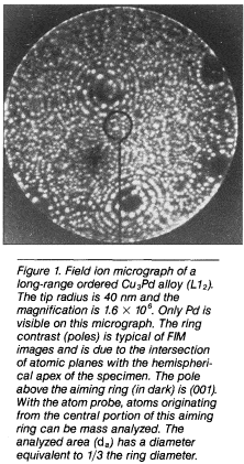
order in dilute alloys or to investigate long-range order (Figure 1). Similarly, in two-phase materials, precipitates often give rise to quite visible contrast. For instance, in nickel-based alloys, aluminum-enriched precipitates appear in bright contrast. It is therefore possible to find the size and also the number density of particles present in the material. However, FIM does not provide any composition data.

Type
Technical Features
Copyright
Copyright © Materials Research Society 1994

Access options

Get access to the full version of this content by using one of the access options below. (Log in options will check for institutional or personal access. Content may require purchase if you do not have access.)

References

1.Müller, E.W., Physik 131 (1951) p. 136.CrossRefGoogle Scholar
2.Wei, C.H. and Seidman, D.N., Philos. Mag. A 43 (1981) p. 1419.CrossRefGoogle Scholar
3.Müller, E. W., J. Phys. Soc. Jpn. 18 (1963) p. 1.Google Scholar
4.Tsong, T.T. and Casanova, R., Phys. Rev. B 24 (1981) p. 3063.CrossRefGoogle Scholar
5.Tsong, T.T., Atom Probe Field Microscopy (Cambridge University Press, New York, 1990).CrossRefGoogle Scholar
6.Miller, M.K. and Smith, G.D.W., Atom Probe Microanalysis: Principles and Applications to Materials Problems (Materials Research Society, Pittsburgh, PA, 1989).Google Scholar
7.Blavette, D. and Menand, A., Ann. Chim. Phys. 11 (1986) p. 321.Google Scholar
8.Blavette, D., Chambreland, S., Loiseau, A., Hanes, J., and Ducastelle, F., J. Phys. (Paris) C8-50 (1989) p. 365; T.T. Tsong and EW. Müller, Appl. Phys. Lett. 9 (1966) p. 7.Google Scholar
9.Müller, E.W., Panitz, J.A., and McLane, S.B., Rev. Sci. Instrum. 38 (1968) p. 83.CrossRefGoogle Scholar
10.Blavette, D. and Chambreland, S., J. Phys. (Paris) C7-47 (1986) p. 503.Google Scholar
11.Ahmad, M. and Tsong, T.T., Appl. Phys. Lett. 44 (1984) p. 40.CrossRefGoogle Scholar
12.Wendt, H. and Haasen, P., Acta Metall. 31 (1983) p. 1649.CrossRefGoogle Scholar
13.Miller, M.K. and Burke, M.G., J. Phys. (Paris) C6-48 (1987) p. 48.Google Scholar
14.Blavette, D. and Bostel, A., Acta Metall. 32 (1984) p. 811.CrossRefGoogle Scholar
15.Karlsson, L. and Norden, N., J. Phys. (Paris) C9-45 (1984) p. 391.Google Scholar
16.Müller, E.W. and Krishnaswamy, S.V., Rev. Sci. Instrum. 45 (1974) p. 1053.CrossRefGoogle Scholar
17.Poschenrieder, W.P., Int. J. Mass Spectrom. Ion Phys. 9 (1972) p. 357.CrossRefGoogle Scholar
18.Kellogg, G.L. and Tsong, T.T., J. Appl. Phys. 51 (1980) p. 1184; T.T. Tsong, T.J. Kinkus, and S.B. McLane, Rev. Sci. Instrum. 53 (1982) p. 1442.CrossRefGoogle Scholar
19.Panitz, J.A., J. Vac. Sci. Technol. 11 (1974) p. 206.CrossRefGoogle Scholar
20.Cerezo, A., Godfrey, T.J., and Smith, G.D.W., J. Phys. (Paris) C6-49 (1988) p. 25.Google Scholar
21.Miller, M.K., Surf. Sci. 266 (1992) p. 454.Google Scholar
22.Blavette, D., Bostel, A., Sarrau, J.M., Deconihout, B., and Menand, A., Nature 363 (1993) p. 432.CrossRefGoogle Scholar
23.Blavette, D., Deconihout, B., Bostel, A., Sarrau, J.M., Bouet, M., and Menand, A., Rev. Sci. Instrum. 64–10 (1993) P. 2911.CrossRefGoogle Scholar
24.Brown, J.E., Cerezo, A., Godefroy, T.J., Hetherington, M.G., and Smith, G.D.W., Mater. Sci. Tech. 6 (1990) p. 293.CrossRefGoogle Scholar
25.Danoix, F., Auger, P., and Blavette, D., Surf. Sci. 266 (1992) p. 364.CrossRefGoogle Scholar
26.Buchon, A. and Blavette, D., C.R.A.S. 315–11 (1992) p. 279.Google Scholar
27.Letellier, L., Bostel, A., and Blavette, D., submitted to Scripta Mat.Google Scholar