Last updated 10th July 2024: Online ordering is currently unavailable due to technical issues. We apologise for any delays responding to customers while we resolve this. For further updates please visit our website https://www.cambridge.org/news-and-insights/technical-incident
We use cookies to distinguish you from other users and to provide you with a better experience on our websites. Close this message to accept cookies or find out how to manage your cookie settings.
To save this undefined to your undefined account, please select one or more formats and confirm that you agree to abide by our usage policies. If this is the first time you used this feature, you will be asked to authorise Cambridge Core to connect with your undefined account.
Find out more about saving content to .
To save this article to your Kindle, first ensure coreplatform@cambridge.org is added to your Approved Personal Document E-mail List under your Personal Document Settings on the Manage Your Content and Devices page of your Amazon account. Then enter the ‘name’ part of your Kindle email address below.
Find out more about saving to your Kindle.
Note you can select to save to either the @free.kindle.com or @kindle.com variations. ‘@free.kindle.com’ emails are free but can only be saved to your device when it is connected to wi-fi. ‘@kindle.com’ emails can be delivered even when you are not connected to wi-fi, but note that service fees apply.
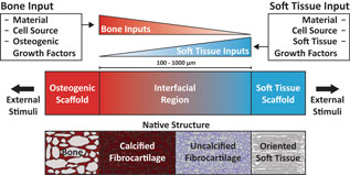
Soft tissue-to-bone interfaces are complex structures that consist of gradients of extracellular matrix materials, cell phenotypes, and biochemical signals. These interfaces, called entheses for ligaments, tendons, and the meniscus, are crucial to joint function, transferring mechanical loads and stabilizing orthopedic joints. When injuries occur to connected soft tissue, the enthesis must be re-established to restore function, but due to structural complexity, repair has proven challenging. Tissue engineering offers a promising solution for regenerating these tissues. This prospective review discusses methodologies for tissue engineering the enthesis, outlined in three key design inputs: materials processing methods, cellular contributions, and biochemical factors.
Neurological and psychiatric disorders account for an increasing proportion of the global disease burden. Correspondingly the neuropharmaceutical industry has experienced a significant contraction in recent years resulting in a poor variety of therapies available to treat an expanding range of conditions. Perhaps the greatest contributor to this failure in drug-discovery is the lack of understanding of the underlying biology of the nervous system and how molecular scale events translate into macroscale pathologies. Due to the unique nature of the human nervous system commonly used model organisms are often poorly representative of human pathologies resulting in a need for the development of advanced in vitro models that are capable of faithfully modeling complex structures within the brain. In this prospective, strategies for the generation of neuronal circuits and cultivation of complex three-dimensional (3D) cultures are explored. Frequently these constructs provide valuable insights into systems and processes that are difficult to explore in vivo due to the isolated and delicate nature of neuronal tissues. New developments are required to assess the physiological functions of 3D tissues in vitro.
To gain a better understanding of the underlying mechanisms of neurological disease, relevant tissue models are imperative. Over the years, this realization has fuelled the development of novel tools and platforms, which aim at capturing in vivo complexity. One example is the field of biofabrication, which focuses on fabrication of three-dimensional (3D) biologically functional products in a controlled and automated manner. Herein, we provide a general overview of classical 3D cell culture platforms, particularly in the context of neurodegenerative disease. Subsequently, the focus is put on bioprinting-based biofabrication, its potential to advance 3D neuronal cell culture and, to conclude, the relevant translational bottlenecks, which will need to be considered as the field evolves.
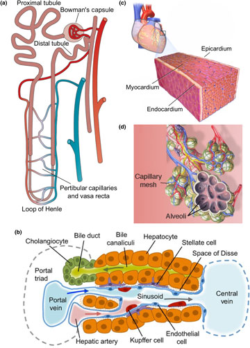
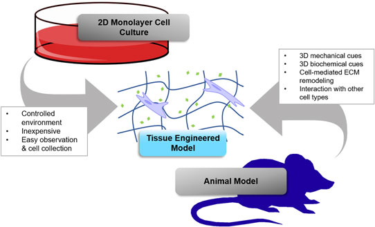
Tissue engineering has been recognized as a translational approach to replace damaged tissue or whole organs. Engineering tissue, however, faces an outstanding knowledge gap in the challenge to fully recapitulate complex organ-specific features. Major components, such as cells, matrix, and architecture, must each be carefully controlled to engineer tissue-specific structure and function that mimics what is found in vivo. Here we review different methods to engineer tissue, and discuss critical challenges in recapitulating the unique features and functional units in four major organs—the kidney, liver, heart, and lung, which are also the top four candidates for organ transplantation in the USA. We highlight advances in tissue engineering approaches to enable the regeneration of complex tissue and organ substitutes, and provide tissue-specific models for drug testing and disease modeling. We discuss the current challenges and future perspectives toward engineering human tissue models.
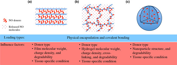
Nitric oxide (NO) acts a pivotal role in regulating various physiological processes of vasodilation, platelet aggregation, and vascular smooth muscle cell mitogenesis and proliferation. This makes NO a promising candidate for the treatment of cardiovascular problems like hypertension and vascular stenosis. However, the high reactivity of NO poses an issue for effective NO delivery. To overcome this limitation, recent developments on three-dimensional (3D) materials have been explored with either physical or chemical incorporation of NO releasing donors, to provide spatiotemporal control over NO-signaling pathways in blood vessels. Here, we offer an overview on the current efforts, and propose future perspectives for precise regulation on NO delivery in advanced 3D materials toward proper vascular functionality.
Three-dimensional (3D) scaffolds composed of poly(ε-caprolactone) and gelatin nanofibers were fabricated by a combination of electrospinning and modified gas-foaming. Arrayed holes throughout the scaffold were created using a punch under cryo conditions. The crosslinking with glutaraldehyde vapor improved the water stability of the scaffolds. Cell spheroids of green fluorescent protein-labeled human dermal fibroblasts were prepared and seeded into the holes. It was found that the fibroblasts adhered well on the surface of nanofibers and migrated into the scaffolds due to the porous structures. The 3D nanofiber scaffolds may hold great potential for engineering tissue constructs for various applications.
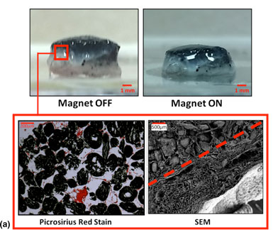
Mesenchymal stem cell behavior can be regulated through mechanical signaling, either by dynamic loading or through biomaterial properties. We developed intrinsically responsive tissue engineering scaffolds that can dynamically load cells. Porous collagen- and alginate-based scaffolds were functionalized with iron oxide to produce magnetically active scaffolds. Reversible deformations in response to magnetic stimulation of up to 50% were recorded by tuning the material properties. Cells could attach to these scaffolds and magnetically induced compressive deformation did not adversely affect viability or cause cell release. This platform should have broad application in the mechanical stimulation of cells for tissue engineering applications.
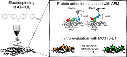
Conjugated polymers have been proposed as promising materials for scaffolds in tissue engineering applications. However, the restricted processability and biodegradability of conjugated polymers limit their use for biomedical applications. Here we synthesized a block-co-polymer of aniline tetramer and PCL (AT–PCL), and processed it into fibrous non-woven scaffolds by electrospinning. We showed that fibronectin (Fn) adhesion was dependent on the AT–PCL oxidative state, with a reduced Fn unfolding length on doped membranes. Furthermore, we demonstrated the cytocompatibility and potential of these membranes to support the growth and osteogenic differentiation of MC3T3-E1 cells over 21 days.
Biomaterials for 3D Cell Biology Prospective Articles
In this work, we investigated the interactions of human mesenchymal stem cells (hMSCs) with three-dimensional (3D) printed scaffolds displaying different scaffold architectures. Pressure-assisted microsyringe system was used to fabricate scaffolds with square (SQR), hexagonal (HEX), and octagonal (OCT) architectures defined by various degrees of curvatures. OCT represents the highest degree of curvature followed by HEX, and SQR is composed of linear struts without curvature. Scaffolds were fabricated from poly(L-lactic acid) and poly(tyrosol carbonate). We found that hMSCs attached and spread by taking the shape of the individual struts, exhibiting high aspect ratios (ARs) and mean cell area when cultured on OCT scaffolds as compared with those cultured on HEX and SQR scaffolds. In contrast, cells appeared bulkier with low AR on SQR scaffolds. These significant changes in cell morphology directly correlate with the stem cell lineage commitment, such that 80 ± 1% of the hMSCs grown on OCT scaffolds differentiated into osteogenic lineage, compared with 70 ± 4% and 62 ± 2% of those grown on HEX and SQR scaffolds, respectively. Cells on OCT scaffolds also showed 2.5 times more alkaline phosphatase activity compared with cells on SQR scaffolds. This study demonstrates the importance of scaffold design to direct stem cell differentiation, and aids in the development of novel 3D scaffolds for bone regeneration.
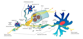
Understanding peripheral nerve repair requires the evaluation of three-dimensional (3D) structures that serve as platforms for 3D cell culture. Multiple platforms for 3D cell culture have been developed, mimicking peripheral nerve growth and function, in order to study tissue repair or diseases. To recreate an appropriate 3D environment for peripheral nerve cells, key factors are to be considered, including selection of cells, polymeric biomaterials to be used, and fabrication techniques to shape and form the 3D scaffolds for cellular culture. This review focuses on polymeric 3D platforms used for the development of 3D peripheral nerve cell cultures.
HYDROGELS
Biomaterials for 3D Cell Biology Prospective Articles
Gelatin-based hydrogels derived from hydrolysis of collagen have been extensively used in pharmaceutical and medical applications because of their biocompatibility and biodegradability. For example, gelatin-based hydrogels are finding use in drug delivery and tissue engineering because they are able to promote cell adhesion and proliferation. In addition, these hydrogels can be used as wound dressings due to their attractive fluid absorbance properties. Manufacturing technologies such as ultraviolet stereolithography and two-photon polymerization can be used to prepare structures containing photosensitive gelatin-based hydrogels. This review describes the preparation of gelatin-based hydrogels and use of these materials for biomedical applications.
Tissue engineering holds great promise for advancing cancer research and achieving the goals of the Cancer Moonshot by providing better models for basic research and testing novel therapeutics. This paper focuses on the use of hydrogel biomaterials due to their unique ability to entrap cells in three-dimensional (3D) matrix that mimics tissues and can be programmed with physical and chemical cues to recreate key aspects of tumor microenvironments. The chemistry of some commonly used hydrogel platforms is discussed, and important examples of their use in tissue engineering 3D cancer models are highlighted. Challenges and opportunities for future research are also discussed.
While preclinical models such as orthotopic tumors generated in mice from patient-derived specimens are widely used to predict sensitivity or therapeutic interventions for cancer, such xenografts can be slow, require extensive infrastructure, and can make in situ assessment difficult. Such concerns are heightened in highly aggressive cancers, such as glioblastoma (GBM), that display genetic diversity and short mean survival. Biomimetic biomaterial technologies offer an approach to create ex vivo models that reflect biophysical features of the tumor microenvironment (TME). We describe a microfluidic templating approach to generate spatially graded hydrogels containing patient-derived GBM cells to explore drug efficacy and resistance mechanisms.
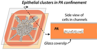
Nuclear translocation of Yes-associated-protein (YAP) in single cells serves as a key sensor of matrix stiffness. On two-dimensional (2D) polyacrylamide (PA) hydrogels, we found that nuclear YAP localization in epithelial clusters increases with gel stiffness and reduces with cell density. To measure YAP activity in 3D-like confinement of tunable stiffness, we fabricated PA-based microchannels. Here, narrower channels enhanced nuclear YAP localization even in softer extracellular matrix and denser epithelial clusters, both of which reduced YAP activation in 2D. Thus, the presented hydrogel microchannel-based platform may reveal new mechanosensitive cellular signatures in 3D-like settings, which cannot be captured on standard 2D hydrogels.
Human mesenchymal stem cells (MSCs) are the most intensely studied and clinically used adult stem cell type. Conventional long-term cultivation of MSCs as a monolayer is known to result in a reduction of their functionality and viability. In addition, large volumes of cell culture medium are required to obtain cell quantities needed for their clinical use. In this proof of concept study, we cultivated human MSCs within a three-dimensional nanofibrillar cellulose (NFC) hydrogel. We show that NFC is biocompatible with human MSCs, and represents a feasible approach to upscaling of their culture.
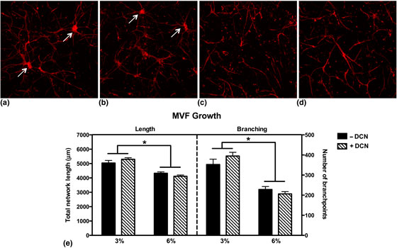
Angiogenesis is a critical component during wound healing, and the process is sensitive to mechanical stimuli. Current in vitro culture environments used to investigate three-dimensional microvascular growth often lack dimensional stability and the ability to withstand compression. We investigated the ability of decorin (DCN), a proteoglycan known to modulate collagen fibrillogenesis, incorporated into a collagen hydrogel to increase construct dimensional stability while maintaining vascular growth. DCN did not affect microvascular growth parameters, while increasing the compressive modulus of collagen gels and significantly reducing the contraction of 3% collagen gels after 16 days in culture.
Biomaterials for 3D Cell Biology Prospective Article
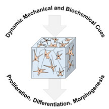
Bioengineered hydrogels enable systematic variation of mechanical and biochemical properties, resulting in the identification of optimal in vitro three-dimensional culture conditions for individual cell types. As the scientific community attempts to mimic and study more complex biologic processes, hydrogel design has become multi-faceted. To mimic organ and tissue heterogeneity in terms of spatial arrangement and temporal changes, hydrogels with spatiotemporal control over mechanical and biochemical properties are needed. In this prospective article, we present studies that focus on the development of hydrogels with dynamic mechanical and biochemical properties, highlighting the discoveries made using these scaffolds.
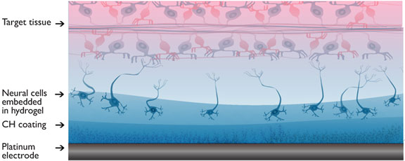
A living electrode construct that enables integration of cells within bionic devices has been developed. The layered construct uses a combination of non-degradable conductive hydrogel and degradable biosynthetic hydrogel to support cell encapsulation at device surfaces. In this study, the material system is designed and analyzed to understand the impact of the cell carrying component on electrode characteristics. The cell carrying layer is shown to provide a soft interface that supports extracellular matrix development within the electrode while not significantly reducing the charge transfer characteristics. The living layer was shown to degrade over 21 days with minimal swelling upon implantation.
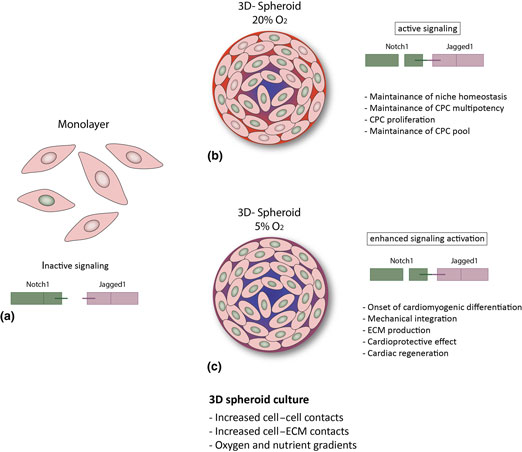
Cardiac progenitor cells (CPCs) are a promising candidate for cardiac regeneration, and the interaction between CPCs and their microenvironment can influence their regenerative response. Notch signaling plays a key role in cell fate decisions in the developing and adult heart. Here, we investigated the effect of three-dimensional (3D) spheroid culture, as a model of the 3D microenvironment, on Notch in fetal and adult human CPCs, under room air (20%) and physiological (5%) oxygen tension. Notch signaling is enhanced in 3D spheroids; spheroid culture under 5% O2 further increases Notch signaling enhancement, and might ultimately improve the regenerative potential of CPCs.
Biomaterials for 3D Cell Biology Prospective Article
Human organoid models recapitulate many aspects of the complex composition and function of native organs. One of the main challenges in developing these models is the growth and maintenance of three-dimensional tissue structures and proper cellular organization that enable function. Biomaterials play an important role by providing a defined and tunable three-dimensional environment that is required for complex cellular organization and organoid growth in vitro or in vivo. This review summarizes organoids of the respiratory and digestive system, and the use of biomaterials to improve upon these model systems.