NenoVision LiteScope™ – Compact AFM for your Scanning Electron Microscope
The NenoVision AFM LiteScope™ can be integrated into many SEMs to extend the capabilities of AFM and SEM instruments. The LiteScope offers precise AFM tip navigation, in situ 3D surface characterization, material and topography contrast, height/depth profiling, and measurement of electrical or mechanical properties. The LiteScope is equipped with a unique CPEM™ technique for true correlative imaging, which enables simultaneous acquisition and direct correlation of SEM and AFM images.
NenoVision
www.nenovision.com/litescope/litescopetm.description

Air Science® Fume Box™ Ductless Enclosures
The Air Science® Fume Box™ ductless enclosures are an effective solution to air filtration needs. Designed to protect the user from chemicals, vapors, or powders during low-volume chemical manipulations, the Fume Box employs advanced carbon filtration technology to offer a safe, high-performance alternative to conventional ducted fume hoods for a wide range of applications in varied industry settings.
Air Science

Leica Microsystems DM6 M LIBS: Visual and Chemical Inspection in One Step
The integrated laser spectroscopy function of the DM6 M LIBS (laser induced breakdown spectroscopy) delivers the chemical composition of the microstructure that you see in the microscope image—within a second. Identify the microstructure composition of interest, and then trigger the LIBS analysis with a single click to visually inspect and chemically analyze a sample. Perform advanced material analysis 90% faster compared to SEM/EDS.
Leica Microsystems

Digital Surf Releases Mountains® 8 Image and Surface Analysis Software
Digital Surf has released its new Mountains® 8 software platform that builds on the Mountains® 7 platform introduced in 2013 that is provided by most microscope and profiler manufacturers with their instruments. Mountains® 8 incorporates significant new contributions from SPIP™ 6 software originally developed by Image Metrology. Mountains® 8 also provides an increase in calculation speed, new types of surface data analysis, greater interactivity, and over a hundred new features.
Digital Surf

Linkam's Optical DSC450
Differential scanning calorimetry (DSC) is a technique used to measure the temperature and heat flow associated with thermal transitions in materials. The optical DSC450 system has been optimized to measure the transition temperatures and enthalpy changes in samples. The design allows mounting of the stage on a microscope, which enables image and time lapse recording of sample transitions at high resolution. Color-coded imaging or morphological changes can be correlated with temperature and phase transitions.
Linkam Scientific

Protochips AXON™ Software
Protochips AXON software changes the in situ TEM experience. It puts the sample front and center as one changes in situ conditions, even at extreme magnifications. Focus is on the sample, at all the key moments when capturing data. AXON is designed to streamline the process of collecting quality data. Experiment conditions are synchronized with images, making data analysis easier before and after the imaging session.
Protochips
www.protochips.com/products/axon

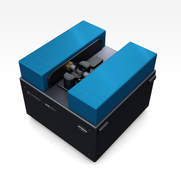
Bruker Introduces Light-Sheet Microscope
Bruker has released the Luxendo LCS SPIM light-sheet fluorescence microscope for 3D imaging of large, optically cleared samples. Light-sheet fluorescence microscopy is a powerful method for high-resolution, cleared-sample imaging. The modular Luxendo LCS SPIM has been designed to be compatible with a broad variety of clearing solutions and sample sizes. Its new sample mounting approach and innovative optical design enables unprecedented acquisition times and minimizes sample distortions while seamlessly integrating into existing clearing and sample preparation pipelines.
Bruker/Luxendo
Four New ZEISS Axiocam Cameras
ZEISS has released four new high-quality CMOS cameras for digital imaging in light microscopy. The ZEISS Axiocam 705 color and 712 color cameras deliver the best possible image quality for histology, pathology, and material research analyses with excellent color rendition and improved dynamic range. The ZEISS Axiocam 705 mono and 712 mono are ideal for fluorescence live-cell imaging with fast frame rates and high dynamic range. Their near-IR sensitivity provides deeper insights into sample structures.
ZEISS Research Microscopy Solutions

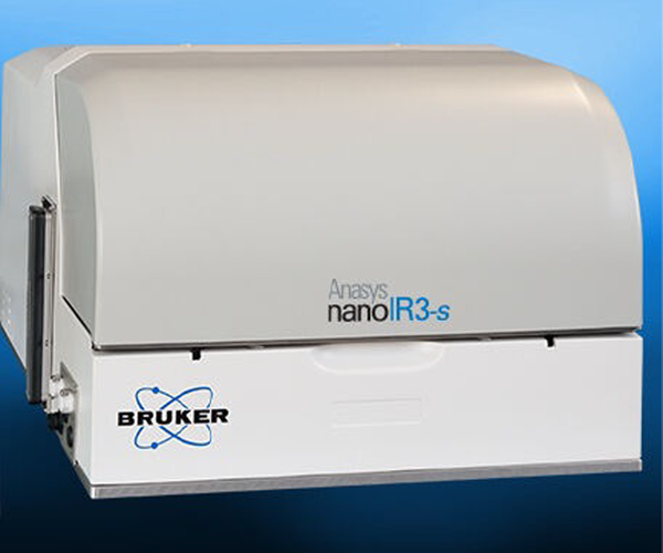
Bruker's Nanoscale Infrared Spectroscopy and Chemical Imaging SNOM/AFM Microscopy System with Broadband Femtosecond IR Laser
The nanoIR3-s Broadband™ nanoscale FTIR (Fourier transform infrared) spectroscopy system combines high-performance, nanoIR3-s s-SNOM (scattering scanning near-field optical microscopy) based on femtosecond IR laser technology. This combination allows researchers to make groundbreaking new discoveries in nanoscale FTIR spectroscopy and chemical imaging for polymeric materials and life science applications. It has applications in nanoscale optical imaging of 2D materials, plasmonic fields, and nanophotonic structures.
Bruker
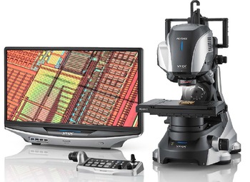
Keyence VHX Series Microscopes
The VHX Series exceeds conventional imaging tools with a depth of field that is 20 times greater than conventional optical microscopes and a wide range of observation modes, including bright-field, dark-field, polarized Light, and differential interference contrast (DIC). With advanced measurement capabilities, this system enables a variety of analyses. Expanded memory capacity allows for storage of millions of images. Its easy-to-use interface can be used effectively by expert and novice users.
Keyence
Rave Scientific's IBSS In Situ Plasma Cleaner
Have you ever de-magnified when imaging in a scanning electron microscope and noticed dark scanlines on your sample? This is hydrocarbon contamination, which can impede the optimum performance of an SEM. Hydrocarbons are generally introduced into the SEM chamber by outgassing samples or by poor specimen handling. As important as it is to change the oil in a car for optimum engine performance, it is just as important to regularly in situ plasma clean an SEM chamber.
Rave Scientific
https://ravescientific.com/sample-preparation/ibss-plasma-cleaner

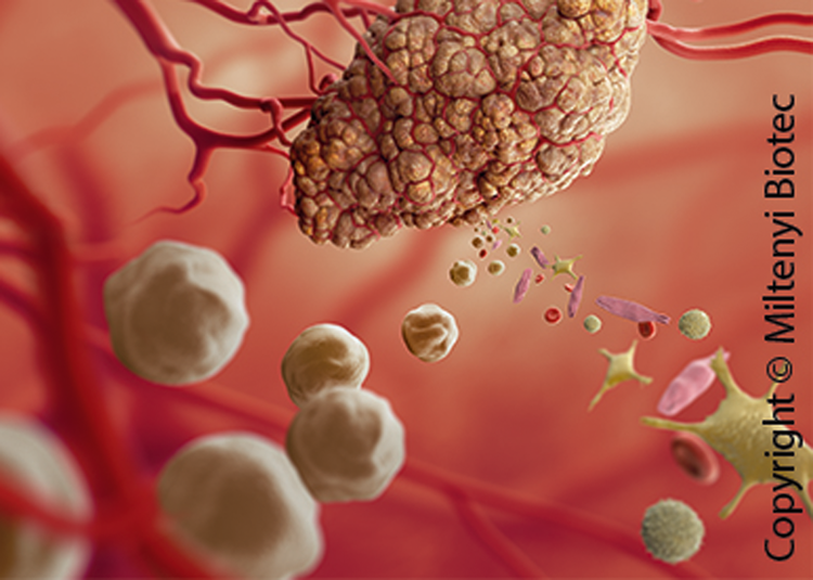
Miltenyi Biotec's FFPE Tissue Dissociation Kit
This kit enables effective dissociation of formalin-fixed paraffin-embedded (FFPE) carcinoma samples for the enrichment and subsequent molecular analysis of carcinoma tumor cells. This enhances the sensitivity in molecular experiments, such as mutation analysis by next-generation sequencing (NGS).
Benefits of the FFPE Tissue Dissociation Kit include effective enrichment of tumor cells from dissociated carcinoma samples based on cytokeratin expression, enhanced sensitivity of DNA analysis from carcinomas with low tumor cell content, and standardized workflow.
Miltenyi Biotec
www.miltenyibiotec.com/upload/assets/IM0020690.pdf
Abberior Instruments Launches the MINFLUX Light Microscope
MINFLUX (minimal photon fluxes) is a cutting-edge method for localizing fluorescent molecules in space with unprecedented precision. MINFLUX probes the location of emitters with an excitation intensity minimum and therefore reduces the number of fluorescent photons required for high-precision localization by a factor of up to 20. For a given number of photons, the precision is improved, and, therefore, localizations below 2 nm are obtained for the first time in a light microscope.
Abberior instruments
www.abberior-instruments.com/products/minflux

PerkinElmer MuviCyte Live-Cell Imaging System
The MuviCyte system is designed to operate inside a cell culture incubator to keep cells healthy and to perform assays over hours, days, or weeks. MuviCyte system can run a variety of assays with three-color fluorescence imaging, z-stacking, and stitching with your preferred culture vessels (chamber slides, Petri dishes, T-flasks, or microplates). Imaging is automatical for walkaway convenience. The MuviCyte live-cell imaging kit consists of a MuviCyte live-cell imaging instrument, objective lenses (4×, 10×, and 20×), PC, and monitor.
Perkin Elmer
www.perkinelmer.com/MuviCyte_Live/Cell_Imaging

Cytation 5 Cell Imaging Multi-Mode Reader
The Biotek Cytation™ 5 combines automated digital microscopy and conventional microplate detection in a configurable, upgradable platform. The microscopy module offers up to 60 × magnification in fluorescence, brightfield, high-contrast brightfield, color brightfield, and phase contrast. The multi-mode detection modules include filter- and monochromator-based fluorescence detection, luminescence, and UV-Vis absorbance detection. Gen5™ software provides complete control over all imaging and data capture, plus powerful image and data analysis.
Biotek

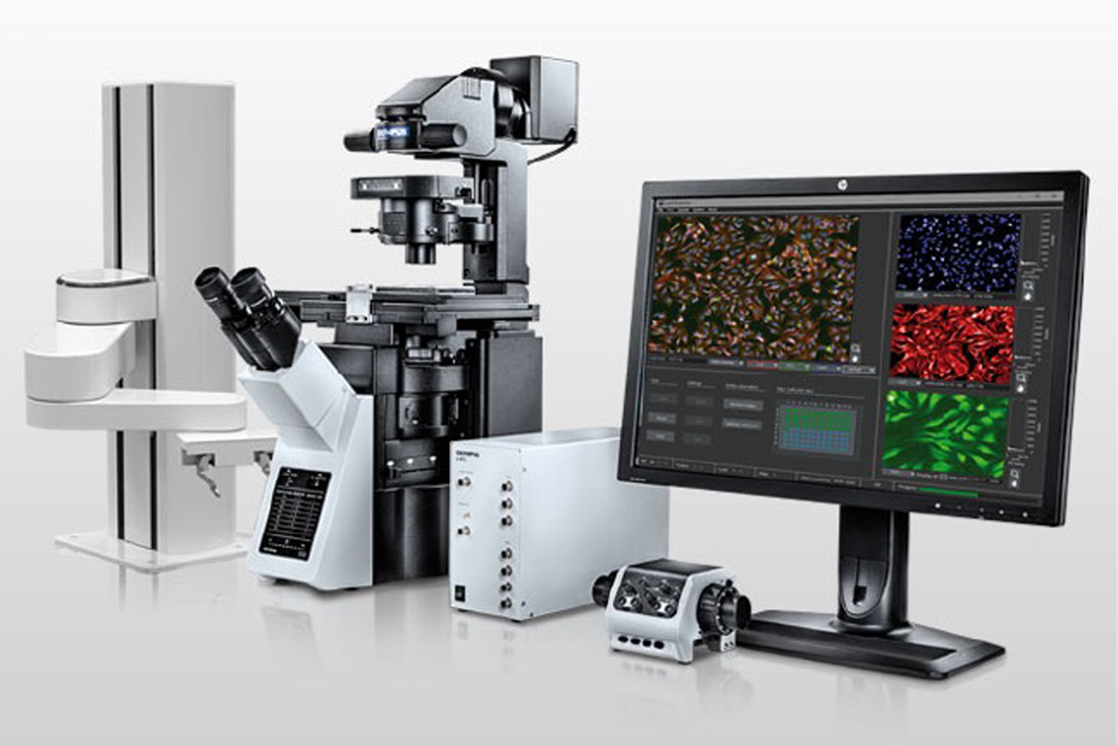
Olympus Announces the scanR High-Content Screening Station
Olympus has launched the scanR high-content screening (HCS) station, a cell imaging solution that uses artificial intelligence (AI) to enable next-generation biological research. It combines the modularity and flexibility of a microscope-based setup with the automation, speed, throughput, and reproducibility of an HCS station. After a one-time training phase, scanR AI enables the system to automatically analyze new data by incorporating the learned analysis protocol into its assay-based workflow.
Olympus
www.olympus-lifescience.com/en/microscopes/inverted/scanr