Skip to main content Accessibility help
×
Hostname: page-component-7479d7b7d-c9gpj Total loading time: 0 Render date: 2024-07-09T19:48:10.244Z Has data issue: false hasContentIssue false

Chapter 2 - Theessential contributions of neuroimaging

Published online by Cambridge University Press:  05 May 2016

Christopher M. Filley
Affiliation:
University of Colorado School of Medicine
Get access

Summary

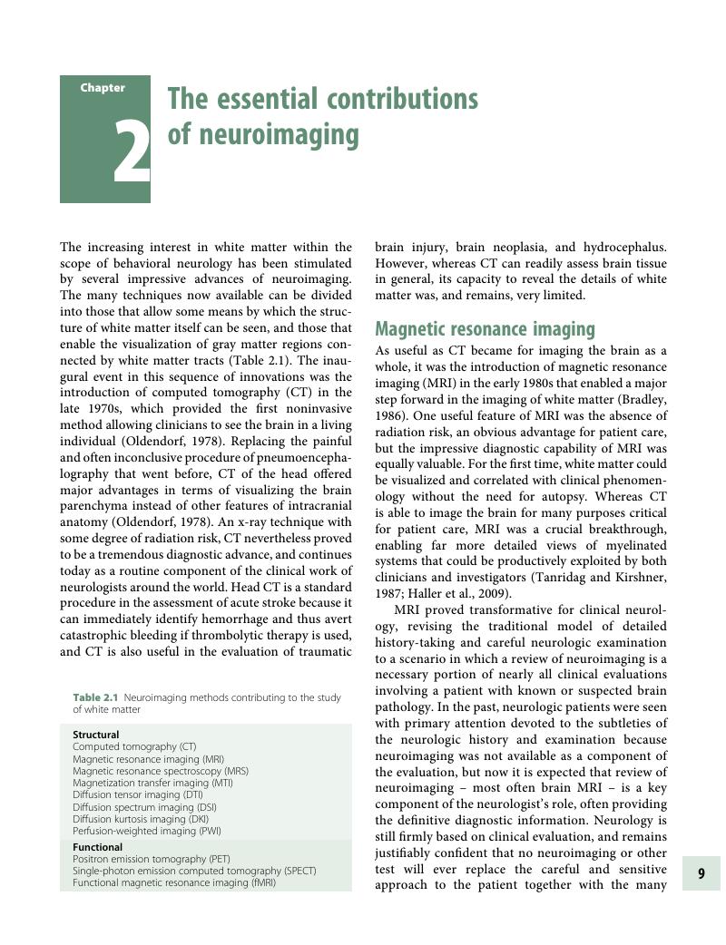
Image of the first page of this content. For PDF version, please use the ‘Save PDF’ preceeding this image.'
Type
Chapter
Information
Publisher: Cambridge University Press
Print publication year: 2016

Access options

Get access to the full version of this content by using one of the access options below. (Log in options will check for institutional or personal access. Content may require purchase if you do not have access.)

References

Alavi, A, Hirsch, LJ. Studies of central nervous system disorders with single photon emission computed tomography and positron emission tomography. Semin Nucl Med 1991; 21: 5881.CrossRefGoogle ScholarPubMed
Alexander, AL, Lee, JE, Lazar, M, Field, AS. Diffusion tensor imaging of the brain. Neurotherapeutics 2007; 4: 316329.CrossRefGoogle ScholarPubMed
Alves, GS, Oertel Knöchel, V, Knöchel, C, et al. Integrating retrogenesis theory to Alzheimer’s disease pathology: insight from DTI-TBSS investigation of the white matter microstructural integrity. Biomed Res Int 2015; 2015: 291658.CrossRefGoogle ScholarPubMed
Bandettini, PA. What’s new in neuroimaging methods? Ann NY Acad Sci 2009; 1156: 260293.CrossRefGoogle ScholarPubMed
Bradley, WG Jr. Magnetic resonance imaging in the central nervous system: comparison with computed tomography. Magn Res Ann 1986; 81122.Google Scholar
Catani, M, Dell’acqua, F, Bizzi, A, et al. Beyond cortical localization in clinico-anatomical correlation. Cortex 2012; 48: 12621287.CrossRefGoogle ScholarPubMed
Catani, M, Thiebaut de Schotten, M. Atlas of human brain connections. Oxford: Oxford University Press, 2012.CrossRefGoogle Scholar
Cercignani, M, Iannucci, G, Rocca, MA, et al. Pathologic damage in MS assessed by diffusion-weighted and magnetization transfer MRI. Neurology 2000; 54: 11391144.CrossRefGoogle ScholarPubMed
Choudhri, AF, Chin, EM, Blitz, AM, Gandhi, D. Diffusion tensor imaging of cerebral white matter: technique, anatomy, and pathologic patterns. Radiol Clin North Am 2014; 52: 413425.CrossRefGoogle ScholarPubMed
Damoiseaux, JS, Greicius, MD. Greater than the sum of its parts: a review of studies combining structural connectivity and resting-state functional connectivity. Brain Struct Funct 2009; 213: 525533.CrossRefGoogle Scholar
DeCarli, C, Fletcher, E, Ramey, V, et al. Anatomical mapping of white matter hyperintensities (WMH): exploring the relationships between periventricular WMH, deep WMH, and total WMH burden. Stroke 2005; 36: 5055.CrossRefGoogle ScholarPubMed
D’Esposito, M. Functional neuroimaging of cognition. Semn Neurol 2000; 20: 487498.CrossRefGoogle ScholarPubMed
Fieremans, E, Jensen, JH, Helpern, JA. White matter characterization with diffusional kurtosis imaging. Neuroimage 2011; 58: 177188.CrossRefGoogle ScholarPubMed
Filley, CM. The behavioral neurology of white matter. 2nd ed. New York: Oxford University Press, 2012.CrossRefGoogle ScholarPubMed
Filley, CM, Kozora, E, Brown, MS, et al. White matter microstructure and cognition in non-neuropsychiatric systemic lupus erythematosus. Cogn Behav Neurol 2009; 22: 3844.CrossRefGoogle ScholarPubMed
Granziera, C, Schmahmann, JD, Hadjikhani, N, et al. Diffusion spectrum imaging shows the structural basis of functional cerebellar circuits in the human cerebellum in vivo. PloS One 2009; 4: e5101.CrossRefGoogle ScholarPubMed
Grossman, RI, Kappos, L, Wolinsky, JS. The contribution of magnetic resonance imaging in the differential diagnosis of the damage of the cerebral hemispheres. J Neurol Sci 2000; 172: S57S62.CrossRefGoogle ScholarPubMed
Haller, S, Pereira, VM, Lazeyras, F, et al. Magnetic resonance imaging techniques in white matter disease: potentials and limitations. Top Magn Reson Imaging 2009; 20: 301312.CrossRefGoogle ScholarPubMed
Horsfield, MA, Jones, DK. Applications of diffusion-weighted and diffusion tensor MRI to white matter diseases – a review. NMR Biomed 2002; 15: 570577.CrossRefGoogle ScholarPubMed
Jensen, JH, Helpern, JA. MRI quantification of non-Gaussian water diffusion by kurtosis analysis. NMR Biomed 2010; 23: 698710.CrossRefGoogle ScholarPubMed
Launer, LJ. Epidemiology of white matter lesions. Top Magn Reson Imaging 2004; 15: 365367.CrossRefGoogle ScholarPubMed
Loevner, LA, Grossman, RI, Cohen, JA, et al. Microscopic disease in normal-appearing white matter on conventional MR imaging in patients with multiple sclerosis: assessment with magnetization-transfer measurements. Radiology 1995; 196: 511515.CrossRefGoogle ScholarPubMed
Mäntylä, R, Erkinjuntti, T, Salonen, O, et al. Variable agreement between visual rating scales for white matter hyperintensities on MRI: comparison of 13 rating scales in a poststroke cohort. Stroke 1997; 28: 16141623.CrossRefGoogle Scholar
Mesulam, M-M. Large-scale neurocognitive networks and distributed processing for attention, language, and memory. Ann Neurol 1990; 28: 597613.CrossRefGoogle ScholarPubMed
Narayana, PA. Magnetic resonance spectroscopy in the monitoring of multiple sclerosis. J Neuroimaging 2005; 15 (4 Suppl): 46S57S.CrossRefGoogle ScholarPubMed
Oldendorf, WH. The quest for an image of brain: a brief historical and technical review of brain imaging techniques. Neurology 1978; 28: 517533.CrossRefGoogle Scholar
Prichard, JW, Cummings, JL. The insistent call from functional MRI. Neurology 1997; 48: 797800.CrossRefGoogle ScholarPubMed
Rademacher, J, Engelbrecht, V, Burgel, U, et al. Measuring in vivo myelination of human white matter fiber tracts with magnetization transfer MR. Neuroimage 1999; 9: 393406.CrossRefGoogle ScholarPubMed
Raichle, ME, MacLeod, AM, Snyder, AZ, et al. A default mode of brain function. Proc Natl Acad Sci 2001; 98: 676882.CrossRefGoogle ScholarPubMed
Rudkin, TM, Arnold, DL. Proton magnetic resonance spectroscopy for the diagnosis and management of cerebral disorders. Arch Neurol 1999; 56: 919926.CrossRefGoogle ScholarPubMed
Schmahmann, JD, Pandya, DN, Wang, R, et al. Association fibre pathways of the brain: parallel observations from diffusion spectrum imaging and autoradiography. Brain 2007; 130: 630653.CrossRefGoogle ScholarPubMed
Seeley, WW, Crawford, RK, Zhou, J, et al. Neurodegenerative diseases target large-scale human brain networks. Neuron 2009; 62: 4252.CrossRefGoogle ScholarPubMed
Simmons, M, Frondoza, C, Coyle, J. Immunocytochemical localization of N-acetyl-aspartate with monoclonal antibodies. Neuroscience 1991; 45: 3745.CrossRefGoogle ScholarPubMed
Simon, JH. MRI outcomes in the diagnosis and disease course of multiple sclerosis. Handb Clin Neurol 2014; 122: 405425.CrossRefGoogle ScholarPubMed
Smith, SM, Jenkinson, M, Johansen-Berg, H, et al. Tract-based spatial statistics: voxelwise analysis of multi-subject diffusion data. Neuroimage 2006; 31: 14871505.CrossRefGoogle ScholarPubMed
Tanabe, JL, Ezekiel, F, Jagust, WJ, et al. Magnetization transfer ratio of white matter hyperintensities in subcortical ischemic vascular dementia. AJNR 1999; 20: 839844.Google ScholarPubMed
Tanridag, O, Kirshner, HS. Magnetic resonance imaging and CT scanning in neurobehavioral syndromes: comparative neuroradiologic findings. Psychosomatics 1987; 28: 517528.CrossRefGoogle ScholarPubMed
Van Buchem, MA. Magnetization transfer: applications in neuroradiology. J Comp Assist Tomogr 1999; 23 (Suppl 1): S9S18.CrossRefGoogle ScholarPubMed
Wedeen, VJ, Wang, RP, Schmahmann, JD, et al. Diffusion spectrum magnetic resonance imaging (DSI) tractography of crossing fibers. Neuroimage 2008; 41: 12671277.CrossRefGoogle ScholarPubMed
Wozniak, JR, Lim, KO. Advances in white matter imaging: a review of in vivo magnetic resonance methodologies and their applicability to the study of development and aging. Neurosci Biobehav Rev 2006; 30: 762774.CrossRefGoogle Scholar

Save book to Kindle

To save this book to your Kindle, first ensure coreplatform@cambridge.org is added to your Approved Personal Document E-mail List under your Personal Document Settings on the Manage Your Content and Devices page of your Amazon account. Then enter the ‘name’ part of your Kindle email address below. Find out more about saving to your Kindle.

Note you can select to save to either the @free.kindle.com or @kindle.com variations. ‘@free.kindle.com’ emails are free but can only be saved to your device when it is connected to wi-fi. ‘@kindle.com’ emails can be delivered even when you are not connected to wi-fi, but note that service fees apply.

Find out more about the Kindle Personal Document Service.

Available formats
×

Save book to Dropbox

To save content items to your account, please confirm that you agree to abide by our usage policies. If this is the first time you use this feature, you will be asked to authorise Cambridge Core to connect with your account. Find out more about saving content to Dropbox.

Available formats
×

Save book to Google Drive

To save content items to your account, please confirm that you agree to abide by our usage policies. If this is the first time you use this feature, you will be asked to authorise Cambridge Core to connect with your account. Find out more about saving content to Google Drive.

Available formats
×