Skip to main content Accessibility help
×
Hostname: page-component-7479d7b7d-q6k6v Total loading time: 0 Render date: 2024-07-12T04:58:00.612Z Has data issue: false hasContentIssue false

Chapter 11 - Brain Imaging in Major Acute Stroke

from Section 2 - Clinical Features, Diagnosis, and Investigation

Published online by Cambridge University Press:  01 August 2018

Gary K. K. Lau
Affiliation:
University of Oxford
Sarah T. Pendlebury
Affiliation:
University of Oxford
Peter M. Rothwell
Affiliation:
University of Oxford
Get access

Summary

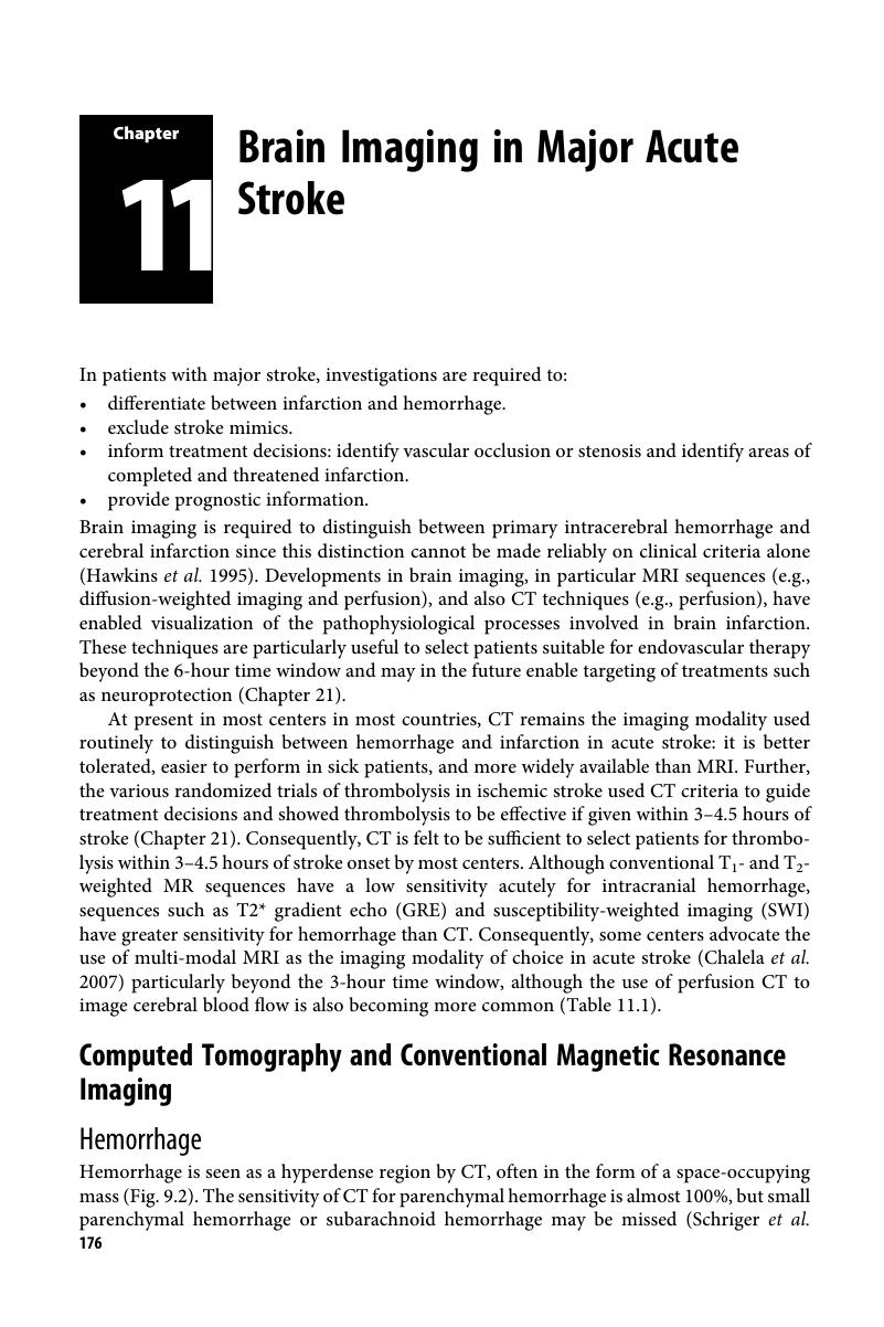
Image of the first page of this content. For PDF version, please use the ‘Save PDF’ preceeding this image.'
Type
Chapter
Information
Transient Ischemic Attack and Stroke
Diagnosis, Investigation and Treatment
, pp. 176 - 190
Publisher: Cambridge University Press
Print publication year: 2018

Access options

Get access to the full version of this content by using one of the access options below. (Log in options will check for institutional or personal access. Content may require purchase if you do not have access.)

References

Albers, GW, Lansberg, MG, Norbash, AM et al. (2000). Yield of diffusion-weighted MRI for detection of potentially relevant findings in stroke patients. Neurology 54:15621567CrossRefGoogle ScholarPubMed
Albers, GW, Marks, MP, Kemp, S et al. (2018). Thrombectomy for stroke at 6 to 16 hours with selection by perfusion imaging. New England Journal of Medicine 378:708718Google Scholar
Allen, LM, Hasso, AN, Handwerker, J et al. (2012). Sequence-specific MR imaging findings that are useful in dating ischemic stroke. Radiographics 32:12851297Google Scholar
Allport, LE, Parsons, MW, Butcher, KS et al. (2005). Elevated hematocrit is associated with reduced reperfusion and tissue survival in acute stroke. Neurology 65:13821387Google Scholar
Ay, H, Koroshetz, WJ, Vangel, M et al. (2005). Conversion of ischemic brain tissue into infarction increases with age. Stroke 36:26322636Google Scholar
Baird, AE, Benfield, A, Schlaug, G et al. (1997). Enlargement of human cerebral ischemic lesion volumes measured by diffusion-weighted magnetic resonance imaging. Annals of Neurology 41:581589Google Scholar
Baird, TA, Parsons, MW, Phanh, T et al. (2003). Persistent poststroke hyperglycemia is independently associated with infarct expansion and worse clinical outcome. Stroke 34:22082214Google Scholar
Bang, OY, Buck, BH, Saver, JL et al. (2007). Prediction of hemorrhagic transformation after recanalization therapy using T*2-permeability magnetic resonance imaging. Annals of Neurology 62:170176CrossRefGoogle ScholarPubMed
Barber, PA, Darby, DG, Desmond, PM et al. (1998). Prediction of stroke outcome with echoplanar perfusion-and diffusion-weighted MRI. Neurology 51:418426Google Scholar
Barber, PA, Demchuk, AM, Zhang, J et al. (2000). Validity and reliability of a quantitative computed tomography score in predicting outcome of hyperacute stroke before thrombolytic therapy. ASPECTS Study Group. Alberta Stroke Programme Early CT Score. Lancet 355:16701674Google Scholar
Beaulieu, C, de Crespigny, A, Tong, DC et al. (1999). Longitudinal magnetic resonance imaging study of perfusion and diffusion in stroke: Evolution of lesion volume and correlation with clinical outcome. Annals in Neurology 46:568578Google Scholar
Bogousslavsky, J (1991). Topographic patterns of cerebral infarcts: Correlation with aetiology. Cerebrovascular Diseases 1:6168Google Scholar
Bryan, RN, Levy, LM, Whitlow, WD et al. (1991). Diagnosis of acute cerebral infarction: Comparison of CT and MR imaging. American Journal of Neuroradiology 12:611620Google Scholar
Campbell, BC, Mitchell, MJ, Kleinig, TJ et al. (2015). Endovascular therapy for ischemic stroke with perfusion-imaging selection. New England Journal of Medicine 372:10091018Google Scholar
Chalela, JA, Kidwell, CS, Nentwich, LM et al. (2007). Magnetic resonance imaging and computed tomography in emergency assessment of patients with suspected acute stroke: A prospective comparison. Lancet 369:293298Google Scholar
Charidimou, A, Pasi, M, Fiorelli, M et al. (2016). Leukoaraiosis, cerebral hemorrhage, and outcome after intravenous thrombolysis for acute ischemic stroke: A meta-analysis. Stroke 47:23642372Google Scholar
Chodosh, EH, Foulkes, MA, Kase, CS et al. (1988). Silent stroke in the NINCDS stroke data bank. Neurology 38:16741679Google Scholar
Cordonnier, C, Al-Shahi Salman, R, Wardlaw, J (2007). Spontaneous brain microbleeds: Systematic review subgroup analyses and standards for study design and reporting. Brain 130:19882003CrossRefGoogle ScholarPubMed
Coutts, SB, Demchuk, AM, Barber, PA et al. (2004). Interobserver variation of ASPECTS in real time. Stroke 35:103105Google Scholar
Derex, L, Hermier, M, Adeleine, P et al. (2005). Clinical and imaging predictors of intracerebral hemorrhage in stroke patients treated with intravenous tissue lasminogen activator. Journal of Neurology, Neurosurgery and Psychiatry 76:7075Google Scholar
Edlow, BL, Hurwitz, S, Edlow, JA (2017). Diagnosis of DWI-negative acute ischaemic stroke. Neurology 89:256262Google Scholar
Essig, M, Shiroishi, MS, Nguyen, TB et al. (2013). Perfusion MRI: The five most frequently asked technical questions. American Journal of Roentgenology 200:2434Google Scholar
Gass, A, Ay, H, Szabo, K et al. (2004). Diffusion-weighted MRI for the “small stuff”: The details of acute cerebral ischemia. Lancet Neurology 3:3945Google Scholar
Goyal, M, Menon, BK, van Zwam, WH et al. (2016). Endovascular thrombectomy after large-vessel ischemic stroke: A meta-analysis of individual patient data from five randomized trials. Lancet 387:17231731CrossRefGoogle Scholar
Grotta, JC, Chiu, D, Lu, M et al. (1999). Agreement and variability in the interpretation of early CT changes in stroke patients qualifying for intravenous rtPA therapy. Stroke 30:15281533Google Scholar
Hand, PJ, Wardlaw, JM, Rivers, CS et al. (2006). MR diffusion-weighted imaging and outcome prediction after ischemic stroke. Neurology 66:11591163Google Scholar
Hawkins, GC, Bonita, R, Broad, JB et al. (1995). Inadequacy of clinical scoring systems to differentiate stroke subtypes in population-based studies. Stroke 26:13381342CrossRefGoogle ScholarPubMed
Hjort, N, Christensen, S, Solling, C et al. (2005a). Ischemic injury detected by diffusion imaging 11 minutes after stroke. Annals of Neurology 58:462465Google Scholar
Hjort, N, Butcher, K, Davis, SM et al. (2005b). Magnetic resonance imaging criteria for thrombolysis in acute cerebral infarct. Stroke 36:388397Google Scholar
Horowitz, SH, Zito, JL, Donnarumma, R et al. (1991). Computed tomographic–angiographic findings within the first five hours of cerebral infarction. Stroke 22:12451253Google Scholar
IST-3 Collaborative Group (2015). Association between brain imaging signs, early and late outcomes, and response to intravenous alteplase after acute ischemic stroke in the third International Stroke Trial (IST-3): Secondary analysis of a randomized controlled trial. Lancet Neurology 14:485496Google Scholar
Karonen, JO, Vanninen, RL, Liu, Y et al. (1999). Combined diffusion and perfusion MRI with correlation to single-photon emission CT in acute ischemic stroke. Ischemic penumbra predicts infarct growth. Stroke 30:15831590Google Scholar
Kidwell, CS, Hsia, AW (2006). Imaging of the brain and cerebral vasculature in patients with suspected stroke: Advantages and disadvantages of CT and MRI. Current Neurology and Neuroscience Reports 6:916Google Scholar
Koennecke, HC (2006). Cerebral microbleeds on MRI: Prevalence associations and potential clinical implications. Neurology 66:165171Google Scholar
Kucinski, T, Vaterlein, O, Glauche, V et al. (2002).Correlation of apparent diffusion coefficient and computed tomography density in acute ischemic stroke. Stroke 33:17861791Google Scholar
Lansberg, MG, Norbash, AM, Marks, MP et al. (2000). Advantages of adding diffusion-weighted magnetic resonance imaging to conventional magnetic resonance imaging for evaluating acute stroke. Archives of Neurology 57:13111316Google Scholar
Lansberg, MG, Thijs, VN, Hamilton, S for the DEFUSE Investigators (2007). Evaluation of the clinical–diffusion and perfusion–diffusion mismatch models in DEFUSE. Stroke 38:18261830Google Scholar
Lee, DK, Kim, JS, Kwon, SU et al. (2005). Lesion patterns and stroke mechanism in atherosclerotic middle cerebral artery disease: Early diffusion-weighted imaging study. Stroke 36:25832588Google Scholar
Lutsep, HL, Albers, GW, DeCrespigny, A et al. (1997). Clinical utility of diffusion-weighted magnetic resonance imaging in the assessment of ischemic stroke. Annals of Neurology 41:574580Google Scholar
Makin, SDJ, Doubal, FN, Dennis, MS et al. (2015). Clinically confirmed stroke with negative diffusion-weighted imaging magnetic resonance imaging. Stroke 46:31423148Google Scholar
Messe, SR, Kasner, SE, Chalela, JA et al. (2007). CT–NIHSS mismatch does not correlate with MRI diffusion–perfusion mismatch. Stroke 38:20792084Google Scholar
Meuli, RA (2004). Imaging viable brain tissue with CT scan during acute stroke. Cerebrovascular Disease 17:2834Google Scholar
Mohr, JP, Biller, J, Hilal, SK et al. (1995). Magnetic resonance versus computed tomographic imaging in acute stroke. Stroke 26:807812Google Scholar
Muir, KW, Buchan, A, von Kummer, R et al. (2006). Imaging of acute stroke. Lancet Neurology 5:755768Google Scholar
Mullins, ME, Schaefer, PW, Sorensen, AG et al. (2002). CT and conventional and diffusion-weighted MR imaging in acute stroke: Study in 691 patients at presentation to the emergency department. Radiology 224:353360Google Scholar
Nabavi, DG, Kloska, SP, Nam, EM et al. (2002). MOSAIC: Multimodal stroke assessment using computed tomography: Novel diagnostic approach for the prediction of infarction size and clinical outcome. Stroke 33:28192826Google Scholar
Nogueira, RG, Jadhav, AP, Haussen, DC et al. (2018). Thrombectomy 6 to 24 hours after stroke with a mismatch between deficit and infarct. New England Journal of Medicine 378:1121Google Scholar
Parsons, MW, Pepper, EM, Chan, V et al. (2005). Perfusion computed tomography: Prediction of final infarct extent and stroke outcome. Annals of Neurology 58:672679Google Scholar
Powers, WJ, Rabinstein, AA, Ackerson, T et al. (2018). 2018 Guidelines for the early management of patients with acute ischemic stroke: A guideline for healthcare professionals from the American Heart Association/American Stroke Association. Stroke 49:e46e110Google Scholar
Rodrigues, MA, Samarasekera, N, Lerpiniere, C et al. (2018). The Edinburgh CT and genetic diagnostic criteria for lobar intracerebral hemorrhage associated with cerebral amyloid angiopathy: Model development and diagnostic test accuracy study. Lancet Neurology 17:232240Google Scholar
Schriger, DL, Kalafut, M, Starkman, S et al. (1998). Cranial computed tomography interpretation in acute stroke: Physician accuracy in determining eligibility for thrombolytic therapy. JAMA 279:12931297Google Scholar
Schulz, UG, Flossman, E, Rothwell, PM (2004). Heritability of ischemic stroke in relation to age, vascular risk factors and subtypes of incident stroke in population-based studies. Stroke 35:819824Google Scholar
Shih, LC, Saver, JL, Alger, JR et al. (2003). Perfusion-weighted magnetic resonance imaging thresholds identifying core, irreversibly infarcted tissue. Stroke 34:14251430Google Scholar
Simmons, Z, Biller, J, Adams, HP Jr. et al. (1986). Cerebellar infarction: Comparison of computed tomography and magnetic resonance imaging. Annals of Neurology 19:291293Google Scholar
Singer, OC, Humpich, MC, Fiehler, J for the MR Stroke Study Group Investigators (2007). Risk for symptomatic intracerebral hemorrhage after thrombolysis assessed by diffusion-weighted magnetic resonance imaging. Annals of Neurology 63:5260Google Scholar
Singhal, AB, Benner, T, Roccatagliata, L et al. (2005). A pilot study of normobaric oxygen therapy in acute ischemic stroke. Stroke 36:797802Google Scholar
Sorensen, AG, Buonanno, FS, Gonzalez, RG et al. (1996). Hyperacute stroke: Evaluation with combined multisection diffusion-weighted and haemodynamically weighted echo-planar MR imaging. Radiology 199:391401Google Scholar
Thijs, VN, Afami, A, Neumann-Haefelin, T et al. (2001). Relationship between severity of MR perfusion deficit and DWI lesion evolution. Neurology 57:12051211Google Scholar
Tsivgoulis, G, Zand, R, Katsanos, AH et al. (2016). Risk of symptomatic intracerebral hemorrhage after intravenous thrombolysis in patients with acute ischemic stroke and high cerebral microbleed burden: A meta-analysis. JAMA Neurology 73:675683Google Scholar
van Everdingen, KJ, van der Grond, J, Kappelle, LJ et al. (1998). Diffusion-weighted magnetic resonance imaging in acute stroke. Stroke 29:17831790Google Scholar
von Kummer, R, Meyding-Lamade, U, Forsting, M et al. (1994). Sensitivity and prognostic value of early CT in occlusion of the middle cerebral artery trunk. American Journal of Neuroradiology 15:915Google Scholar
von Kummer, R, Holle, R, Gizyska, U et al. (1996). Interobserver agreement in assessing early CT signs of middle cerebral artery infarction. American Journal of Neuroradiology 17:17431748Google Scholar
Wintermark, M, Bogousslavsky, J (2003). Imaging of acute ischemic brain injury: The return of computed tomography. Current Opinions in Neurology 16:5963Google Scholar
Wintermark, M, Meuli, R, Browaeys, P et al. (2007). Comparison of CT perfusion and angiography and MRI in selecting stroke patients for acute treatment. Neurology 68:694697Google Scholar
Yuh, WT, Crain, MR, Loes, DJ et al. (1991). MR imaging of cerebral ischemia: Findings in the first 24 hours. American Journal of Neuroradiology 12:621629Google Scholar
Zaharchuk, G (2014). Arterial spin labeled perfusion imaging in acute ischemic stroke. Stroke 45:12021207Google Scholar

Save book to Kindle

To save this book to your Kindle, first ensure coreplatform@cambridge.org is added to your Approved Personal Document E-mail List under your Personal Document Settings on the Manage Your Content and Devices page of your Amazon account. Then enter the ‘name’ part of your Kindle email address below. Find out more about saving to your Kindle.

Note you can select to save to either the @free.kindle.com or @kindle.com variations. ‘@free.kindle.com’ emails are free but can only be saved to your device when it is connected to wi-fi. ‘@kindle.com’ emails can be delivered even when you are not connected to wi-fi, but note that service fees apply.

Find out more about the Kindle Personal Document Service.

Available formats
×

Save book to Dropbox

To save content items to your account, please confirm that you agree to abide by our usage policies. If this is the first time you use this feature, you will be asked to authorise Cambridge Core to connect with your account. Find out more about saving content to Dropbox.

Available formats
×

Save book to Google Drive

To save content items to your account, please confirm that you agree to abide by our usage policies. If this is the first time you use this feature, you will be asked to authorise Cambridge Core to connect with your account. Find out more about saving content to Google Drive.

Available formats
×