Skip to main content Accessibility help
×
Hostname: page-component-5c6d5d7d68-sv6ng Total loading time: 0 Render date: 2024-08-14T23:56:39.274Z Has data issue: false hasContentIssue false

Chapter 21 - Timing Perinatal Hypoxic-Ischemic Brain Injury

from Section 3 - Diagnosis of the Infant with Brain Injury

Published online by Cambridge University Press:  13 December 2017

David K. Stevenson
Affiliation:
Stanford University, California
William E. Benitz
Affiliation:
Stanford University, California
Philip Sunshine
Affiliation:
Stanford University, California
Susan R. Hintz
Affiliation:
Stanford University, California
Maurice L. Druzin
Affiliation:
Stanford University, California
Get access

Summary

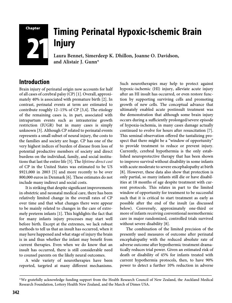
Image of the first page of this content. For PDF version, please use the ‘Save PDF’ preceeding this image.'
Type
Chapter
Information
Publisher: Cambridge University Press
Print publication year: 2017

Access options

Get access to the full version of this content by using one of the access options below. (Log in options will check for institutional or personal access. Content may require purchase if you do not have access.)

References

Reid, SM, Carlin, JB, Reddihough, DS. Rates of cerebral palsy in Victoria, Australia, 1970 to 2004: has there been a change? Dev Med Child Neurol 2011; 53: 907–12.Google Scholar
Behrman, RE, Butler, AS, eds. Preterm Birth: Causes, Consequences, and Prevention. Washington, DC: Institute of Medicine of the National Academies, 2007 Available at http://books.nap.edu/openbook.php?record_id=11622&page=1.Google Scholar
McIntyre, S, Blair, E, Badawi, N, et al. Antecedents of cerebral palsy and perinatal death in term and late preterm singletons. Obstet Gynecol 2013; 122: 869–77.CrossRefGoogle ScholarPubMed
Himmelmann, K, Hagberg, G, Beckung, E, et al. The changing panorama of cerebral palsy in Sweden. IX. Prevalence and origin in the birth-year period 1995–1998. Acta Paediatr 2005; 94: 287–94.Google Scholar
Centers for Disease Control and Prevention (CDC). Economic costs associated with mental retardation, cerebral palsy, hearing loss, and vision impairment–United States, 2003. MMWR Morb Mortal Wkly Rep 2004; 53: 57–9.Google Scholar
Kruse, M, Michelsen, SI, Flachs, EM, et al. Lifetime costs of cerebral palsy. Dev Med Child Neurol 2009; 51: 622–8.CrossRefGoogle ScholarPubMed
Gunn, AJ, Bennet, L. Timing of injury in the fetus and neonate. Curr Opin Obstet Gynecol 2008; 20: 175–81.CrossRefGoogle ScholarPubMed
Jacobs, SE, Berg, M, Hunt, R, et al. Cooling for newborns with hypoxic ischaemic encephalopathy. Cochrane Database Syst Rev 2013; 1: CD003311.Google Scholar
Gunn, AJ, Thoresen, M. Hypothermic neuroprotection. NeuroRx 2006; 3: 154–69.CrossRefGoogle ScholarPubMed
Beilharz, EJ, Williams, CE, Dragunow, M, et al. Mechanisms of delayed cell death following hypoxic-ischemic injury in the immature rat: evidence for apoptosis during selective neuronal loss. Brain Res Mol Brain Res 1995; 29: 114.Google Scholar
Geddes, R, Vannucci, RC, Vannucci, SJ. Delayed cerebral atrophy following moderate hypoxia-ischemia in the immature rat. Dev Neurosci 2001; 23: 180–5.Google Scholar
Roth, SC, Baudin, J, Cady, E, et al. Relation of deranged neonatal cerebral oxidative metabolism with neurodevelopmental outcome and head circumference at 4 years. Dev Med Child Neurol 1997; 39: 718–25.CrossRefGoogle ScholarPubMed
Shankaran, S, Laptook, AR, Ehrenkranz, RA, et al. Whole-body hypothermia for neonates with hypoxic-ischemic encephalopathy. N Engl J Med 2005; 353: 1574–84.Google Scholar
Gluckman, PD, Wyatt, JS, Azzopardi, D, et al. Selective head cooling with mild systemic hypothermia after neonatal encephalopathy: multicentre randomised trial. Lancet 2005; 365: 663–70.Google Scholar
Westgate, JA, Wibbens, B, Bennet, L, et al. The intrapartum deceleration in center stage: a physiological approach to interpretation of fetal heart rate changes in labor. Am J Obstet Gynecol 2007; 197: e1e11.236.CrossRefGoogle Scholar
de Haan, HH, Gunn, AJ, Williams, CE, Gluckman, PD. Brief repeated umbilical cord occlusions cause sustained cytotoxic cerebral edema and focal infarcts in near-term fetal lambs. Pediatr Res 1997; 41: 96104.CrossRefGoogle ScholarPubMed
Graham, EM, Ruis, KA, Hartman, AL, et al. A systematic review of the role of intrapartum hypoxia-ischemia in the causation of neonatal encephalopathy. Am J Obstet Gynecol 2008; 199: 587–95.CrossRefGoogle ScholarPubMed
MacLennan, A, International Cerebral Palsy Task Force, et al. A template for defining a causal relation between acute intrapartum events and cerebral palsy: international consensus statement. BMJ 1999; 319: 1054–9.Google Scholar
Cowan, F, Rutherford, M, Groenendaal, F, et al. Origin and timing of brain lesions in term infants with neonatal encephalopathy. Lancet 2003; 361: 736–42.CrossRefGoogle ScholarPubMed
Iwata, O, Iwata, S, Bainbridge, A, et al. Supra- and sub-baseline phosphocreatine recovery in developing brain after transient hypoxia-ischaemia: relation to baseline energetics, insult severity and outcome. Brain 2008; 131: 2220–6.Google Scholar
Szeto, HH. Mitochondria-targeted cytoprotective peptides for ischemia-reperfusion injury. Antioxid Redox Signal 2008; 10: 601–19.Google Scholar
Edwards, AD, Brocklehurst, P, Gunn, AJ, et al. Neurological outcomes at 18 months of age after moderate hypothermia for perinatal hypoxic ischaemic encephalopathy: synthesis and meta-analysis of trial data. BMJ 2010; 340: c363.Google Scholar
Nelson, KB, Dambrosia, JM, Ting, TY, Grether, JK. Uncertain value of electronic fetal monitoring in predicting cerebral palsy. N Engl J Med 1996; 334: 613–8.CrossRefGoogle ScholarPubMed
Murray, DM, O’Riordan, MN, Horgan, R, et al. Fetal heart rate patterns in neonatal hypoxic-ischemic encephalopathy: relationship with early cerebral activity and neurodevelopmental outcome. Am J Perinatol 2009; 26: 605–12.Google Scholar
Wayenberg, JL. Threshold of metabolic acidosis associated with neonatal encephalopathy in the term newborn. J Matern Fetal Neonatal Med 2005; 18: 381–5.Google Scholar
Low, JA, Lindsay, BG, Derrick, EJ. Threshold of metabolic acidosis associated with newborn complications. Am J Obstet Gynecol 1997; 177: 1391–4.CrossRefGoogle ScholarPubMed
Shah, S, Tracy, M, Smyth, J. Postnatal lactate as an early predictor of short-term outcome after intrapartum asphyxia. J Perinatol 2004; 24: 1620.Google Scholar
Boardman, JP, Hawdon, JM. Hypoglycaemia and hypoxic-ischaemic encephalopathy. Dev Med Child Neurol 2015; 57: 2933.CrossRefGoogle ScholarPubMed
Nadeem, M, Murray, DM, Boylan, GB, et al. Early blood glucose profile and neurodevelopmental outcome at two years in neonatal hypoxic-ischaemic encephalopathy. BMC Pediatr 2011; 11: 10.Google Scholar
Chouthai, NS, Sobczak, H, Khan, R, et al. Hyperglycemia is associated with poor outcome in newborn infants undergoing therapeutic hypothermia for hypoxic ischemic encephalopathy. J Neonatal Perinatal Med 2015; 8: 125–31.Google Scholar
Basu, SK, Kaiser, JR, Guffey, D, et al. Hypoglycaemia and hyperglycaemia are associated with unfavourable outcome in infants with hypoxic ischaemic encephalopathy: a post hoc analysis of the CoolCap Study. Arch Dis Child Fetal Neonatal Ed 2016; 101:F149–55.CrossRefGoogle ScholarPubMed
Shelley, HJ. Glycogen reserves and their changes at birth and in anoxia. Br Med Bull 1961; 17: 137–43.Google Scholar
Dawes, GS, Mott, JC, Shelley, HJ. The importance of cardiac glycogen for the maintenance of life in foetal lambs and newborn animals during anoxia. J Physiol 1959; 146: 516–38.Google Scholar
Wassink, G, Bennet, L, Booth, LC, et al. The ontogeny of hemodynamic responses to prolonged umbilical cord occlusion in fetal sheep. J Appl Physiol 2007; 103: 1311–7.Google Scholar
Laptook, AR, Shankaran, S, Ambalavanan, N, et al. Outcome of term infants using Apgar scores at 10 minutes following hypoxic-ischemic encephalopathy. Pediatrics 2009; 124: 1619–26.Google Scholar
Natarajan, G, Shankaran, S, Laptook, AR, et al. Apgar scores at 10 min and outcomes at 6–7 years following hypoxic-ischaemic encephalopathy. Arch Dis Child Fetal Neonatal Ed 2013; 98: F473–9.CrossRefGoogle ScholarPubMed
Bayram, F, Ozerkan, K, Cengiz, C, et al. Perinatal asphyxia is associated with the umbilical cord nucleated red blood cell count in pre-eclamptic pregnancies. J Obstet Gynaecol 2010; 30: 383–6.Google Scholar
Li, J, Kobata, K, Kamei, Y, et al. Nucleated red blood cell counts: an early predictor of brain injury and 2-year outcome in neonates with hypoxic-ischemic encephalopathy in the era of cooling-based treatment. Brain Dev 2014; 36: 472–8.CrossRefGoogle ScholarPubMed
Christensen, RD, Lambert, DK, Richards, DS. Estimating the nucleated red blood cell ‘emergence time’ in neonates. J Perinatol 2014; 34: 116–9.Google Scholar
Ferber, A, Fridel, Z, Weissmann-Brenner, A, et al. Are elevated fetal nucleated red blood cell counts an indirect reflection of enhanced erythropoietin activity? Am J Obstet Gynecol 2004; 190: 1473–5.Google Scholar
Christensen, RD, Henry, E, Andres, RL, Bennett, ST. Reference ranges for blood concentrations of nucleated red blood cells in neonates. Neonatology 2011; 99: 289–94.CrossRefGoogle ScholarPubMed
Bennet, L, Booth, L, Gunn, AJ. Potential biomarkers for hypoxic-ischemic encephalopathy. Semin Fetal Neonatal Med 2010; 15: 253–60.Google Scholar
Gazzolo, D, Abella, R, Marinoni, E, et al. New markers of neonatal neurology. J Matern Fetal Neonatal Med 2009; 2(Suppl 3): 5761.Google Scholar
Ramaswamy, V, Horton, J, Vandermeer, B, et al. Systematic review of biomarkers of brain injury in term neonatal encephalopathy. Pediatr Neurol 2009; 40: 215–26.CrossRefGoogle ScholarPubMed
Chalak, LF, Sanchez, PJ, Adams-Huet, B, et al. Biomarkers for severity of neonatal hypoxic-ischemic encephalopathy and outcomes in newborns receiving hypothermia therapy. J Pediatr 2014; 164: 468–74.Google Scholar
Loukovaara, M, Teramo, K, Alfthan, H, et al. Amniotic fluid S100B protein and erythropoietin in pregnancies at risk for fetal hypoxia. Eur J Obstet Gynecol Reprod Biol 2009; 142: 115–8.Google Scholar
Routsi, C, Stamataki, E, Nanas, S, et al. Increased levels of serum S100B protein in critically ill patients without brain injury. Shock 2006; 26: 20–4.CrossRefGoogle ScholarPubMed
Garnier, Y, Frigiola, A, Li Volti, G, et al. Increased maternal/fetal blood S100B levels following systemic endotoxin administration and periventricular white matter injury in preterm fetal sheep. Reprod Sci 2009; 16: 758–66.Google Scholar
Friel, LA, Romero, R, Edwin, S, et al. The calcium binding protein, S100B, is increased in the amniotic fluid of women with intra-amniotic infection/inflammation and preterm labor with intact or ruptured membranes. J Perinat Med 2007; 35: 385–93.Google Scholar
Schulpis, KH, Margeli, A, Akalestos, A, et al. Effects of mode of delivery on maternal-neonatal plasma antioxidant status and on protein S100B serum concentrations. Scand J Clin Lab Invest 2006; 66: 733–42.Google Scholar
Bokesch, PM, Appachi, E, Cavaglia, M, et al. A glial-derived protein, S100B, in neonates and infants with congenital heart disease: evidence for preexisting neurologic injury. Anesth Analg 2002; 95: 889–92.Google Scholar
Bruschettini, M, van den Hove, DL, Gazzolo, D, et al. A single course of antenatal betamethasone reduces neurotrophic factor S100B concentration in the hippocampus and serum in the neonatal rat. Brain Res Dev Brain Res 2005; 159: 113–8.Google Scholar
Vicente, E, Tramontina, F, Leite, MC, et al. S100B levels in the cerebrospinal fluid of rats are sex and anaesthetic dependent. Clin Exp Pharmacol Physiol 2007; 34: 1126–30.Google Scholar
Gazzolo, D, Vinesi, P, Marinoni, E, et al. S100B protein concentrations in cord blood: correlations with gestational age in term and preterm deliveries. Clin Chem 2000; 46: 9981000.Google Scholar
Wijnberger, LD, Nikkels, PG, van Dongen, AJ, et al. Expression in the placenta of neuronal markers for perinatal brain damage. Pediatr Res 2002; 51: 492–6.Google Scholar
Zaigham, M, Lundberg, F, Hayes, R, et al. Umbilical cord blood concentrations of ubiquitin carboxy-terminal hydrolase L1 (UCH-L1) and glial fibrillary acidic protein (GFAP) in neonates developing hypoxic-ischemic encephalopathy. J Matern Fetal Neonatal Med 2015:1–7.Google Scholar
Looney, AM, Walsh, BH, Moloney, G, et al. Downregulation of umbilical cord blood levels of miR-374a in neonatal hypoxic ischemic encephalopathy. J Pediatr 2015; 167: 269–73.CrossRefGoogle ScholarPubMed
Whitehead, CL, Teh, WT, Walker, SP, et al. Circulating microRNAs in maternal blood as potential biomarkers for fetal hypoxia in-utero. PloS One 2013; 8: e78487.Google Scholar
Qu, Y, Wu, J, Chen, D, et al. MiR-139-5p inhibits HGTD-P and regulates neuronal apoptosis induced by hypoxia-ischemia in neonatal rats. Neurobiol Dis 2014; 63: 184–93.Google Scholar
Sarnat, HB, Sarnat, MS. Neonatal encephalopathy following fetal distress: a clinical and electroencephalographic study. Arch Neurol 1976; 33: 696705.CrossRefGoogle ScholarPubMed
Wyatt, JS, Gluckman, PD, Liu, PY, et al. Determinants of outcomes after head cooling for neonatal encephalopathy. Pediatrics 2007; 119: 912–21.Google Scholar
Nagdyman, N, Komen, W, Ko, HK, et al. Early biochemical indicators of hypoxic-ischemic encephalopathy after birth asphyxia. Pediatr Res 2001; 49: 502–6.Google Scholar
Nagdyman, N, Grimmer, I, Scholz, T, et al. Predictive value of brain-specific proteins in serum for neurodevelopmental outcome after birth asphyxia. Pediatr Res 2003; 54: 270–5.CrossRefGoogle ScholarPubMed
Thorngren-Jerneck, K, Alling, C, Herbst, A, et al. S100 protein in serum as a prognostic marker for cerebral injury in term newborn infants with hypoxic ischemic encephalopathy. Pediatr Res 2004; 55: 406–12.CrossRefGoogle ScholarPubMed
Martins, RO, Rotta, NT, Portela, LV, Souza, DO. S100B protein related neonatal hypoxia. Arq Neuropsiquiatr 2006; 64: 24–9.Google Scholar
Murabayashi, M, Minato, M, Okuhata, Y, et al. Kinetics of serum S100B in newborns with intracranial lesions. Pediatr Int 2008; 50: 1722.Google Scholar
Bashir, M, Frigiola, A, Iskander, I, et al. Urinary S100A1B and S100BB to predict hypoxic ischemic encephalopathy at term. Front Biosci (Elite Ed) 2009; 1: 560–7.Google Scholar
Giuseppe, D, Sergio, C, Pasqua, B, et al. Perinatal asphyxia in preterm neonates leads to serum changes in protein S-100 and neuron specific enolase. Curr Neurovasc Res 2009; 6: 110–6.Google Scholar
Giussani, DA, Thakor, AS, Frulio, R, Gazzolo, D. Acute hypoxia increases S100beta protein in association with blood flow redistribution away from peripheral circulations in fetal sheep. Pediatr Res 2005; 58: 179–84.Google Scholar
Fujii, EY, Kozuki, M, Mu, J, et al. Correlation of neuron-specific enolase and S100B with histological cerebral damage in fetal sheep after severe asphyxia. Brain Res 2004; 1018: 136–40.Google Scholar
Nylen, K, Ost, M, Csajbok, LZ, et al. Serum levels of S100B, S100A1B and S100BB are all related to outcome after severe traumatic brain injury. Acta Neurochir 2008; 150: 221–7.CrossRefGoogle ScholarPubMed
Berger, RP, Adelson, PD, Pierce, MC, et al. Serum neuron-specific enolase, S100B, and myelin basic protein concentrations after inflicted and noninflicted traumatic brain injury in children. J Neurosurg 2005; 103: 61–8.Google Scholar
Denihan, NM, Boylan, GB, Murray, DM. Metabolomic profiling in perinatal asphyxia: a promising new field. Biomed Res Int 2015; 2015: 254076.Google Scholar
Shalak, LF, Laptook, AR, Velaphi, SC, Perlman, JM. Amplitude-integrated electroencephalography coupled with an early neurologic examination enhances prediction of term infants at risk for persistent encephalopathy. Pediatrics 2003; 111: 351–7.Google Scholar
van Rooij, LG, Toet, MC, Osredkar, D, et al. Recovery of amplitude integrated electroencephalographic background patterns within 24 hours of perinatal asphyxia. Arch Dis Child Fetal Neonatal Ed 2005; 90: F245–51.Google Scholar
Murray, DM, Boylan, GB, Ryan, CA, Connolly, S. Early EEG findings in hypoxic-ischemic encephalopathy predict outcomes at 2 years. Pediatrics 2009; 124: e459–67.CrossRefGoogle ScholarPubMed
Pezzani, C, Radvanyi-Bouvet, MF, Relier, JP, Monod, N. Neonatal electroencephalography during the first twenty-four hours of life in full-term newborn infants. Neuropediatrics 1986; 17: 11–8.Google Scholar
George, S, Gunn, AJ, Westgate, JA, et al. Fetal heart rate variability and brainstem injury after asphyxia in preterm fetal sheep. Am J Physiol Regul Integr Comp Physiol 2004; 287: R925–33.CrossRefGoogle ScholarPubMed
Gunn, AJ, Wyatt, JS, Whitelaw, A, et al. Therapeutic hypothermia changes the prognostic value of clinical evaluation of neonatal encephalopathy. J Pediatr 2008; 152: 55–8.Google Scholar
Azzopardi, D. Predictive value of the amplitude integrated EEG in infants with hypoxic ischaemic encephalopathy: data from a randomised trial of therapeutic hypothermia. Arch Dis Child Fetal Neonatal Ed 2014; 99: F80–2.CrossRefGoogle ScholarPubMed
Cseko, AJ, Bango, M, Lakatos, P, et al. Accuracy of amplitude-integrated electroencephalography in the prediction of neurodevelopmental outcome in asphyxiated infants receiving hypothermia treatment. Acta Paediatr 2013; 102: 707–11.Google Scholar
Hallberg, B, Grossmann, K, Bartocci, M, Blennow, M. The prognostic value of early aEEG in asphyxiated infants undergoing systemic hypothermia treatment. Acta Paediatr 2010; 99: 531–6.Google Scholar
Thoresen, M, Hellstrom-Westas, L, Liu, X, de Vries, LS. Effect of hypothermia on amplitude-integrated electroencephalogram in infants with asphyxia. Pediatrics 2010; 126: e131–9.Google Scholar
Padden, B, Scheer, I, Brotschi, B, et al. Does amplitude-integrated electroencephalogram background pattern correlate with cerebral injury in neonates with hypoxic-ischaemic encephalopathy? J Paediatr Child Health 2015; 51: 180–5.Google Scholar
Wong, FY, Barfield, CP, Walker, AM. Power spectral analysis of two-channel EEG in hypoxic-ischaemic encephalopathy. Early Hum Dev 2007; 83: 379–83.Google Scholar
Doyle, OM, Greene, BR, Murray, DM, et al. The effect of frequency band on quantitative EEG measures in neonates with hypoxic-ischaemic encephalopathy. In Conference Proceedings of the IEEE Engineering in Medicine and Biology Society (Piscataway, NJ: IEEE, 2007): 717–21.Google Scholar
Jiang, D, Wu, W, Jia, X, et al. Scaling exponents of EEG are related to the temporal process of the therapeutic hypothermia following ischemic brain injury. In Conference Proceedings of the IEEE Engineering in Medicine and Biology Society, vol. 1 (Piscataway, NJ: IEEE, 2009): 2192–5.Google Scholar
Tichauer, KM, Elliott, JT, Hadway, JA, Lee, TY, St Lawrence, K. Cerebral metabolic rate of oxygen and amplitude-integrated electroencephalography during early reperfusion after hypoxia-ischemia in piglets. J Appl Physiol 2009; 106: 1506–12.Google Scholar
Bennet, L, Roelfsema, V, Pathipati, P, et al. Relationship between evolving epileptiform activity and delayed loss of mitochondrial activity after asphyxia measured by near-infrared spectroscopy in preterm fetal sheep. J Physiol 2006; 572: 141–54.CrossRefGoogle ScholarPubMed
Bennet, L, Roelfsema, V, Dean, J, et al. Regulation of cytochrome oxidase redox state during umbilical cord occlusion in preterm fetal sheep. Am J Physiol Regul Integr Comp Physiol 2007; 292: R1569–76.Google Scholar
Dean, JM, George, SA, Wassink, G, et al. Suppression of post hypoxic-ischemic EEG transients with dizocilpine is associated with partial striatal protection in the preterm fetal sheep. Neuropharmacology 2006; 50: 491503.Google Scholar
Scher, MS. Controversies regarding neonatal seizure recognition. Epileptic Disord 2002; 4: 139–58.Google Scholar
Dean, JM, George, S, Naylor, AS, et al. Partial neuroprotection with low-dose infusion of the 2-adrenergic receptor agonist clonidine after severe hypoxia in preterm fetal sheep. Neuropharmacology 2008; 55: 166–74.Google Scholar
Bennet, L, Dean, JM, Wassink, G, Gunn, AJ. Differential effects of hypothermia on early and late epileptiform events after severe hypoxia in preterm fetal sheep. J Neurophysiol 2007; 97: 572–8.Google Scholar
Dean, JM, Gunn, AJ, Wassink, G, et al. Endogenous alpha(2)-adrenergic receptor-mediated neuroprotection after severe hypoxia in preterm fetal sheep. Neuroscience 2006; 142: 615–28.Google Scholar
Dean, JM, Gunn, AJ, Wassink, G, Bennet, L. Transient NMDA receptor-mediated hypoperfusion following umbilical cord occlusion in preterm fetal sheep. Exp Physiol 2006; 91: 423–29.Google Scholar
Scher, MS, Bova, JM, Dokianakis, SG, Steppe, DA. Physiological significance of sharp wave transients on EEG recordings of healthy pre-term and full-term neonates. Electroencephalogr Clin Neurophysiol 1994; 90: 179–85.Google ScholarPubMed
Vecchierini-Blineau, MF, Nogues, B, Louvet, S, Desfontaines, O. Positive temporal sharp waves in electroencephalograms of the premature newborn. Neurophysiol Clin 1996; 26: 350–62.Google Scholar
Marics, G, Cseko, A, Vasarhelyi, B, et al. Prevalence and etiology of false normal aEEG recordings in neonatal hypoxic-ischaemic encephalopathy. BMC Pediatr 2013; 13: 194.Google Scholar
Lear, CA, Koome, MM, Davidson, JO, et al. The effects of dexamethasone on post-asphyxial cerebral oxygenation in the preterm fetal sheep. J Physiol 2014; 592: 5493–505.Google Scholar
Davidson, JO, Quaedackers, JS, George, SA, et al. Maternal dexamethasone and EEG hyperactivity in preterm fetal sheep. J Physiol 2011; 589: 3823–35.Google Scholar
Counsell, SJ, Ball, G, Edwards, AD. New imaging approaches to evaluate newborn brain injury and their role in predicting developmental disorders. Curr Opin Neurol 2014; 27: 168–75.Google Scholar
Robertson, NJ, Thayyil, S, Cady, EB, Raivich, G. Magnetic resonance spectroscopy biomarkers in term perinatal asphyxial encephalopathy: from neuropathological correlates to future clinical applications. Curr Pediatr Rev 2014; 10: 3747.Google Scholar
Martin, E, Buchli, R, Ritter, S, et al. Diagnostic and prognostic value of cerebral 31P magnetic resonance spectroscopy in neonates with perinatal asphyxia. Pediatr Res 1996; 40: 749–58.Google Scholar
Thayyil, S, Chandrasekaran, M, Taylor, A, et al. Cerebral magnetic resonance biomarkers in neonatal encephalopathy: a meta-analysis. Pediatrics 2010; 125: e382–95.Google Scholar
Vannucci, RC, Towfighi, J, Vannucci, SJ. Secondary energy failure after cerebral hypoxia-ischemia in the immature rat. J Cereb Blood Flow Metab 2004; 24: 1090–7.CrossRefGoogle ScholarPubMed
Cady, EB, Iwata, O, Bainbridge, A, et al. Phosphorus magnetic resonance spectroscopy 2 h after perinatal cerebral hypoxia-ischemia prognosticates outcome in the newborn piglet. J Neurochem 2008; 107: 1027–35.Google Scholar
Alderliesten, T, de Vries, LS, Benders, MJ, et al. MR imaging and outcome of term neonates with perinatal asphyxia: value of diffusion-weighted MR imaging and 1H MR spectroscopy. Radiology 2011; 261: 235–42.Google Scholar
Alderliesten, T, Nikkels, PG, Benders, MJ, et al. Antemortem cranial MRI compared with postmortem histopathologic examination of the brain in term infants with neonatal encephalopathy following perinatal asphyxia. Arch Dis Child Fetal Neonatal Ed 2013; 98: F304–9.Google Scholar
Shankaran, S, McDonald, SA, Laptook, AR, et al. Neonatal magnetic resonance imaging pattern of brain injury as a biomarker of childhood outcomes following a trial of hypothermia for neonatal hypoxic-ischemic encephalopathy. J Pediatr 2015; 167: 987–93.Google Scholar
Wilkinson, D. MRI and withdrawal of life support from newborn infants with hypoxic-ischemic encephalopathy. Pediatrics 2010; 126: e451–8.Google Scholar
Wisnowski, JL, Wu, TW, Reitman, AJ, et al. The effects of therapeutic hypothermia on cerebral metabolism in neonates with hypoxic-ischemic encephalopathy: an in vivo 1H-MR spectroscopy study. J Cereb Blood Flow Metab 2016; 36:1075–86.Google Scholar
Liao, SM, Culver, JP. Near infrared optical technologies to illuminate the status of the neonatal brain. Current Pediatr Rev 2014; 10: 7386.Google Scholar
Liu, P, Chalak, LF, Lu, H. Non-invasive assessment of neonatal brain oxygen metabolism: a review of newly available techniques. Early Hum Dev 2014; 90: 695701.Google Scholar
Bale, G, Mitra, S, Meek, J, et al. A new broadband near-infrared spectroscopy system for in-vivo measurements of cerebral cytochrome-c-oxidase changes in neonatal brain injury. Biomed Optics Express 2014; 5: 3450–66.Google Scholar
Drury, PP, Bennet, L, Booth, LC, et al. Maturation of the mitochondrial redox response to profound asphyxia in fetal sheep. PLoS One 2012; 7: e39273.Google Scholar
van Bel, F, Lemmers, P, Naulaers, G. Monitoring neonatal regional cerebral oxygen saturation in clinical practice: value and pitfalls. Neonatology 2008; 94: 237–44.Google Scholar
Bainbridge, A, Tachtsidis, I, Faulkner, SD, et al. Brain mitochondrial oxidative metabolism during and after cerebral hypoxia-ischemia studied by simultaneous phosphorus magnetic-resonance and broadband near-infrared spectroscopy. Neuroimage 2014; 102: 173–83.Google Scholar
Matcher, SJ, Elwell, CE, Cooper, CE, et al. Performance comparison of several published tissue near-infrared spectroscopy algorithms. Anal Biochem 1995; 227: 5468.Google Scholar
Cooper, CE, Cope, M, Springett, R, et al. Use of mitochondrial inhibitors to demonstrate that cytochrome oxidase near-infrared spectroscopy can measure mitochondrial dysfunction noninvasively in the brain. J Cereb Blood Flow Metab 1999; 19: 2738.Google Scholar
Peeters-Scholte, C, van den Tweel, E, Groenendaal, F, van Bel, F. Redox state of near infrared spectroscopy-measured cytochrome aa(3) correlates with delayed cerebral energy failure following perinatal hypoxia-ischaemia in the newborn pig. Exp Brain Res 2004; 156: 20–6.Google Scholar
Jensen, EC, Bennet, L, Hunter, CJ, et al. Post-hypoxic hypoperfusion is associated with suppression of cerebral metabolism and increased tissue oxygenation in near-term fetal sheep. J Physiol 2006; 572: 131–9.Google Scholar
Winter, JD, Tichauer, KM, Gelman, N, et al. Changes in cerebral oxygen consumption and high-energy phosphates during early recovery in hypoxic-ischemic piglets: a combined near-infrared and magnetic resonance spectroscopy study. Pediatr Res 2009; 65: 181–7.Google Scholar
Kurth, CD, McCann, JC, Wu, J, et al. Cerebral oxygen saturation-time threshold for hypoxic-ischemic injury in piglets. Anesth Analg 2009; 108: 1268–77.Google Scholar
Perlman, JM. Summary proceedings from the neurology group on hypoxic-ischemic encephalopathy. Pediatrics 2006; 117: S2833.Google Scholar
Greisen, G. Cerebral blood flow and oxygenation in infants after birth asphyxia: clinically useful information? Early Hum Dev 2014; 90: 703–5.Google Scholar
Meek, JH, Elwell, CE, McCormick, DC, et al. Abnormal cerebral haemodynamics in perinatally asphyxiated neonates related to outcome. Arch Dis Child Fetal Neonatal Ed 1999; 81: F110–15.CrossRefGoogle ScholarPubMed
Pryds, O, Greisen, G, Lou, H, Friis-Hansen, B. Vasoparalysis associated with brain damage in asphyxiated term infants. J Pediatr 1990; 117: 119–25.Google Scholar
Marks, KA, Mallard, CE, Roberts, I, et al. Nitric oxide synthase inhibition attenuates delayed vasodilation and increases injury after cerebral ischemia in fetal sheep. Pediatr Res 1996; 40: 185–91.Google Scholar
Wintermark, P, Hansen, A, Warfield, SK, et al. Near-infrared spectroscopy versus magnetic resonance imaging to study brain perfusion in newborns with hypoxic-ischemic encephalopathy treated with hypothermia. Neuroimage 2014; 85: 287–93.Google Scholar
Shellhaas, RA, Thelen, BJ, Bapuraj, JR, et al. Limited short-term prognostic utility of cerebral NIRS during neonatal therapeutic hypothermia. Neurology 2013; 81: 249–55.Google Scholar
Ancora, G, Maranella, E, Locatelli, C, et al. Changes in cerebral hemodynamics and amplitude integrated EEG in an asphyxiated newborn during and after cool cap treatment. Brain Dev 2009; 31: 442–4.Google Scholar
Massaro, AN, Govindan, RB, Vezina, G, et al. Impaired cerebral autoregulation and brain injury in newborns with hypoxic-ischemic encephalopathy treated with hypothermia. J Neurophysiol 2015; 114: 818–24.Google Scholar
Burton, VJ, Gerner, G, Cristofalo, E, et al. A pilot cohort study of cerebral autoregulation and 2-year neurodevelopmental outcomes in neonates with hypoxic-ischemic encephalopathy who received therapeutic hypothermia. BMC Neurol 2015; 15: 209.Google Scholar
Wiberg-Itzel, E, Lipponer, C, Norman, M, et al. Determination of pH or lactate in fetal scalp blood in management of intrapartum fetal distress: randomized, controlled multicentre trial. BMJ 2008; 336: 1284–7.Google Scholar
Borruto, F, Comparetto, C, Treisser, A. Prevention of cerebral palsy during labour: role of foetal lactate. Arch Gynecol Obstet 2008; 278: 1722.Google Scholar
Ramanah, R, Martin, A, Clement, MC, et al. Fetal scalp lactate microsampling for non-reassuring fetal status during labor: a prospective observational study. Fetal Diagn Ther 2010; 27: 14–9.Google Scholar
Wiberg, N, Kallen, K, Herbst, A, Olofsson, P. Relation between umbilical cord blood pH, base deficit, lactate, 5-minute Apgar score and development of hypoxic ischemic encephalopathy. Acta Obstet Gynaecol Scand 2010; 89: 1263–9.Google Scholar
Hogan, L, Ingemarsson, I, Thorngren-Jerneck, K, Herbst, A. How often is a low 5-min Apgar score in term newborns due to asphyxia? Eur J Obstet Gynecol Reprod Biol 2007; 130: 169–75.Google Scholar
Thorngren-Jerneck, K, Herbst, A. Low 5-minute Apgar score: a population-based register study of 1 million term births. Obstet Gynecol 2001; 98: 6570.Google Scholar
Kovalak, EE, Dede, FS, Gelisen, O, et al. Nonreassuring fetal heart rate patterns and nucleated red blood cells in term neonates. Arch Gynecol Obstet 2011; 283: 1005–9.CrossRefGoogle ScholarPubMed
Gazzolo, D, Marinoni, E, Di Iorio, R, et al. Urinary S100B protein measurements: a tool for the early identification of hypoxic-ischemic encephalopathy in asphyxiated full-term infants. Crit Care Med 2004; 32: 131–6.Google Scholar
Florio, P, Luisi, S, Moataza, B, et al. High urinary concentrations of activin A in asphyxiated full-term newborns with moderate or severe hypoxic ischemic encephalopathy. Clin Chem 2007; 53: 520–2.Google Scholar
Aly, H, Hassanein, S, Nada, A, et al. Vascular endothelial growth factor in neonates with perinatal asphyxia. Brain Dev 2009; 31: 600–4.Google Scholar
Reinke, SN, Walsh, BH, Boylan, GB, et al. 1H NMR derived metabolomic profile of neonatal asphyxia in umbilical cord serum: implications for hypoxic ischemic encephalopathy. J Proteome Res 2013; 12: 4230–9.Google Scholar
Thoresen, M. Patient selection and prognostication with hypothermia treatment. Semin Fetal Neonatal Med 2010; 15: 247–52.Google Scholar
Ambalavanan, N, Carlo, WA, Shankaran, S, et al. Predicting outcomes of neonates diagnosed with hypoxemic-ischemic encephalopathy. Pediatrics 2006; 118: 2084–93.Google Scholar
Haiju, Z, Suyuan, H, Xiufang, F, et al. The combined detection of umbilical cord nucleated red blood cells and lactate: early prediction of neonatal hypoxic ischemic encephalopathy. J Perinat Med 2008; 36: 240–7.Google Scholar
Bednarek, N, Svedin, P, Garnotel, R, et al. Increased MMP-9 and TIMP-1 in mouse neonatal brain and plasma and in human neonatal plasma after hypoxia-ischemia: a potential marker of neonatal encephalopathy. Pediatr Res 2012; 71: 6370.Google Scholar
Kirimi, E, Peker, E, Tuncer, O, et al. Increased serum malondialdehyde level in neonates with hypoxic-ischaemic encephalopathy: prediction of disease severity. J Int Med Res 2010; 38: 220–6.Google Scholar
van Laerhoven, H, de Haan, TR, Offringa, M, et al. Prognostic tests in term neonates with hypoxic-ischemic encephalopathy: a systematic review. Pediatrics 2013; 131: 8898.Google Scholar
Horn, AR, Swingler, GH, Myer, L, et al. Early clinical signs in neonates with hypoxic ischemic encephalopathy predict an abnormal amplitude-integrated electroencephalogram at age 6 hours. BMC Pediatr 2013; 13: 52.Google Scholar
Jose, A, Matthai, J, Paul, S. Correlation of EEG, CT, and MRI brain with neurological outcome at 12 months in term newborns with hypoxic ischemic encephalopathy. J Clin Neonatol 2013; 2: 125–30.Google Scholar
Ancora, G, Soffritti, S, Lodi, R, et al. A combined a-EEG and MR spectroscopy study in term newborns with hypoxic-ischemic encephalopathy. Brain Dev 2010; 32: 835–42.Google Scholar
Twomey, E, Twomey, A, Ryan, S, et al. MR imaging of term infants with hypoxic-ischaemic encephalopathy as a predictor of neurodevelopmental outcome and late MRI appearances. Pediatr Radiol 2010; 40: 1526–35.Google Scholar
Garcia-Alix, A, Cabanas, F, Pellicer, A, et al. Neuron-specific enolase and myelin basic protein: relationship of cerebrospinal fluid concentrations to the neurologic condition of asphyxiated full-term infants. Pediatrics 1994; 93: 234–40.Google Scholar

Save book to Kindle

To save this book to your Kindle, first ensure coreplatform@cambridge.org is added to your Approved Personal Document E-mail List under your Personal Document Settings on the Manage Your Content and Devices page of your Amazon account. Then enter the ‘name’ part of your Kindle email address below. Find out more about saving to your Kindle.

Note you can select to save to either the @free.kindle.com or @kindle.com variations. ‘@free.kindle.com’ emails are free but can only be saved to your device when it is connected to wi-fi. ‘@kindle.com’ emails can be delivered even when you are not connected to wi-fi, but note that service fees apply.

Find out more about the Kindle Personal Document Service.

Available formats
×

Save book to Dropbox

To save content items to your account, please confirm that you agree to abide by our usage policies. If this is the first time you use this feature, you will be asked to authorise Cambridge Core to connect with your account. Find out more about saving content to Dropbox.

Available formats
×

Save book to Google Drive

To save content items to your account, please confirm that you agree to abide by our usage policies. If this is the first time you use this feature, you will be asked to authorise Cambridge Core to connect with your account. Find out more about saving content to Google Drive.

Available formats
×