Skip to main content Accessibility help
×
Hostname: page-component-848d4c4894-tn8tq Total loading time: 0 Render date: 2024-06-30T08:28:58.408Z Has data issue: false hasContentIssue false

4 - Identification and Quantification of Clays

Published online by Cambridge University Press:  30 August 2017

Markus Gräfe
Affiliation:
Emirates Global Aluminium (EGA)
Craig Klauber
Affiliation:
Curtin University of Technology, Perth
Angus J. McFarlane
Affiliation:
Commonwealth Scientific and Industrial Research Organisation, Canberra
David J. Robinson
Affiliation:
Commonwealth Scientific and Industrial Research Organisation, Canberra
Get access

Summary

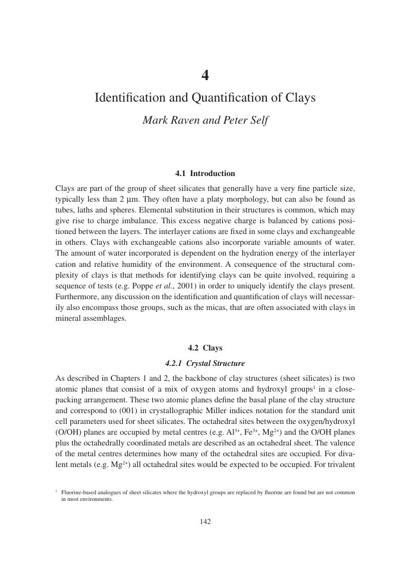
Image of the first page of this content. For PDF version, please use the ‘Save PDF’ preceeding this image.'
Type
Chapter
Information
Publisher: Cambridge University Press
Print publication year: 2017

Access options

Get access to the full version of this content by using one of the access options below. (Log in options will check for institutional or personal access. Content may require purchase if you do not have access.)

References

Alcover, J. F., Gatineau, L. & Mering, J. 1973. Exchangeable cation distribution in nickel-vermiculites and magnesium-vermiculites. Clays and Clay Minerals, 21 (2), 131136.Google Scholar
American Petroleum Institute. 2009. ANSI/API RP 13B-1. Recommended Practice for Field Testing Water-Based Drilling Fluids, 4th edition (Identical to ISO 10414-1:2008). Washington, DC: American Petroleum Institute.Google Scholar
Bailey, S. W. 1988a. Polytypism of 1:1 layer silicates. In: Bailey, S. W. (ed.) Hydrous Phyllosilicates (Exclusive of Micas). Washington, DC: Mineralogical Society of America.CrossRefGoogle Scholar
Bailey, S. W. 1988b. Chlorites: Structure and crystal chemistry. In: Bailey, S. W. (ed.) Hydrous Phyllosilicates (Exclusive of Micas). Washington, DC: Mineralogical Society of America.Google Scholar
Bailey, S. W. 1980. Structures of layer silicates. In: Brindley, G. W. & Brown, G. (eds) Crystal Structures of Clay Minerals and their X-ray Identification, new edition. London: Mineralogical Society.Google Scholar
Bailey, S. W., Hurley, P. M., Fairbairn, H. W. & Pinson, W. H. 1962. K–Ar dating of sedimentary illite polytypes. Geological Society of America Bulletin, 73 (9), 11671170.CrossRefGoogle Scholar
Bailey, S. W., Brindley, G. W., Fanning, D. S., Kodama, H. & Martin, R. T. 1984. Report of the Clay Mineral Society Nomenclature Committee for 1982 and 1983. Clays and Clay Minerals, 32 (3), 239240.CrossRefGoogle Scholar
Bain, D. C., McHardy, W. J. & Lachowski, E. E. 1994. X-ray fluorescence spectroscopy and microanalysis. In: Wilson, M. J. (ed.) Clay Mineralogy: Spectroscopic and Chemical Determinative Methods. Amsterdam: Springer.Google Scholar
Baker, J. C., Uwins, P. J. R. & MacKinnon, I. D. R. 1993. ESEM study of illite/smectite freshwater sensitivity in sandstone reservoirs. Journal of Petroleum Science and Engineering, 9 (2), 8394.CrossRefGoogle Scholar
Baronnet, A. 1992. Polytypism and stacking disorder. In: Buseck, P. R. (ed.) Minerals and Reactions at the Atomic Scale: Transmission Electron Microscopy. Washington, DC: Mineralogical Society of America.Google Scholar
Barshard, I. 1948. Vermiculite and its relation to biotite as revealed by base exchange reactions, X-ray analyses, differential thermal curves and water content. American Mineralogist, 33 (11–12), 655678.Google Scholar
Battaglia, S., Leoni, L. & Sartori, F. 2006. A method for determining the CEC and chemical composition of clays via XRF. Clay Minerals, 41 (3), 717725.CrossRefGoogle Scholar
Bergaya, F. & Lagaly, G. (eds) 2013. Handbook of Clay Science. Amsterdam: Elsevier B.V.Google Scholar
Bergaya, F., Theng, B. K. G. & Lagaly, G. (eds) 2006. Handbook of Clay Science. Amsterdam: Elsevier.Google Scholar
Bergmann, J. & Kleeberg, R. 1998. Rietveld analysis of disordered layer silicates. Materials Science Forum, 278 (2), 300305.Google Scholar
Bergmann, K. & O’Konski, C. T. 1963. A spectroscopic study of methylene blue monomer, dimer and complexes with montmorillonite. Journal of Physical Chemistry, 67, 21692177.Google Scholar
Bergmann, J., Friedel, P. & Kleeberg, R. 1998. BGMN a new fundamental parameter based Rietveld program for laboratory X-ray sources, its use in quantitative analysis and structure investigations. Commission of Powder Diffraction: International Union of Crystallography, CPD Newsletter, 20, 58.Google Scholar
Bertaux, J., Frohlich, F. & Ildefonse, P. 1998. Multicomponent analysis of FTIR spectra: Quantification of amorphous and crystallized mineral phases in synthetic and natural sediments. Journal of Sedimentary Research, 68 (3), 440447.Google Scholar
Bertrand, I., Janik, L. J., Holloway, R. E., Armstrong, R. D. & McLaughlin, M. J. 2002. The rapid assessment of concentrations and solid phase associations of macro- and micronutrients in alkaline soils by mid-infrared diffuse reflectance spectroscopy. Soil Research, 40 (8), 13391356.CrossRefGoogle Scholar
Biscoe, J. & Warren, B. E. 1942. An X-ray study of carbon black. Journal of Applied Physics, 13 (6), 364371.CrossRefGoogle Scholar
Bradley, W. F. 1950. The alternating layer sequence of rectorite. American Mineralogist, 35 (3–4), 278.Google Scholar
Brindley, G. W. 1945. The effect of grain or particle size on x-ray reflections from mixed powders and alloys, considered in relation to the quantitative determination of crystalline substances by x-ray methods. Philosophical Magazine Series 7, 36 (256), 347369.CrossRefGoogle Scholar
Brindley, G. W. 1956. Allevardite, a swelling double-layer mica mineral. American Mineralogist, 41 (1–2), 91103.Google Scholar
Brindley, G. W. & Brown, G. (eds) 1980. Crystal Structures of Clay Minerals and Their X-ray Identification. London: Mineralogical Society.Google Scholar
Brown, B. E. & Bailey, S. W. 1962. Chlorite polytypism:.1. Regular and semi-random 1-layer structures. American Mineralogist, 47 (7–8), 819850.Google Scholar
Bruker AXS. 2009. Topas v4.2: General Profile and Structure Analysis Software for Powder Diffraction Data. Karlsruhe: Bruker AXS.Google Scholar
Brunauer, S., Emmett, P. H. & Teller, E. A. 1938. Adsorption of gases in multimolecular layers. Journal of the American Chemical Society, 60, 309319.CrossRefGoogle Scholar
Buseck, P. R. & Self, P. G. 1992. Electron energy-loss spectroscopy (EELS) and electron channelling (ALCHEMI). In: Buseck, P. R. (ed.) Minerals and Reactions at the Atomic Scale: Transmission Electron Microscopy. Washington, DC: Mineralogical Society of America.Google Scholar
Capitani, G. C. & Mellini, M. 2007. High-resolution transmission electron microscopy (HRTEM) investigation of antigorite polysomes (m = 15 to 18). American Mineralogist, 92 (1), 6471.Google Scholar
Carrado, K. A., Decarreau, A., Petit, S., Bergaya, F. & Lagaly, G. 2006. Synthetic clay minerals and the purification of natural clays. In: Bergaya, F., Theng, B. K. G. & Lagaly, G. (eds) Handbook of Clay Science. Amsterdam: Elsevier.Google Scholar
Cases, J. M., Grillet, Y., François, M., et al. 1991. Evolution of the porous structure and surface area of palygorskite under vacuum thermal treatment. Clays and Clay Minerals, 39, 191201.Google Scholar
Chabrillat, S., Goetz, A. F. H., Krosley, L. & Olsen, H. W. 2002. Use of hyperspectral images in the identification and mapping of expansive clay soils and the role of spatial resolution. Remote Sensing of Environment, 82 (2), 431445.Google Scholar
Chao, T. T. 1984. Use of partial dissolution techniques in geochemical-exploration. Journal of Geochemical Exploration, 20 (2), 101135.Google Scholar
Chao, T. T. & Sanzolone, R. F. 1977. Chemical dissolution of sulphide minerals. Journal of Research of the US Geological Survey, 5, 409412.Google Scholar
Cheary, R. W. & Coelho, A. 1992. A fundamental parameters approach to x-ray line-profile fitting. Journal of Applied Crystallography, 25 (2), 109121.CrossRefGoogle Scholar
Chipera, S. J. & Bish, D. L. 2002. FULLPAT: A full-pattern quantitative analysis program for X-ray powder diffraction using measured and calculated patterns. Journal of Applied Crystallography, 35 (6), 744749.CrossRefGoogle Scholar
Chukhrov, F. V., Gorshkov, A. I., Rudnitskaya, E. S., Beresovskaya, V. V. & Sivtsov, A. V. 1980. Manganese minerals in clays: A review. Clays and Clay Minerals, 28 (5), 346354.CrossRefGoogle Scholar
Chung, F. H. 1974a. Quantitative interpretation of X-ray-diffraction patterns of mixtures: 1. Matrix-flushing method for quantitative multicomponent analysis. Journal of Applied Crystallography, 7 (6), 519525.Google Scholar
Chung, F. H. 1974b. Quantitative interpretation of X-ray-diffraction patterns of mixtures: 2. Adiabatic principle of X-ray-diffraction analysis of mixtures. Journal of Applied Crystallography, 7 (6), 526531.Google Scholar
Churchman, G. J., Whitton, J. S., Claridge, G. G. C. & Theng, B. K. G. 1984. Intercalation method using formamide for differentiating halloysite from kaolinite. Clays and Clay Minerals, 32 (4), 241248.Google Scholar
Churchman, G. J., Pasbakhsh, P., Lowe, D. J. & Theng, B. K. G. 2016. Unique but diverse: Some observations on the formation, structure and morphology of halloysite. Clay Minerals, 51 (3), 395416.CrossRefGoogle Scholar
Clinard, C., Mandalia, T., Tchoubar, D. & Bergaya, F. 2003. HRTEM image filtration: Nanostructural analysis of a pillared clay. Clays and Clay Minerals, 51 (4), 421.CrossRefGoogle Scholar
Cocks, T., Jenssen, R., Stewart, A., Wilson, I. & Shields, T. 1998. The HyMap airborne hyperspectral sensor: The system, calibration and performance. In: Schaepman, M., Schläpfer, D. R. & Itten, K. (eds) 1st EARSEL Workshop on Imaging Spectroscopy, 6–8 October 1998. Zurich: SUI. EARSel, 17.Google Scholar
Cornell, R. M. & Schwertmann, U. 2003. The Iron Oxides: Structure, Properties, Reactions, Occurrences, and Uses. Weinheim: Wiley-VCH.Google Scholar
Cullity, B. D. 1978. Elements of X-ray Diffraction. Reading, MA: Addison-Wesley.Google Scholar
Danilatos, G. D. 1991. Review and outline of environmental SEM at present. Journal of Microscopy, 162 (3), 391402.CrossRefGoogle Scholar
Danilatos, G. D. 1994. Environmental scanning electron microscopy and microanalysis. Mikrochimica Acta, 114–115, 143155.Google Scholar
de la Calle, C. & Suquet, H. 1988. Vermiculite. In: Bailey, S. W. (ed.) Hydrous Phyllosilicates (Exclusive of Micas). Madison, WI: Mineralogical Society.Google Scholar
Dinnebier, R. E. & Billinge, S. J. L. (eds) 2008. Powder Diffraction: Theory and Practice. Cambridge: Royal Society of Chemistry.Google Scholar
Dixon, J. B. & Weed, S. B. (eds) 1989. Minerals in Soil Environments. Madison, WI: Soil Science Society of America.CrossRefGoogle Scholar
Dódony, I. & Buseck, P. R. 2004. Serpentines close-up and intimate: An HRTEM view. International Geology Review, 46 (6), 507527.CrossRefGoogle Scholar
Dódony, I., Pósfai, M. & Buseck, P. R. 2002. Revised structure models for antigorite: An HRTEM study. American Mineralogist, 87 (10), 14431457.Google Scholar
Drever, J. I. 1973. The preparation of oriented clay mineral specimens for X-ray diffraction analysis by a filter-membrane peel technique. American Mineralogist, 58 (5–6), 553554.Google Scholar
Drits, V. A. & Tchoubar, C. 1990. X-ray Diffraction by Disordered Lamellar Structures: Theory and Applications to Microdivided Silicates and Carbons. Berlin: Springer-Verlag.Google Scholar
Duan, X. & Evans, D. G. (eds) 2006. Layered Double Hydroxides. Berlin: Springer.Google Scholar
Eberl, D. D. 2003. User’s guide to ROCKJOCK – a program for determining quantitative mineralogy from powder X-ray diffraction data. US Geological Survey Open-File Report [Online]. Available: ftp://brrcrftp.cr.usgs.gov/pub/ddeberl.CrossRefGoogle Scholar
Echlin, P. 1992. Low-Temperature Microscopy and Analysis. New York: Springer.CrossRefGoogle Scholar
Egerton, R. F. 2011. Electron Energy-loss Spectroscopy in the Electron Microscope. New York: Springer.Google Scholar
Elsass, F., Chenu, C. & Tessier, D. 2008. Transmission electron microscopy for soil samples: Preparation methods and use. In: Ulery, A. L. & Richard Drees, L. (eds) Methods of Soil Analysis, Part 5 – Mineralogical Methods, 5.5. Madison, WI: Soil Science Society of America.Google Scholar
Farmer, V. C. (ed.) 1974. The Infrared Spectra of Minerals. London: Mineralogical Society.Google Scholar
Frost, R. L. 1995. Fourier transform Raman spectroscopy of kaolinite, dickite and halloysite. Clays and Clay Minerals, 43 (2), 191195.CrossRefGoogle Scholar
Frost, R. L. & Kloprogge, J. T. 2000. Vibrational spectroscopy of ferruginous smectite and nontronite. Spectrochimica Acta Part A: Molecular and Biomolecular Spectroscopy, 56 (11), 21772189.Google Scholar
Frost, R. L. & Martens, W. N. 2005. Raman spectroscopy of kaolinite and selected intercalates. In: Kloprogge, J. T. (ed.) The Application of Vibrational Spectroscopy to Clay Minerals and Layered Double Hydroxides. Aurora, CO: The Clay Mineral Society.Google Scholar
Gates, W. P. 2006. X-ray absorption spectroscopy. In: Bergaya, F., Theng, B. K. G. & Lagaly, G. (eds) Handbook of Clay Science. Amsterdam: Elsevier.Google Scholar
Gates, W. P., Slade, P. G., Manceau, A. & Lanson, B. 2002. Site occupancies by iron in nontronites. Clays and Clay Minerals, 50 (2), 223239.Google Scholar
Ghabru, S. K., Mermut, A. R. & St. Arnaud, R. J. 1989. Layer-charge and cation-exchange capacity characteristics of vermiculite (weathered biotite) isolated from a gray luvisol in northeastern Saskatchewan. Clays and Clay Minerals, 37 (2), 164172.CrossRefGoogle Scholar
GieseJr., R. F. & Costanzo, P. M. 1987. Behavior of water on the surface of kaolin minerals. In: Davis, J. A. & Hayes, K. F. (eds) Geochemical Processes at Mineral Surfaces. Washington, DC: American Chemical Society.Google Scholar
Goldstein, J. I., Newbury, D. E., Echlin, P., et al. 1992. Scanning Electron Microscopy and X-ray Microanalysis: A Text for Biologists, Material Scientists, and Geologists. New York: Plenum Press.Google Scholar
Gražulis, S., Chateigner, D., Downs, R. T., et al. 2009. Crystallography Open Database: An open-access collection of crystal structures. Journal of Applied Crystallography, 42 (4), 726729.Google Scholar
Grim, R. E. 1968. Clay Mineralogy. New York: McGraw-Hill.Google Scholar
Grim, R. E., Bray, R. H. & Bradley, W. F. 1937. The mica in argillaceous sediments. American Mineralogist, 22 (7), 813829.Google Scholar
Gu, Y. 2003. Automated scanning electron microscope based mineral liberation analysis. Journal of Minerals and Materials Characterization and Engineering, 2 (1), 3341.CrossRefGoogle Scholar
Guggenheim, S., Adams, J. M., Bain, D. C., et al. 2006. Summary of recommendations of nomenclature committees relevant to clay mineralogy: Report of the Association Internationale pour l’Etude des Argiles (AIPEA) Nomenclature Committee for 2006. Clay Minerals, 41 (4), 863877.Google Scholar
Guggenheim, S., Brown, R., Daniels, E., et al. 2011. The Clay Minerals Society Glossary for Clay Science Project. The Clay Minerals Society [Online]. Available: www.clays.org/GLOSSARY/GlossIntro.html.Google Scholar
Guillemette, R. N. 2008. Electron microprobe techniques. In: Ulery, A. L. & Richard Drees, L. (eds) Methods of Soil Analysis Part 5 – Mineralogical Methods, 5.5. Madison, WI: Soil Science Society of America.Google Scholar
GutherieJr, G. D. & Veblen, D. R. 1990. High-resolution transmission electron microscopy applied to clay minerals. In: Coyne, L. M., Mckeever, S. W. S. & Blake, D. F. (eds) Spectroscopic Characterisation of Minerals and Their Surfaces. Washington, DC: American Chemical Society.Google Scholar
Güven, N. 1988. Smectites. In: Bailey, S. W. (ed.) Hydrous Phyllosilicates (Exclusive of Micas). Washington, DC: Mineralogical Society of America.Google Scholar
Hagni, A. M. 2008. Phase identification, phase quantification, and phase association determinations utilizing automated mineralogy technology. JOM, 60 (4), 3337.CrossRefGoogle Scholar
Haynes, W. M., Lide, D. R. & Bruno, T. J. (eds) 2012. CRC Handbook of Chemistry and Physics. Boca Raton, FL: CRC Press.Google Scholar
Hill, R. J. & Howard, C. J. 1987. Quantitative phase-analysis from neutron powder diffraction data using the Rietveld method. Journal of Applied Crystallography, 20 (6), 467474.Google Scholar
Hillier, S. 1999. Use of an air brush to spray dry samples for X-ray powder diffraction. Clay Minerals, 34 (1), 127135.Google Scholar
Hubbard, C. R. & Snyder, R. L. 1988. RIR: Measurement and use in quantitative XRD. Powder Diffraction, 3 (2), 7477.Google Scholar
Hubbard, C. R., Evans, E. H. & Smith, D. K. 1976. The reference intensity ratio, I/Ic, for computer simulated powder patterns. Journal of Applied Crystallography, 9 (2), 169174.CrossRefGoogle Scholar
Hudson, L. K., Misra, C., Perrotta, A. J., Wefers, K. & Williams, F. S. 2000. Aluminum oxide. In: Ullmann’s Encyclopedia of Industrial Chemistry. New York: Wiley & Sons.Google Scholar
Huggett, J. M. & Uwins, P. J. R. 1994. Observations of water–clay reactions in water-sensitive sandstone and mudrocks using an environmental scanning electron microscope. Journal of Petroleum Science and Engineering, 10 (3), 211222.CrossRefGoogle Scholar
Huggett, J. M., McCarty, D. K., Calvert, C. C., Gale, A. S. & Kirk, C. 2006. Serpentine–nontronite–vermiculite mixed-layer clay from the Weches formation, Claiborne group, middle Eocene, northeast Texas. Clays and Clay Minerals, 54 (1), 101.CrossRefGoogle Scholar
Hunt, G. R. 1977. Spectral signatures of particulate minerals in the visible and near infrared. Geophysics, 42 (3), 501513.CrossRefGoogle Scholar
Huntington, J. F., Mauger, A. J., Skirrow, R. G., et al. 2006. Automated mineralogical core logging at the Emmie Bluff iron oxide–copper–gold prospect. MESA Journal, 41, 3844.Google Scholar
Ibers, J. A. & Hamilton, W. C. 1974. International Tables for X-ray Crystallography. Birmingham: Kynoch Press.Google Scholar
Jackson, M. L. 1956. Soil Chemical Analysis: Advanced Course. Madison, WI: Department of Soils, University of Wisconsin.Google Scholar
Janik, L. J. & Keeling, J. L. 1993. FT-IR partial least-squares analysis of tubular halloysite in kaolin samples from the Mount Hope kaolin deposit. Clay Minerals, 28 (3), 365378.Google Scholar
Janik, L. J., Merry, R. H. & Skjemstad, J. O. 1998. Can mid infrared diffuse reflectance analysis replace soil extractions? Australian Journal of Experimental Agriculture, 38 (7), 681696.Google Scholar
Jenkins, R. & Snyder, R. L. 1996. Introduction to X-ray Powder Diffractometry. New York: Wiley & Sons.Google Scholar
Johnston, C. T. 1990. Raman and FT-IR spectra of kaolinite–hydrazine intercalate. In: Coyne, L. M., Mckeever, S. W. S. & Blake, D. F. (eds) Spectroscopic Characterization of Minerals and Their Surfaces. Washington, DC: American Chemical Society.Google Scholar
Jones, B. F. & Galan, E. 1988. Sepiolite and palygorskite. In: Bailey, S. W. (ed.) Hydrous Phyllosilicates (Exclusive of Micas). Madison, WI: Mineralogical Society.Google Scholar
Joussein, E., Petit, S., Churchman, J., et al. 2005. Halloysite clay minerals: A review. Clay Minerals, 40 (4), 383426.Google Scholar
Kleeberg, R. 2005. Results of the second Reynolds Cup contest in quantitative mineral analysis. IUCr CPD Newsletter, 30, 2226.Google Scholar
Klimentidis, R. E. & MacKinnon, I. D. R. 1986. High-resolution imaging of ordered mixed-layer clays. Clays and Clay Minerals, 34 (2), 155164.Google Scholar
Kloprogge, J. T. (ed.) 2005. The Application of Vibrational Spectroscopy to Clay Minerals and Layered Double Hydroxides. Aurora, CO: The Clay Minerals Society.Google Scholar
Klug, H. P. & Alexander, L. E. 1974. X-ray Diffraction Procedures for Polycrystalline and Amorphous Materials. New York: Wiley.Google Scholar
Kruse, F. A. 2010. Mineral mapping using spectroscopy: From field measurements to airborne and satellite-based imaging spectrometry. Art, Science and Applications of Reflectance Spectroscopy Symposium, 23–25 February 2010. Boulder, CO: ASD Inc., 115.Google Scholar
Kruse, F. A., Boardman, J. W., Lefkoff, A. B., et al. 2000. HyMap: An Australian hyperspectral sensor solving global problems – results from USA HyMap data acquisitions. 10th Australasian Remote Sensing and Photogrammetry Conference, 21–25 August 2000. Adelaide, SA: Causal Productions, 116.Google Scholar
Laird, D. A. & Nater, E. A. 1993. Nature of the illitic phase associated with randomly interstratified smectite/illite in soils. Clays and Clay Minerals, 41 (3), 280287.Google Scholar
Laribi, S., Jouffrey, B. & Fleureau, J. M. 2007. Experimental electron energy loss spectroscopy of clays. European Physical Journal: Applied Physics, 39 (3), 257265.Google Scholar
Larkin, P. 2011. Infrared and Raman Spectroscopy: Principles and Spectral Interpretation. Boston, MA: Elsevier.Google Scholar
MacEwan, D. M. C. & Wilson, M. J. 1984. Interlayer and intercalation of complexes of clay minerals. In: Brindley, G. W. & Brown, G. (eds) Crystal Structures of Clay Minerals and Their X-ray Identification. London: Mineralogical Society.Google Scholar
MacKinnon, I. D. R. & Mumpton, F. A. (eds) 1990. Electron-Optical Methods in Clay Science. Boulder, CO: The Clay Minerals Society.Google Scholar
Madsen, I. C. & Scarlett, N. V. Y. 2008. Quantitative phase analysis. In: Dinnebier, R. E. & Billinge, S. J. L. (eds) Powder Diffraction: Theory and Practice. London: The Royal Society of Chemistry.Google Scholar
Madsen, I. C., Scarlett, N. V. Y., Cranswick, L. M. D. & Lwin, T. 2001. Outcomes of the International Union of Crystallography Commission on Powder Diffraction round robin on quantitative phase analysis: Samples 1a to 1h. Journal of Applied Crystallography, 34 (4), 409426.CrossRefGoogle Scholar
Mauger, A. J., Keeling, J. L. & Huntington, J. F. 2007. Alteration mapping of the Tarcoola Goldfield (South Australia) using a suite of hyperspectral methods. Applied Earth Science, 116 (1), 212.Google Scholar
McBride, M. B. 1994. Environmental Chemistry of Soils. New York: Oxford University Press.Google Scholar
McCarty, D. K. 2005. Quantitative mineral analysis of clay-bearing mixtures: The ‘Reynolds Cup’ contest. IUCr CPD Newsletter, 27, 1216.Google Scholar
McFarlane, A., Bremmell, K. & Addai-Mensah, J. 2006. Improved dewatering behavior of clay minerals dispersions via interfacial chemistry and particle interactions optimization. Journal of Colloid and Interface Science, 293 (1), 116127.Google Scholar
McKenzie, R. M. 1989. Manganese oxides and hydroxides. In: Dixon, J. B. & Weed, S. B. (eds) Minerals in Soil Environments, 2nd edition. Madison, WI: Soil Science Society of America.Google Scholar
Mehra, O. P. & Jackson, M. L. 1958. Iron oxide removal from soils and clays by a dithionite–citrate system buffered with sodium bicarbonate. In: Swineford, A. (ed.) Clays and Clay Minerals: Proceedings of the Seventh National Conference on Clays and Clay Minerals. Washington, DC: Pergamon Press, 317327.Google Scholar
Méring, J. 1975. Smectites. In: Gieseking, J. E. (ed.) Soil Components. Berlin: Springer-Verlag.Google Scholar
Mermut, A. R. & Lagaly, G. 2001. Baseline studies of the Clay Minerals Society source clays: Layer-charge determination and characteristics of those minerals containing 2:1 layers. Clays and Clay Minerals, 49 (5), 393397.Google Scholar
Michot, L. J. & Villiéras, F. 2006. Surface area and porosity. In: Bergaya, F., Theng, B. K. G. & Lagaly, G. (eds) Handbook of Clay Science. Amsterdam: Elsevier.Google Scholar
Mielenz, R. C. & King, M. E. 1955. Physical-chemical properties and engineering performance of clays. In: Pask, J. A. & Turner, M. D. (eds) Clays and Clay Technology: Proceedings of the First National Conference on Clays and Clay Technology. San Francisco, CA. Division of Mines & Geology, 196254.Google Scholar
Mikutta, R., Kleber, M., Kaiser, K. & Jahn, R. 2005. Review: Organic matter removal from soils using hydrogen peroxide, sodium hypochlorite, and disodium peroxodisulfate. Soil Science Society of America Journal, 69 (1), 120135.Google Scholar
Miller, P. R., Reid, A. F. & Zuiderwyk, M. A. 1983. QEM* SEM image analysis in the determination of modal assays, mineral associations, and mineral liberation. In: Maltby, P. D. R. (ed.) Proceedings of the XIV International Mineral Processing Congress, 17–23 October, 1982. Toronto, ON: Canadian Institute of Mining and Metallurgy, 3.Google Scholar
Milnes, A. R. & Fitzpatrick, R. W. 1989. Titanium and zirconium minerals. In: Dixon, J. B. & Weed, S. B. (eds) Minerals in Soil Environments, 2nd edition. Madison, WI: Soil Science Society of America.Google Scholar
Montes-H, G., Geraud, Y., Duplay, J. & Reuschlé, T. 2005. ESEM observations of compacted bentonite submitted to hydration/dehydration conditions. Colloids and Surfaces A: Physicochemical and Engineering Aspects, 262 (1), 1422.Google Scholar
Mooney, R. W., Keenan, A. G. & Wood, L. A. 1952. Adsorption of water vapor by montmorillonite: II. Effect of exchangeable ions and lattice swelling as measured by X-ray diffraction. Journal of the American Chemical Society, 74, 13711374.Google Scholar
Moore, D. M. & Reynolds, R. C. 1997. X-ray Diffraction and the Identification and Analysis of Clay Minerals. Oxford: Oxford University Press.Google Scholar
Morris, G. E. & Żbik, M. S. 2009. Smectite suspension structural behaviour. International Journal of Mineral Processing, 93 (1), 2025.CrossRefGoogle Scholar
Nagy, K. L. & Blum, A. E. (eds) 1994. Scanning Probe Microscopy of Clay Minerals. Boulder, CO: The Clay Minerals Society.Google Scholar
Nieto, F. & Arroyo, X. 2010. Identification and characterisation with microscopic methods. In: Fiore, S., Cuadros, J. & Huertas, F. J. (eds) AIPEA Educational Series, Pub. No. 1. Bari, IT: Digilabs.Google Scholar
Norrish, K. 1954. The swelling of montmorillonite. Discussions of the Faraday Society, 18, 120134.Google Scholar
Norrish, K. & Hutton, J. T. 1969. An accurate X-ray spectrographic method for the analysis of a wide range of geological samples. Geochimica et Cosmochimica Acta, 33 (4), 431453.CrossRefGoogle Scholar
Norrish, K. & Chappell, B. W. 1977. X-ray fluorescence spectrometry. In: Zussman, J. (ed.) Physical Methods in Determinative Mineralogy. London: Academic Press.Google Scholar
O’Connor, B. H. & Chang, W. J. 1986. The amorphous character and particle-size distributions of powders produced with the micronizing mill for quantitative X-ray-powder diffractometry. X-Ray Spectrometry, 15 (4), 267270.Google Scholar
O’Connor, B. H. & Raven, M. D. 1988. Application of the Rietveld refinement procedure in assaying powdered mixtures. Powder Diffraction, 3 (1), 26.CrossRefGoogle Scholar
Omotoso, O., McCarty, D. K., Hillier, S. & Kleeberg, R. 2006. Some successful approaches to quantitative mineral analysis as revealed by the 3rd Reynolds Cup contest. Clays and Clay Minerals, 54(6), 748760.Google Scholar
Otten, M. T. & Buseck, P. R. 1987. The oxidation state of Ti in hornblende and biotite determined by electron energy-loss spectroscopy, with inferences regarding the Ti substitution. Physics and Chemistry of Minerals, 14 (1), 4551.Google Scholar
Ottner, F., Gier, S., Kuderna, M. & Schwaighofer, B. 2000. Results of an inter-laboratory comparison of methods for quantitative clay analysis. Applied Clay Science, 17 (5–6), 223243.Google Scholar
Pansu, M. & Gautheyrou, J. 2006. Handbook of Soil Analysis: Mineralogical, Organic and Inorganic Methods. Berlin: Springer-Verlag.Google Scholar
Pearlman, J., Carman, S., Segal, C., et al. 2001. Overview of the Hyperion Imaging Spectrometer for the NASA EO-1 mission. In: Geoscience and Remote Sensing Symposium, 2001. IGARSS ‘01. IEEE 2001 International, 2001, 30363038.Google Scholar
Plançon, A. & Tchoubar, C. 1977. Determination of structural defects in phyllosilicates by X-ray diffraction: Part II. Nature and proportions of defects in natural kaolinites. Clays and Clay Minerals, 25 (6), 436450.Google Scholar
Poppe, L. J. & Eliason, A. E. 2009. A BASIC program to calculate gravitational and centrifugal parameters. In Geological Society of America, Northeastern Section – 44th Annual Meeting. Portland, ME: Geological Society of America, 26.Google Scholar
Poppe, L. J., Paskevich, V. F., Hathaway, J. C. & Blackwood, D. S. 2001. A laboratory manual for X-ray powder diffraction. US Geological Survey Open-File Report [Online]. Available: http://pubs.usgs.gov/of/2001/of01-041/index.htm.Google Scholar
Post, J. E. & Heaney, P. J. 2008. Synchrotron powder X-ray diffraction study of the structure and dehydration behavior of palygorskite. American Mineralogist, 93 (4), 667675.Google Scholar
Post, J. E., Bish, D. L. & Heaney, P. J. 2007. Synchrotron powder X-ray diffraction study of the structure and dehydration behavior of sepiolite. American Mineralogist, 92 (1), 9197.Google Scholar
Radoslovich, E. W. 1960. The structure of muscovite, KAl2(Si3Al)O10(OH)2. Acta Crystallographica, 13 (11), 919932.Google Scholar
Raven, M. D. & Self, P. G. 2011. Secrets to winning the 5th Reynolds Cup competition. In: 48th Annual Meeting of The Clay Minerals Society. Lake Tahoe, NV: The Clay Minerals Society, 85.Google Scholar
Rayment, G. E. & Lyons, D. J. 2011. Soil Chemical Methods : Australasia. Collingwood, VIC: CSIRO Publishing.Google Scholar
Reynolds, R. C. 1980. Interstratified clays. In: Brindley, G. W. & Brown, G. (eds) Crystal Structures of Clay Minerals and Their X-ray Identification, new edition. London: Mineralogical Society.Google Scholar
Rieder, M. G. C., D’yakonov, Y. S., Frank-Kamenetskii, V. A., et al. 1998. Nomenclature of micas. The Canadian Mineralogist, 36 (2), 905912.Google Scholar
Rietveld, H. M. 1967. Line profiles of neutron powder-diffraction peaks for structure refinement. Acta Crystallographica, 22, 151.Google Scholar
Rietveld, H. M. 1969. A profile refinement method for nuclear and magnetic structures. Journal of Applied Crystallography, 2 (2), 6571.Google Scholar
Ritz, M., Vaculikova, L. & Plevova, E. 2010. Identification of clay minerals by infrared spectroscopy and discriminant analysis. Applied Spectroscopy, 64 (12), 13791387.Google Scholar
Rueda, E. H., Ballesteros, M. C., Grassi, R. L. & Blesa, M. A. 1992. Dithionite as a dissolving reagent for goethite in the presence of EDTA and citrate: Application to soil analysis. Clays and Clay Minerals, 40 (5), 575585.Google Scholar
Russell, J. D. & Fraser, A. R. 1994. Infrared methods. In: Wilson, M. J. (ed.) Clay Mineralogy: Spectroscopic and Chemical Determinative Methods. Amsterdam: Springer.Google Scholar
Salvo, L., Cloetens, P., Maire, E., et al. 2003. X-ray micro-tomography an attractive characterisation technique in materials science. Nuclear Instruments and Methods in Physics Research Section B: Beam Interactions with Materials and Atoms, 200, 273286.Google Scholar
Sawhney, B. L. 1989. Interstratification of layer silicates. In: Dixon, J. B., Weed, S. B. & Dinauer, R. C. (eds) Minerals in Soil Environments, 2nd edition. Madison, WI: Soil Science Society of America.Google Scholar
Scarlett, N. V. Y. & Madsen, I. C. 2006. Quantification of phases with partial or no known crystal structures. Powder Diffraction, 21 (4), 278284.Google Scholar
Scarlett, N. V. Y., Madsen, I. C., Cranswick, L. M. D., et al. 2002. Outcomes of the International Union of Crystallography Commission on powder diffraction round robin on quantitative phase analysis: Samples 2, 3, 4, synthetic bauxite, natural granodiorite and pharmaceuticals. Journal of Applied Crystallography, 35 (4), 383400.Google Scholar
Sergeev, Y. M., Spivak, G. V., Sasov, A. Y., et al. 1984a. Quantitative morphological analysis in a SEM-microcomputer system – I: Quantitative shape analysis of single objects. Journal of Microscopy, 135 (1), 112.Google Scholar
Sergeev, Y. M., Spivak, G. V., Sasov, A. Y., et al. 1984b. Quantitative morphological analysis in a SEM-microcomputer system – II: Morphological analysis of complex SEM images. Journal of Microscopy, 135 (1), 1324.Google Scholar
Shirozu, H. & Bailey, S. W. 1966. Crystal structure of a 2-layer Mg-vermiculite. American Mineralogist, 51 (7), 1124.Google Scholar
Singh, B. 1996. Why does halloysite roll? A new model. Clays and Clay Minerals, 44 (2), 191196.Google Scholar
Skinner, H. C. W. & Fitzpatrick, R. W. 1992. Biomineralization: Processes of Iron and Manganese – Modern and Ancient Environments. Cremlingen: Catena Verlag.Google Scholar
Skipper, N. T., Soper, A. K. & McConnell, J. D. C. 1991. The structure of interlayer water in vermiculite. Journal of Chemical Physics, 94 (8), 57515760.CrossRefGoogle Scholar
Smith, D. K. 2001/1992. Particle statistics and whole-pattern methods in quantitative X-ray powder diffraction analysis. Powder Diffraction, 16 (4), 186191. (Reprinted from Advances in X-ray Analysis, 35A)Google Scholar
Smith, J. V. & Yoder, H. S. 1955. Experimental and theoretical studies of the mica polymorphs. American Mineralogist, 40 (3–4), 343344.Google Scholar
Środoń, J. 1999. Nature of mixed-layer clays and mechanisms of their formation and alteration. Annual Review of Earth and Planetary Sciences, 27, 19.CrossRefGoogle Scholar
Środoń, J., Andreoli, C., Elsass, F. & Robert, M. 1990. Direct high-resolution transmission electron microscopic measurement of expandability of mixed-layer illite/smectite in bentonite rock. Clays and Clay Minerals, 38 (4), 373379.Google Scholar
Sudo, T., Shimoda, S., Yotsumoto, H. & Aita, S. (eds) 1981. Electron Micrographs of Clay Minerals. Amsterdam: Elsevier.Google Scholar
Sumner, M. E. (ed.) 2000. Handbook of Soil Science. Washington, DC: CRC Press.Google Scholar
Sumner, M. E. & Miller, W. P. 1996. Cation exchange capacity and exchange coefficients. In: Sparks, D. L., Page, A. L., Helmke, P. A., et al. (eds) Methods of Soil Analysis Part 3 – Chemical Methods. Madison, WI: Soil Science Society of America.Google Scholar
Svedberg, T. & Nichols, J. B. 1923. Determination of size and distribution of size of particle by centrifugal methods. Journal of the American Chemical Society, 45, 29102917.Google Scholar
Tanner, C. B. & Jackson, M. L. 1948. Nomographs of sedimentation times for soil particles under gravity or centrifugal acceleration. Soil Science Society of America Journal, 12 (C), 6065.Google Scholar
Tappert, M., Rivard, B., Giles, D., Tappert, R. & Mauger, A. J. 2011. Automated drill core logging using visible and near-infrared reflectance spectroscopy: A case study from Olympic Dam IOGG deposit, South Australia. Economic Geology, 106 (2), 289296.Google Scholar
Taylor, J. C. 1991. Computer programs for standardless quantitative analysis of minerals using the full powder diffraction profile. Powder Diffraction, 6 (1), 29.Google Scholar
Taylor, J. C. & Matulis, C. E. 1994. A new method for Rietveld clay analysis: Part I. Use of a universal measured standard profile for Rietveld quantification of montmorillonites. Powder Diffraction, 9 (2), 119123.Google Scholar
Tchoubar, D. & Cohaut, N. 2006. Small-angle scattering techniques. In: Bergaya, F., Theng, B. K. G. & Lagaly, G. (eds) Handbook of Clay Science. Amsterdam: Elsevier.Google Scholar
Theng, B. K. G., Churchman, G. J., Whitton, J. S. & Claridge, G. G. C. 1984. Comparison of intercalation methods for differentiating halloysite from kaolinite. Clays and Clay Minerals, 32 (4), 249258.Google Scholar
Treacy, M. M. J., Newsam, J. M. & Deem, M. W. 1991. A general recursion method for calculating diffracted intensities from crystals containing planar faults. Proceedings of the Royal Society of London Series A: Mathematical Physical and Engineering Sciences, 433 (1889), 499520.Google Scholar
Tsipursky, S. I. & Drits, V. A. 1984. The distribution of octahedral cations in the 2:1 layers of dioctahedral smectites studied by oblique-texture electron diffraction. Clay Minerals, 19(2), 177.Google Scholar
Ufer, K., Roth, G., Kleeberg, R., et al. 2004. Description of X-ray powder pattern of turbostratically disordered layer structures with a Rietveld compatible approach. Zeitschrift Fur Kristallographie, 219 (9), 519527.Google Scholar
Vali, H. & Hesse, R. 1990. Alkylammonium ion treatment of clay minerals in ultrathin section: A new method for HRTEM examination of expandable layers. American Mineralogist, 75 (11–12), 14431446.Google Scholar
Vali, H. & Hesse, R. 1992. Arrangement of n-alkylammonium ions in phlogopite and vermiculite: An XRD and TEM study. Clays and Clay Minerals, 40 (2), 240245.CrossRefGoogle Scholar
Vali, H. & Köster, H. M. 1986. Expanding behaviour, structural disorder, regular and random irregular interstratification of 2:1 layer-silicates studied by high-resolution images of transmission electron microscopy. Clay Minerals, 21 (5), 827859.CrossRefGoogle Scholar
van Aken, P. A., Liebscher, B. & Styrsa, V. J. 1998. Quantitative determination of iron oxidation states in minerals using Fe L2,3-edge electron energy-loss near-edge structure spectroscopy. Physics and Chemistry of Minerals, 25 (5), 323327.Google Scholar
van der Marel, H. W. & Beutelspacher, H. 1976. Atlas of Infrared Spectroscopy of Clay Minerals and Their Admixtures. Amsterdam: Elsevier.Google Scholar
Velde, B. 1984. Electron microprobe analysis of clay minerals. Clay Minerals, 19 (2), 243247.Google Scholar
Verburg, K. & Baveye, P. 1994. Hysteresis in the binary exchange of cations on 2:1 clay minerals: A critical review. Clays and Clay Minerals, 42 (2), 207220.Google Scholar
Viani, A., Gaultieri, A. F. & Artioli, G. 2002. The nature of disorder in montmorillonite by simulation of X-ray powder patterns. American Mineralogist, 87 (7), 966975.CrossRefGoogle Scholar
Viscarra Rossel, R. A. 2011. Fine-resolution multiscale mapping of clay minerals in Australian soils measured with near infrared spectra. Journal of Geophysical Research: Earth Surface, 116 (F4), F04023.Google Scholar
Viscarra Rossel, R. A., Walvoort, D. J. J., McBratney, A. B., Janik, L. J. & Skjemstad, J. O. 2006. Visible, near infrared, mid infrared or combined diffuse reflectance spectroscopy for simultaneous assessment of various soil properties. Geoderma, 131 (1), 5975.Google Scholar
Wada, K. 1989. Allophane and imogolite. In: Dixon, J. B., Weed, S. B. & Dinauer, R. C. (eds) Minerals in Soil Environments, 2nd edition. Madison, WI: Soil Science Society of America.Google Scholar
Wada, S. I. & Wada, K. 1977. Density and structure of allophane. Clay Minerals, 12 (4), 289298.Google Scholar
Wada, K. & Yoshinaga, N. 1969. Structure of imogolite. American Mineralogist, 54 (1–2), 50.Google Scholar
Wang, M. K., Wang, S. L. & Wang, W. M. 1996. Rapid estimation of cation-exchange capacities of soils and clays with methylene blue exchange. Soil Science Society of America Journal, 60 (1), 138141.Google Scholar
Wang, X. D., Li, J., Hart, R. D., van Riessen, A. & McDonald, R. 2011. Quantitative X-ray diffraction phase analysis of poorly ordered nontronite clay in nickel laterites. Journal of Applied Crystallography, 44 (5), 902910.Google Scholar
Wang, X. D., Hart, R. D., Li, J., McDonald, R. G. & van Riessen, A. 2012. Quantitative analysis of turbostratically disordered nontronite with a supercell model calibrated by the PONKCS method. Journal of Applied Crystallography, 45 (6), 12951302.Google Scholar
Warren, B. E. 1941. X-ray diffraction in random layer lattices. Physical Review, 59 (9), 693698.Google Scholar
Wicks, F. J. & O’Hanley, D. S. 1988. Serpentine minerals: Structure and petrology. In: Bailey, S. W. (ed.) Hydrous Phyllosilicates (Exclusive of Micas). Madison, WI: Mineralogical Society.Google Scholar
Willis, J., Feather, C. & Turner, K. 2014. Guidelines for XRF Analysis. Cape Town: James Willis Consultants.Google Scholar
Wilson, M. J. 1994. Clay Mineralogy: Spectroscopic and Chemical Determinative Methods. Dordrecht, NL: Springer Netherlands.Google Scholar
Wilson, M. J. 2013. Sheet Silicates: Clay Minerals. London: The Geological Society.Google Scholar
Yang, D. & Frindt, R. F. 1996. Powder X-ray diffraction of turbostratically stacked layer systems. Journal of Materials Research, 11 (7), 17331738.Google Scholar
Yitagesu, F. A., van der Meer, F., van der Werff, H. & Hecker, C. 2011. Spectral characteristics of clay minerals in the 2.5–14μm wavelength region. Applied Clay Science, 53 (4), 581591.Google Scholar
Young, R. A. 1980. Structural analysis from X-ray powder diffraction with the Rietveld method. In: Block, S. & Hubbard, C. R. (eds) Accuracy in Powder Diffraction. Gaithersburg, MD: National Bureau of Standards, 143162.Google Scholar
Yu, X., Zhao, L., Gao, X., Zhang, X. & Wu, N. 2006. The intercalation of cetyltrimethylammonium cations into muscovite by a two-step process: II. The intercalation of cetyltrimethylammonium cations into Li-muscovite. Journal of Solid State Chemistry, 179 (5), 15251535.CrossRefGoogle Scholar
Żbik, M. S. & Frost, R. L. 2009. Micro-structure differences in kaolinite suspensions. Journal of Colloid and Interface Science, 339 (1), 110116.Google Scholar
Żbik, M. S., Smart, R. C. & Morris, G. E. 2008. Kaolinite flocculation structure. Journal of Colloid and Interface Science, 328 (1), 7380.Google Scholar
Żbik, M. S., Song, Y.-F. & Frost, R. L. 2010. Kaolinite flocculation induced by smectite addition: A transmission X-ray microscopic study. Journal of Colloid and Interface Science, 349 (1), 8692.Google Scholar
Żbik, M. S., Song, Y.-F., Frost, R. L. & Wang, C.-C. 2012. Transmission X-ray microscopy: A new tool in clay mineral floccules characterization. Minerals, 2 (4), 283299.Google Scholar
Zevin, L. S., Kimmel, G. & Mureinik, I. 1995. Quantitative X-ray Diffractometry. New York: Springer.Google Scholar

Save book to Kindle

To save this book to your Kindle, first ensure coreplatform@cambridge.org is added to your Approved Personal Document E-mail List under your Personal Document Settings on the Manage Your Content and Devices page of your Amazon account. Then enter the ‘name’ part of your Kindle email address below. Find out more about saving to your Kindle.

Note you can select to save to either the @free.kindle.com or @kindle.com variations. ‘@free.kindle.com’ emails are free but can only be saved to your device when it is connected to wi-fi. ‘@kindle.com’ emails can be delivered even when you are not connected to wi-fi, but note that service fees apply.

Find out more about the Kindle Personal Document Service.

Available formats
×

Save book to Dropbox

To save content items to your account, please confirm that you agree to abide by our usage policies. If this is the first time you use this feature, you will be asked to authorise Cambridge Core to connect with your account. Find out more about saving content to Dropbox.

Available formats
×

Save book to Google Drive

To save content items to your account, please confirm that you agree to abide by our usage policies. If this is the first time you use this feature, you will be asked to authorise Cambridge Core to connect with your account. Find out more about saving content to Google Drive.

Available formats
×