Hostname: page-component-78c5997874-m6dg7 Total loading time: 0 Render date: 2024-11-18T05:55:49.225Z Has data issue: false hasContentIssue false

Use of ultrasound with magnetic field for enhanced in vitro drug delivery in colon cancer treatment

Published online by Cambridge University Press:  28 March 2018

Somoshree Sengupta
Affiliation:
Bioceramics & Coating Division, CSIR-Central Glass & Ceramic Research Institute, Kolkata-700032, India; and Academy of Scientific and Innovative Research (AcSIR), CSIR-Central Glass & Ceramic Research Institute Campus, Kolkata-700032, India
Chandra Khatua
Affiliation:
Bioceramics & Coating Division, CSIR-Central Glass & Ceramic Research Institute, Kolkata-700032, India; and Academy of Scientific and Innovative Research (AcSIR), CSIR-Central Glass & Ceramic Research Institute Campus, Kolkata-700032, India
Anuradha Jana
Affiliation:
Bioceramics & Coating Division, CSIR-Central Glass & Ceramic Research Institute, Kolkata-700032, India; and Academy of Scientific and Innovative Research (AcSIR), CSIR-Central Glass & Ceramic Research Institute Campus, Kolkata-700032, India
Vamsi Krishna Balla*
Affiliation:
Bioceramics & Coating Division, CSIR-Central Glass & Ceramic Research Institute, Kolkata-700032, India; and Academy of Scientific and Innovative Research (AcSIR), CSIR-Central Glass & Ceramic Research Institute Campus, Kolkata-700032, India
*
a) Address all correspondence to this author. e-mail: vamsiballa@cgcri.res.in
Get access

Abstract

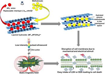
Drug delivery systems (DDSs) have been developed to target tumor cells by releasing active biomolecules at the specific site of infection, thus eliminating the side effects of anticancer drugs. However, DDSs are generally limited by high drug dosage, biobarriers, poor target recognition, etc. To address these deficiencies, we propose a new noninvasive method consisting of exposing the cancer cells to a combination of low-intensity pulsed ultrasound (LIPUS) and static magnetic field (SMF). This combined treatment found to negatively regulate colon cancer cell (HCT116) activities in vitro by altering their cell membrane potential and permeability thus increased the DDS efficacy by 40%. The treated cancer cell membrane became hyperpolarized leading to cancer cell death. The combination treatment (LIPUS + SMF) restricted the cancer cell proliferation to 16 and 5% in the presence of bare anticancer drug and DDS, respectively, in 72 h, which is almost 40% higher than that observed without the treatment. The acceleration of cancer cellular inhibition was confirmed by the significant increase in the apoptosis of the cell exposed to the LIPUS + SMF treatment. The observed improvement is believed to be due to changes in the cell membrane stability/permeability as a result of mechanical (20–22 kPa) and electrical (19–23 µV/cm) stimuli generated during the LIPUS + SMF treatment.

Type
Articles
Copyright
Copyright © Materials Research Society 2018 

Access options

Get access to the full version of this content by using one of the access options below. (Log in options will check for institutional or personal access. Content may require purchase if you do not have access.)

References

REFERENCES

Markasz, L., Stuber, G., Vanherberghen, B., Flaberg, E., Olah, E., Carbone, E., Eksborg, S., Klein, E., Skribek, H., and Szekely, L.: Effect of frequently used chemotherapeutic drugs on the cytotoxic activity of human natural killer cells. Mol. Cancer Ther. 6, 644 (2007).Google Scholar
Duménil, D., Sainteny, F., and Frindel, E.: Some effects of chemotherapeutic drugs on bone marrow stem cells—II. Effect of non-hodgkin lymphoma chemotherapy on various hemopoietic compartments of the mouse. Canc. Chemother. Pharmacol. 2, 203 (1979).Google Scholar
Bracci, L., Schiavoni, G., Sistigu, A., and Belardelli, F.: Immune-based mechanisms of cytotoxic chemotherapy: Implications for the design of novel and rationale-based combined treatments against cancer. Cell Death Differ. 21, 15 (2014).Google Scholar
Yao, J., Feng, J., and Chen, J.: External-stimuli responsive systems for cancer theranostic. Asian J. Pharm. Sci. 11, 585 (2016).Google Scholar
Miller, D.L., Smith, N.B., Bailey, M.R., Czarnota, G.J., Hynynen, K., Makin, I.R.S., and Bioeffects Committee of the American Institute of Ultrasound in Medicine: Overview of therapeutic ultrasound applications and safety considerations. J. Ultrasound Med. 31, 623 (2012).Google Scholar
Varshney, A. and Kumar, G.: Effects of magnetic field on cancer cell line. J. Exp. Biol. Agric. Sci. 1, 91 (2013).Google Scholar
You, D.G., Deepagan, V.G., Um, W., Jeon, S., Son, S., Chang, H., Yoon, H.I., Cho, Y.W., Swierczewska, M., Lee, S., Pomper, M.G., Kwon, I.C., Kim, K., and Park, J.H.: ROS-generating TiO2 nanoparticles for non-invasive sonodynamic therapy of cancer. Sci. Rep. 6, 23200 (2016).Google Scholar
Nuccitelli, R., Tran, K., Sheikh, S., Athos, B., Kreis, M., and Nuccitelli, P.: Optimized nanosecond pulsed electric field therapy can cause murine malignant melanomas to self-destruct with a single treatment. Int. J. Cancer 127, 1727 (2010).Google Scholar
Kirson, E.D., Gurvich, Z., Schneiderman, R., Dekel, E., Itzhaki, A., Wasserman, Y., Schatzberger, R., and Palti, Y.: Disruption of cancer cell replication by alternating electric fields. Cancer Res. 64, 3288 (2004).Google Scholar
Utreja, P., Jain, S., and Tiwary, A.K.: Novel drug delivery systems for sustained and targeted delivery of anti-cancer drugs: Current status and future prospects. Curr. Drug Deliv. 7, 152 (2010).Google Scholar
Alexis, F., Pridgen, E.M., Langer, R., and Farokhzad, O.C.: Nanoparticle technologies for cancer therapy. Handb. Exp. Pharmacol. 197, 55 (2010).CrossRefGoogle Scholar
Blanco, E., Shen, H., and Ferrari, M.: Principles of nanoparticle design for overcoming biological barriers to drug delivery. Nat. Biotechnol. 33, 941 (2015).Google Scholar
Oupicky, D., Bisht, H., Manickam, D., and Zhou, Q.: Stimulus-controlled delivery of drugs and genes. Expet Opin. Drug Deliv. 2, 653 (2005).Google Scholar
Mura, S., Nicolas, J., and Couvreur, P.: Stimuli-responsive nanocarriers for drug delivery. Nat. Mater. 12, 991 (2013).Google Scholar
Mo, S., Coussios, C., Seymour, L., and Carlisle, R.: Ultrasound-enhanced drug delivery for cancer. Expet Opin. Drug Deliv. 9, 1525 (2012).Google Scholar
Tardoski, S., Ngo, J., Gineyts, E., Roux, J-P., Clézardin, P., and Melodelima, D.: Low-intensity continuous ultrasound triggers effective bisphosphonate anticancer activity in breast cancer. Sci. Rep. 5, 16354 (2015).Google Scholar
Gebauer, D., Mayr, E., Orthner, E., and Ryaby, J.P.: Low-intensity pulsed ultrasound: Effects on nonunions. Ultrasound Med. Biol. 31, 1391 (2005).Google Scholar
Heckman, J.D.: Acceleration by non-invasive, of tibial low-intensity fracture-healing pulsed ultrasound. J. Bone Jt. Surg. Am. 76, 26 (1994).CrossRefGoogle ScholarPubMed
Yang, M. and Brackenbury, W.: Membrane potential and cancer progression. Front. Physiol. 4, 185 (2013).Google Scholar
Hanahan, D. and Weinberg, R.A.: Hallmarks of cancer: The next generation. Cell 144, 646 (2011).Google Scholar
Lejbkowicz, F. and Salzberg, S.: Distinct sensitivity of normal and malignant cells to ultrasound in vitro. Environ. Health Perspect. 105, 1575 (1997).Google Scholar
Raylman, R.R., Clavo, C., and Wahl, R.L.: Exposure to strong static magnetic field slows the growth of human cancer cells in vitro. Bioelectromagnetics 17, 358 (1996).Google Scholar
Ghibelli, L., Cerella, C., Cordisco, S., Clavarino, G., Marazzi, S., De Nicola, M., Nuccitelli, S., D’Alessio, M., Magrini, A., Bergamaschi, A., Guerrisi, V., and Porfiri, L.M.: NMR exposure sensitizes tumor cells to apoptosis. Apoptosis 11, 359 (2006).Google Scholar
Strieth, S., Strelczyk, D., Eichhorn, M.E., Dellian, M., Luedemann, S., Griebel, J., Bellemann, M., Berghaus, A., and Brix, G.: Static magnetic fields induce blood flow decrease and platelet adherence in tumor microvessels. Canc. Biol. Ther. 7, 814 (2008).Google Scholar
Teodori, L., Grabarek, J., Smolewski, P., Ghibelli, L., Bergamaschi, A., De Nicola, M., and Darzynkiewicz, Z.: Exposure of cells to static magnetic field accelerates loss of integrity of plasma membrane during apoptosis. Cytometry 49, 113 (2002).CrossRefGoogle ScholarPubMed
Soukup, D., Moise, S., Cespedes, E., Dobson, J., and Telling, N.D.: In situ measurement of magnetization relaxation of internalized nanoparticles in live cells. ACS Nano 9, 231 (2015).Google Scholar
Szakacs, G., Paterson, J.K., Ludwig, J.A., Booth-Genthe, C., and Gottesman, M.M.: Targeting multidrug resistance in cancer. Nat. Rev. Drug Discovery 5, 219 (2006).Google Scholar
McCaig, C.D., Rajnicek, A.M., Song, B., and Zhao, M.: Controlling cell behavior electrically: Current views and future potential. Physiol. Rev. 85, 943 (2005).CrossRefGoogle ScholarPubMed
Zhao, M.: Electrical fields in wound healing—An overriding signal that directs cell migration. Semin. Cell Dev. Biol. 20, 674 (2009).Google Scholar
Batista Napotnik, T., Reberšek, M., Vernier, P.T., Mali, B., and Miklavčič, D.: Effects of high voltage nanosecond electric pulses on eukaryotic cells (in vitro): A systematic review. Bioelectrochemistry 110, 1 (2016).Google Scholar
Nuccitelli, R., Pliquett, U., Chen, X., Ford, W., James Swanson, R., Beebe, S.J., Kolb, J.F., and Schoenbach, K.H.: Nanosecond pulsed electric fields cause melanomas to self-destruct. Biochem. Biophys. Res. Commun. 343, 351 (2006).Google Scholar
Giladi, M., Weinberg, U., Schneiderman, R.S., Porat, Y., Munster, M., Voloshin, T., Blatt, R., Cahal, S., Itzhaki, A., Onn, A., Kirson, E.D., and Palti, Y.: Alternating electric fields (tumor-treating fields therapy) can improve chemotherapy treatment efficacy in non-small cell lung cancer both in vitro and in vivo. Semin. Oncol. 41, S35 (2014).Google Scholar
Nuccitelli, R., Wood, R., Kreis, M., Athos, B., Huynh, J., Lui, K., Nuccitelli, P., and Epstein, E.H.: First-in-human trial of nanoelectroablation therapy for basal cell carcinoma: Proof of method. Exp. Dermatol. 23, 135 (2014).Google Scholar
Montalibet, A., Jossinet, J., Matias, A., and Cathignol, D.: Electric current generated by ultrasonically induced Lorentz force in biological media. Med. Biol. Eng. Comput. 39, 15 (2001).Google Scholar
Nuccitelli, R.: Physiological electric fields can influence cell motility, growth, and polarity. Adv. Mol. Cell. Biol. 2, 213233 (1988).Google Scholar
Furusawa, Y., Fujiwara, Y., Campbell, P., Zhao, Q.L., Ogawa, R., Hassan, M.A., Tabuchi, Y., Takasaki, I., Takahashi, A., and Kondo, T.: DNA double-strand breaks induced by cavitational mechanical effects of ultrasound in cancer cell lines. PLoS One 7, e29012 (2012).Google Scholar
Chakraborty, J., Roychowdhury, S., Sengupta, S., and Ghosh, S.: Mg–Al layered double hydroxide–methotrexate nanohybrid drug delivery system: Evaluation of efficacy. Mater. Sci. Eng. C 33, 2168 (2013).Google Scholar
Chakraborty, M., Dasgupta, S., Sengupta, S., Chakraborty, J., and Basu, D.: Layered double hydroxides based ceramic nanocapsules as reservoir and carrier of functional anions. Trans. Indian Ceram. Soc. 69, 153 (2010).Google Scholar
Shirasaka, T.: Ultrasonic imaging apparatus. US Patent No. 4,815,043, 1989.Google Scholar
Savage, J.R.K. and Prasad, R.: Generalized blocking in S phase by methotrexate. Mutat. Res. Fund Mol. Mech. Mutagen 201, 195 (1988).Google Scholar
Li, S.D. and Huang, L.: Pharmacokinetics and biodistribution of nanoparticles. Mol. Pharm. 5, 496 (2008).Google Scholar
Soto-Cerrato, V., Manuel-Manresa, P., Hernando, E., Calabuig-Fariñas, S., Martínez-Romero, A., Fernández-Dueñas, V., Sahlholm, K., Knöpfel, T., García-Valverde, M., Rodilla, A.M., Jantus-Lewintre, E., Farrás, R., Ciruela, F., Pérez-Tomás, R., and Quesada, R.: Facilitated anion transport induces hyperpolarization of the cell membrane that triggers differentiation and cell death in cancer stem cells. J. Am. Chem. Soc. 137, 15892 (2015).Google Scholar
Bolander, M.E., Greenleaf, J.F., Bronk, J.T., and Kinnick, R.R.: Generation of micromotion in soft tissue adjacent to fractured bone by pulsed ultrasound-generated pressure waves. Bone 36, S192 (2005).Google Scholar
Okkenhaug, K., Bilancio, A., Emery, J.L., and Vanhaesebroeck, B.: Phosphoinositide 3-kinase in T cell activation and survival. Biochem. Soc. Trans. 32, 332 (2004).Google Scholar
Hoffman, B.D., Grashoff, C., and Schwartz, M.A.: Dynamic molecular processes mediate cellular mechano transduction. Nature 475, 316 (2011).Google Scholar
Binggeli, R. and Weinstein, R.C.: Membrane potentials and sodium channels: Hypotheses for growth regulation and cancer formation based on changes in sodium channels and gap junctions. J. Theor. Biol. 123, 377 (1986).CrossRefGoogle ScholarPubMed
Lobikin, M., Chernet, B., Lobo, D., and Levin, M.: Resting potential, oncogene-induced tumorigenesis, and metastasis: The bioelectric basis of cancer in vivo. Phys. Biol. 9, 65002 (2012).Google Scholar
Cone, C.D. and Tongier, M.: Contact inhibition of division: Involvement of the electrical transmembrane potential. J. Cell. Physiol. 82, 373 (1973).Google Scholar
World Health Organization: Static Fields, Environmental Health Criteria Monograph No. 232 (WHO, Geneva, Switzerland, 2006).Google Scholar
Ghodbane, S., Lahbib, A., Sakly, M., and Abdelmelek, H.: Bioeffects of static magnetic fields: Oxidative stress, genotoxic effects, and cancer studies. BioMed Res. Int. 2013, 602987 (2013).Google Scholar
Nathan, C.: Nitric oxide as a secretory product of mammalian cells. FASEB J. 6, 3051 (1992).Google Scholar
Lander, H.M.: An essential role for free radicals and derived species in signal transduction. FASEB J. 11, 118 (1997).Google Scholar
Berk, M., Dodd, S., and Henry, M.: Do ambient electromagnetic fields affect behaviour? A demonstration of the relationship between geomagnetic storm activity and suicide. Bioelectromagnetics 27, 151 (2006).Google Scholar
Tachibana, K., Uchida, T., Ogawa, K., Yamashita, N., and Tamura, K.: Induction of cell-membrane porosity by ultrasound. Lancet 353, 1409 (1999).Google Scholar
Baker, K.G., Robertson, V.J., and Duck, F.A.: A review of therapeutic ultrasound: Biophysical effects. Phys. Ther. 81, 1351 (2001).Google Scholar
Nuccitelli, R., Huynh, J., Lui, K., Wood, R., Kreis, M., Athos, B., and Nuccitelli, P.: Nanoelectroablation of human pancreatic carcinoma in a murine xenograft model without recurrence. Int. J. Cancer 132, 1933 (2013).Google Scholar