Skip to main content Accessibility help
×
Hostname: page-component-78c5997874-g7gxr Total loading time: 0 Render date: 2024-11-19T12:04:18.420Z Has data issue: false hasContentIssue false

Section 4 - Imaging and Biomarker Development in Alzheimer’s Disease Drug Discovery

Published online by Cambridge University Press:  03 March 2022
Jeffrey Cummings
Affiliation:
University of Nevada, Las Vegas
Jefferson Kinney
Affiliation:
University of Nevada, Las Vegas
Howard Fillit
Affiliation:
Alzheimer’s Drug Discovery Foundation
Get access

Summary

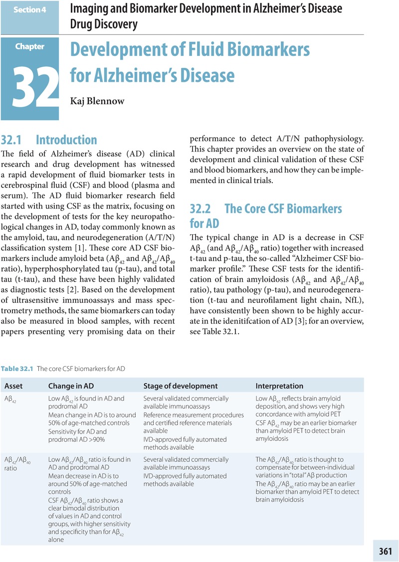
Image of the first page of this content. For PDF version, please use the ‘Save PDF’ preceeding this image.'
Type
Chapter
Information
Alzheimer's Disease Drug Development
Research and Development Ecosystem
, pp. 361 - 428
Publisher: Cambridge University Press
Print publication year: 2022

Access options

Get access to the full version of this content by using one of the access options below. (Log in options will check for institutional or personal access. Content may require purchase if you do not have access.)

References

References

Jack, CR Jr., Bennett, DA, Blennow, K, et al. A/T/N: an unbiased descriptive classification scheme for Alzheimer disease biomarkers. Neurology 2016; 87: 539–47.Google Scholar
Blennow, K. Biomarkers in Alzheimer’s disease drug development. Nat Med 2010; 16: 1218–22.CrossRefGoogle ScholarPubMed
Olsson, B, Lautner, R, Andreasson, U, et al. CSF and blood biomarkers for the diagnosis of Alzheimer’s disease: a systematic review and meta-analysis. Lancet Neurol 2016; 15: 673–84.CrossRefGoogle ScholarPubMed
Portelius, E, Tran, AJ, Andreasson, U, et al. Characterization of amyloid beta peptides in cerebrospinal fluid by an automated immunoprecipitation procedure followed by mass spectrometry. J Proteome Res 2007; 6: 4433–9.CrossRefGoogle ScholarPubMed
Andreasen, N, Minthon, L, Vanmechelen, E, et al. Cerebrospinal fluid tau and Abeta42 as predictors of development of Alzheimer’s disease in patients with mild cognitive impairment. Neurosci Lett 1999; 273: 58.Google Scholar
Blennow, K, Mattsson, N, Scholl, M, Hansson, O, Zetterberg, H. Amyloid biomarkers in Alzheimer’s disease. Trends Pharmacol Sci 2015; 36: 297309.Google Scholar
Lewczuk, P, Lelental, N, Spitzer, P, Maler, JM, Kornhuber, J. Amyloid-beta 42/40 cerebrospinal fluid concentration ratio in the diagnostics of Alzheimer’s disease: validation of two novel assays. J Alzheimers Dis 2015; 43: 183–91.Google ScholarPubMed
Janelidze, S, Zetterberg, H, Mattsson, N, et al. CSF Abeta42/Abeta40 and Abeta 42/Abeta 38 ratios: better diagnostic markers of Alzheimer disease. Ann Clin Transl Neurol 2016; 3: 154–65.Google Scholar
Sato, C, Barthelemy, NR, Mawuenyega, KG, et al. Tau kinetics in neurons and the human central nervous system. Neuron 2018; 98: 861–4.CrossRefGoogle ScholarPubMed
Mudher, A, Colin, M, Dujardin, S, et al. What is the evidence that tau pathology spreads through prion-like propagation? Acta Neuropathol Commun 2017; 5: 99.CrossRefGoogle ScholarPubMed
Skillback, T, Rosen, C, Asztely, F, et al. Diagnostic performance of cerebrospinal fluid total tau and phosphorylated tau in Creutzfeldt–Jakob disease: results from the Swedish Mortality Registry. JAMA Neurol 2014; 71: 476–83.Google Scholar
Blennow, K, Hampel, H. CSF markers for incipient Alzheimer’s disease. Lancet Neurol 2003; 2: 605–13.Google Scholar
Hampel, H, Buerger, K, Zinkowski, R, et al. Measurement of phosphorylated tau epitopes in the differential diagnosis of Alzheimer disease: a comparative cerebrospinal fluid study. Arch Gen Psychiatry 2004; 61: 95102.Google Scholar
Hanes, J, Kovac, A, Kvartsberg, H, et al. Evaluation of a novel immunoassay to detect p-tau Thr127 in the CSF to distinguish Alzheimer disease from other dementias. Neurology 2020; 95: e3026–35.Google Scholar
Hesse, C, Rosengren, L, Andreasen, N, et al. Transient increase in total tau but not phospho-tau in human cerebrospinal fluid after acute stroke. Neurosci Lett 2001; 297: 187–90.CrossRefGoogle Scholar
Suarez-Calvet, M, Karikari, TK, Ashton, NJ, et al. Novel tau biomarkers phosphorylated at T181, T217 or T231 rise in the initial stages of the preclinical Alzheimer’s continuum when only subtle changes in Abeta pathology are detected. EMBO Mol Med 2020; 12: e12921.CrossRefGoogle ScholarPubMed
Mattsson-Carlgren, N, Andersson, E, Janelidze, S, et al. Abeta deposition is associated with increases in soluble and phosphorylated tau that precede a positive tau PET in Alzheimer’s disease. Sci Adv 2020; 6: eaaz2387.Google Scholar
Meredith, JE Jr., Sankaranarayanan, S, Guss, V, et al. Characterization of novel CSF tau and p-tau biomarkers for Alzheimer’s disease. PloS One 2013; 8: e76523.CrossRefGoogle Scholar
Zhang, Z, Song, M, Liu, X, et al. Cleavage of tau by asparagine endopeptidase mediates the neurofibrillary pathology in Alzheimer’s disease. Nat Med 2014; 20: 1254–62.Google Scholar
Blennow, K, Chen, C, Cicognola, C, et al. Cerebrospinal fluid tau fragment correlates with tau PET: a candidate biomarker for tangle pathology. Brain 2020; 143: 650–60.CrossRefGoogle ScholarPubMed
Hansson, O, Zetterberg, H, Buchhave, P, et al. Association between CSF biomarkers and incipient Alzheimer’s disease in patients with mild cognitive impairment: a follow-up study. Lancet Neurol 2006; 5: 228–34.Google Scholar
Shaw, LM, Vanderstichele, H, Knapik-Czajka, M, et al. Cerebrospinal fluid biomarker signature in Alzheimer’s Disease Neuroimaging Initiative subjects. Ann Neurol 2009; 65: 403–13.Google Scholar
Kuhlmann, J, Andreasson, U, Pannee, J, et al. CSF Abeta 1–42: an excellent but complicated Alzheimer’s biomarker – a route to standardisation. Clin Chim Acta 2017; 467: 2733.Google Scholar
Hansson, O, Seibyl, J, Stomrud, E, et al. CSF biomarkers of Alzheimer’s disease concord with amyloid-beta PET and predict clinical progression: a study of fully automated immunoassays in BioFINDER and ADNI cohorts. Alzheimers Dement 2018; 14: 1470–81.Google Scholar
Kaplow, J, Vandijck, M, Gray, J, et al. Concordance of Lumipulse cerebrospinal fluid t-tau/Abeta 42 ratio with amyloid PET status. Alzheimers Dement 2020; 16: 144–52.CrossRefGoogle Scholar
Boulo, S, Kuhlmann, J, Andreasson, U, et al. First amyloid beta 1–42 certified reference material for re-calibrating commercial immunoassays. Alzheimers Dement 2020; 16 :1493–503.CrossRefGoogle Scholar
Shaw, LM, Arias, J, Blennow, K, et al. Appropriate use criteria for lumbar puncture and cerebrospinal fluid testing in the diagnosis of Alzheimer’s disease. Alzheimers Dement 2018; 14: 1505–21.Google Scholar
Janelidze, S, Stomrud, E, Palmqvist, S, et al. Plasma beta-amyloid in Alzheimer’s disease and vascular disease. Sci Rep 2016; 6: 26801.CrossRefGoogle ScholarPubMed
Kaneko, N, Yamamoto, R, Sato, TA, Tanaka, K. Identification and quantification of amyloid beta-related peptides in human plasma using matrix-assisted laser desorption/ionization time-of-flight mass spectrometry. Proc Jpn Acad Ser B Phys Biol Sci 2014; 90: 104–17.Google Scholar
Pannee, J, Tornqvist, U, Westerlund, A, et al. The amyloid-beta degradation pattern in plasma: a possible tool for clinical trials in Alzheimer’s disease. Neurosci Lett 2014; 573: 712.Google Scholar
Ovod, V, Ramsey, KN, Mawuenyega, KG, et al. Amyloid beta concentrations and stable isotope labeling kinetics of human plasma specific to central nervous system amyloidosis. Alzheimers Dement 2017; 13: 841–9.Google ScholarPubMed
Nakamura, A, Kaneko, N, Villemagne, VL, et al. High performance plasma amyloid-beta biomarkers for Alzheimer’s disease. Nature 2018; 554: 249–54.Google Scholar
Schindler, SE, Bollinger, JG, Ovod, V, et al. High-precision plasma beta-amyloid 42/40 predicts current and future brain amyloidosis. Neurology 2019; 93: e1647–59.Google Scholar
Palmqvist, S, Mattsson, N, Hansson, O, Alzheimer’s Disease Neuroimaging Initiative. Cerebrospinal fluid analysis detects cerebral amyloid-beta accumulation earlier than positron emission tomography. Brain 2016; 139: 1226–36.Google Scholar
Tatebe, H, Kasai, T, Ohmichi, T, et al. Quantification of plasma phosphorylated tau to use as a biomarker for brain Alzheimer pathology: pilot case–control studies including patients with Alzheimer’s disease and Down syndrome. Mol Neurodegener 2017; 12: 63.Google Scholar
Mielke, MM, Hagen, CE, Xu, J, et al. Plasma phospho-tau 181 increases with Alzheimer’s disease clinical severity and is associated with tau- and amyloid-positron emission tomography. Alzheimers Dement 2018; 14: 989–97.Google Scholar
Thijssen, EH, La Joie, R, Wolf, A, et al. Diagnostic value of plasma phosphorylated tau 181 in Alzheimer’s disease and frontotemporal lobar degeneration. Nat Med 2020; 26: 387–97.Google Scholar
Janelidze, S, Mattsson, N, Palmqvist, S, et al. Plasma p-tau181 in Alzheimer’s disease: relationship to other biomarkers, differential diagnosis, neuropathology and longitudinal progression to Alzheimer’s dementia. Nat Med 2020; 26: 379–86.CrossRefGoogle ScholarPubMed
Karikari, TK, Pascoal, TA, Ashton, NJ, et al. Blood phosphorylated tau 181 as a biomarker for Alzheimer’s disease: a diagnostic performance and prediction modelling study using data from four prospective cohorts. Lancet Neurol 2020; 19: 422–33.Google Scholar
Ashton, NJ, Pascoal, TA, Karikari, TK, et al. Plasma p-tau231: a new biomarker for incipient Alzheimer’s disease pathology. Acta Neuropathol 2021; 141: 709–24.CrossRefGoogle ScholarPubMed
Barthelemy, NR, Horie, K, Sato, C, Bateman, RJ. Blood plasma phosphorylated-tau isoforms track CNS change in Alzheimer’s disease. J Exp Med 2020; 217;DOI: http://doi.org/10.1084/jem.20200861.Google Scholar
Barthelemy, NR, Bateman, RJ, Hirtz, C, et al. Cerebrospinal fluid phospho-tau T217 outperforms T181 as a biomarker for the differential diagnosis of Alzheimer’s disease and PET amyloid-positive patient identification. Alzheimers Res Ther 2020; 12: 26.Google Scholar
Palmqvist, S, Janelidze, S, Quiroz, YT, et al. Discriminative accuracy of plasma phospho-tau 217 for Alzheimer disease vs other neurodegenerative disorders. JAMA 2020; 24: 772–81.Google Scholar
O’Connor, A, Karikari, TK, Poole, T, et al. Plasma phospho-tau 181 in presymptomatic and symptomatic familial Alzheimer’s disease: a longitudinal cohort study. Mol Psychiatry 2020;DOI: https://doi.org/10.1038/s41380-020-0838-x.Google Scholar
Lantero Rodriguez, J, Karikari, TK, Suarez-Calvet, M, et al. Plasma p-tau181 accurately predicts Alzheimer’s disease pathology at least 8 years prior to post-mortem and improves the clinical characterisation of cognitive decline. Acta Neuropathol 2020; 140: 267–78.CrossRefGoogle ScholarPubMed
Kovacs, GG. Invited review: neuropathology of tauopathies: principles and practice. Neuropathol Appl Neurobiol 2015; 41: 323.CrossRefGoogle ScholarPubMed
Rubenstein, R, Chang, B, Yue, JK, et al. Comparing plasma phospho tau, total tau, and phospho tau–total tau ratio as acute and chronic traumatic brain injury biomarkers. JAMA Neurol 2017; 74: 1063–72.Google Scholar
Palmqvist, S, Insel, PS, Stomrud, E, et al. Cerebrospinal fluid and plasma biomarker trajectories with increasing amyloid deposition in Alzheimer’s disease. EMBO Mol Med 2019; 11: e11170.Google Scholar
Randall, J, Mortberg, E, Provuncher, GK, et al. Tau proteins in serum predict neurological outcome after hypoxic brain injury from cardiac arrest: results of a pilot study. Resuscitation 2013; 84: 351–6.CrossRefGoogle ScholarPubMed
Zetterberg, H, Wilson, D, Andreasson, U, et al. Plasma tau levels in Alzheimer’s disease. Alzheimers Res Ther 2013; 5: 9.CrossRefGoogle ScholarPubMed
Mattsson, N, Zetterberg, H, Janelidze, S, et al. Plasma tau in Alzheimer disease. Neurology 2016; 87: 1827–35.CrossRefGoogle ScholarPubMed
Mattsson, N, Zetterberg, H, Nielsen, N, et al. Serum tau and neurological outcome in cardiac arrest. Ann Neurol 2017; 82: 665–75.CrossRefGoogle ScholarPubMed
Shahim, P, Tegner, Y, Wilson, DH, et al. Blood biomarkers for brain injury in concussed professional ice hockey players. JAMA Neurol 2014; 71: 684–92.CrossRefGoogle ScholarPubMed
Vacchi, E, Kaelin-Lang, A, Melli, G. Tau and alpha synuclein synergistic effect in neurodegenerative diseases: when the periphery is the core. Int J Mol Sci 2020; 21: 5030.Google Scholar
Gisslen, M, Price, RW, Andreasson, U, et al. plasma concentration of the neurofilament light protein (NfL) is a biomarker of CNS injury in HIV infection: a cross-sectional study. EBioMedicine 2016; 3: 135–40.Google Scholar
Mattsson, N, Andreasson, U, Zetterberg, H, Blennow, K. Association between longitudinal plasma neurofilament light and neurodegeneration in patients with Alzheimer disease. JAMA Neurol 2019; 76: 791–9.CrossRefGoogle ScholarPubMed
Weston, PSJ, Poole, T, Ryan, NS, et al. Serum neurofilament light in familial Alzheimer disease: a marker of early neurodegeneration. Neurology 2017; 89: 2167–75.Google Scholar
Preische, O, Schultz, SA, Apel, A, et al. Serum neurofilament dynamics predicts neurodegeneration and clinical progression in presymptomatic Alzheimer’s disease. Nat Med 2019; 25: 277–83.Google Scholar
Khalil, M, Teunissen, CE, Otto, M, et al. Neurofilaments as biomarkers in neurological disorders. Nat Rev Neurol 2018; 14: 577–89.CrossRefGoogle ScholarPubMed
Schindler, SE, Bollinger, JG, Ovod, V, et al. High-precision plasma beta-amyloid 42/40 predicts current and future brain amyloidosis. Neurology 2019; 93: e1647–59.Google Scholar
Hansson, O, Janelidze, S, Hall, S, et al. Blood-based NfL: a biomarker for differential diagnosis of parkinsonian disorder. Neurology 2017; 88: 930–7.Google Scholar
Illan-Gala, I, Lleo, A, Karydas, A, et al. Plasma tau and neurofilament light in frontotemporal lobar degeneration and Alzheimer’s disease. Neurology 2021; 96: e671–83.Google Scholar
Palmqvist, S, Janelidze, S, Stomrud, E, et al. Performance of fully automated plasma assays as screening tests for Alzheimer disease-related β-amyloid status. JAMA Neurol 2019; 76: 1060–9.Google Scholar
Hampel, H, O’Bryant, SE, Molinuevo, JL, et al. Blood-based biomarkers for Alzheimer disease: mapping the road to the clinic. Nat Rev Neurol 2018; 14: 639–52.Google Scholar
Karikari, TK, Benedet, AL, Ashton, NJ, et al. Diagnostic performance and prediction of clinical progression of plasma phospho-tau 181 in the Alzheimer’s disease neuroimaging initiative. Mol Psychiatry 2020; 26: 429–42.Google Scholar
Mattke, S, Cho, SK, Bittner, T, Hlavka, J, Hanson, M. Blood-based biomarkers for Alzheimer’s pathology and the diagnostic process for a disease-modifying treatment: projecting the impact on the cost and wait times. Alzheimers Dement (Amst) 2020; 12: e12081.Google ScholarPubMed
Beach, TG, Monsell, SE, Phillips, LE, Kukull, W. Accuracy of the clinical diagnosis of Alzheimer disease at National Institute on Aging Alzheimer Disease Centers, 2005–2010. J Neuropathol Exp Neurol 2012; 71: 266–73.Google Scholar
Knopman, DS, DeKosky, ST, Cummings, JL, et al. Practice parameter: diagnosis of dementia (an evidence-based review). Report of the Quality Standards Subcommittee of the American Academy of Neurology. Neurology 2001; 56: 1143–53.CrossRefGoogle ScholarPubMed
Portelius, E, Olsson, B, Hoglund, K, et al. Cerebrospinal fluid neurogranin concentration in neurodegeneration: relation to clinical phenotypes and neuropathology. Acta Neuropathol 2018; 136: 363–76.Google Scholar
Blennow, K, de Leon, MJ, Zetterberg, H. Alzheimer’s disease. Lancet 2006; 368: 387403.CrossRefGoogle ScholarPubMed
Kennedy, ME, Stamford, AW, Chen, X, et al. The BACE1 inhibitor verubecestat (MK-8931) reduces CNS beta-amyloid in animal models and in Alzheimer’s disease patients. Sci Transl Med 2016; 8: 363ra150.CrossRefGoogle ScholarPubMed
Wessels, AM, Lines, C, Stern, RA, et al. Cognitive outcomes in trials of two BACE inhibitors in Alzheimer’s disease. Alzheimers Dement 2020; 16: 1483–92.CrossRefGoogle ScholarPubMed
Masters, CL, Bateman, R, Blennow, K, et al. Alzheimer’s disease. Nat Rev Dis Primers 2015; 1: 15056.Google Scholar
Olsson, B, Alberg, L, Cullen, NC, et al. NfL is a marker of treatment response in children with SMA treated with nusinersen. J Neurol 2019; 266: 2129–36.Google Scholar
Piehl, F, Kockum, I, Khademi, M, et al. Plasma neurofilament light chain levels in patients with MS switching from injectable therapies to fingolimod. Mult Scler 2018; 24: 1046–54.Google Scholar
Blennow, K, Zetterberg, H, Rinne, JO, et al. Effect of immunotherapy with bapineuzumab on cerebrospinal fluid biomarker levels in patients with mild to moderate Alzheimer disease. Arch Neurol 2012; 69: 1002–10.Google Scholar
Ostrowitzki, S, Lasser, RA, Dorflinger, E, et al. A Phase III randomized trial of gantenerumab in prodromal Alzheimer’s disease. Alzheimers Res Ther 2017; 9: 95.Google Scholar
Salloway, S, Sperling, R, Fox, NC, et al. Two Phase 3 trials of bapineuzumab in mild-to-moderate Alzheimer’s disease. N Engl J Med 2014; 370: 322–33.CrossRefGoogle ScholarPubMed
Tolar, M, Abushakra, S, Hey, JA, Porsteinsson, A, Sabbagh, M. Aducanumab, gantenerumab, BAN2401, and ALZ-801-the first wave of amyloid-targeting drugs for Alzheimer’s disease with potential for near term approval. Alzheimers Res Ther 2020; 12: 95.CrossRefGoogle ScholarPubMed
Blennow, K, Zetterberg, H, Minthon, L, et al. Longitudinal stability of CSF biomarkers in Alzheimer’s disease. Neurosci Lett 2007; 419: 1822.Google Scholar
Zetterberg, H, Pedersen, M, Lind, K, et al. Intra-individual stability of CSF biomarkers for Alzheimer’s disease over two years. J Alzheimers Dis 2007; 12: 255–60.CrossRefGoogle ScholarPubMed
Sevigny, J, Chiao, P, Bussiere, T, et al. The antibody aducanumab reduces Abeta plaques in Alzheimer’s disease. Nature 2016; 537: 50–6.Google Scholar
Sperling, RA, Jack, CR Jr., Black, SE, et al. Amyloid-related imaging abnormalities in amyloid-modifying therapeutic trials: recommendations from the Alzheimer’s Association Research Roundtable Workgroup. Alzheimers Dement 2011; 7: 367–85.CrossRefGoogle ScholarPubMed

References

Khachaturian, ZS. Revised criteria for diagnosis of Alzheimer’s disease: National Institute on Aging–Alzheimer’s Association diagnostic guidelines for Alzheimer’s disease. Alzheimers Dement 2011; 7: 253–6.CrossRefGoogle ScholarPubMed
Jack, CR Jr., Bennett, DA, Blennow, K, et al. A/T/N: an unbiased descriptive classification scheme for Alzheimer disease biomarkers. Neurology 2016; 87: 539–47.CrossRefGoogle ScholarPubMed
Magistretti, PJ, Pellerin, L. Cellular mechanisms of brain energy metabolism and their relevance to functional brain imaging. Philos Trans R Soc Lond B Biol Sci 1999; 354: 1155–63.Google Scholar
Minter, MR, Taylor, JM, Crack, PJ. The contribution of neuroinflammation to amyloid toxicity in Alzheimer’s disease. J Neurochem 2016; 136: 457–74.Google Scholar
Janelidze, S, Mattsson, N, Palmqvist, S, et al. Plasma p-tau181 in Alzheimer’s disease: relationship to other biomarkers, differential diagnosis, neuropathology and longitudinal progression to Alzheimer’s dementia. Nat Med 2020; 26: 379–86.Google Scholar
Rogers, MB. Tau PET scans turn positive when amyloid does; symptoms follow. AlzForum series: Clinical Trials on Alzheimer’s Disease 2019, Part 8 of 9. January, 2020. Available at: www.alzforum.org/news/conference-coverage/tau-pet-scans-turn-positive-when-amyloid-does-symptoms-follow (accessed November 15, 2020).Google Scholar
Palmqvist, S, Schöll, M, Strandberg, O, et al. Earliest accumulation of β-amyloid occurs within the default-mode network and concurrently affects brain connectivity. Nat Comm 2017; 8: 1214.Google Scholar
Ossenkoppele, R, Schonhaut, DR, Schöll, M, et al. Tau PET patterns mirror clinical and neuroanatomical variability in Alzheimer’s disease. Brain 2016; 139: 1551–67.Google Scholar
Koychev, I, Gunn, RN, Firouzian, A, et al. PET tau and amyloid-β burden in mild Alzheimer’s disease: divergent relationship with age, cognition, and cerebrospinal fluid biomarkers. J Alzheimers Dis 2017; 60: 283–93.Google Scholar
Guerrero-Muñoz, MJ, Gerson, J, Castillo-Carranza, DL. Tau oligomers: the toxic player at synapses in Alzheimer’s disease. Front Cell Neurosci 2015; 9: 464.CrossRefGoogle ScholarPubMed
Vargas, LM, Cerpa, W, Muñoz, FJ, Zanlungo, S, Alvarez, AR. Amyloid-β oligomers synaptotoxicity: the emerging role of EphA4/c-Abl signaling in Alzheimer’s disease. Biochim Biophys Acta Mol Basis Dis 2018; 1864A: 1148–59.Google Scholar
Insel, PS, Ossenkoppele, R, Gessert, D, et al. Time to amyloid positivity and preclinical changes in brain metabolism, atrophy, and cognition: evidence for emerging amyloid pathology in Alzheimer’s disease. Front Neurosci 2017; 11: 281.Google Scholar
La Joie, R, Visani, AV, Baker, SL, et al. Prospective longitudinal atrophy in Alzheimer’s disease correlates with the intensity and topography of baseline tau-PET. Sci Transl Med 2020; 12: eaau5732.Google Scholar
Timmers, T, Ossenkoppele, R, Wolters, EE, et al. Associations between quantitative [18F]flortaucipir tau PET and atrophy across the Alzheimer’s disease spectrum. Alzheimers Res Ther 2019; 11: 60.Google Scholar
Salloway, S, Sperling, R, Fox, NC, et al. Two phase 3 trials of bapineuzumab in mild-to-moderate Alzheimer’s disease. N Engl J Med 2014; 370: 322–33.Google Scholar
Weintraub, S, Carrillo, MC, Farias, ST, et al. Measuring cognition and function in the preclinical stage of Alzheimer’s disease. Alzheimers Dement (N Y) 2018; 4: 6475.Google Scholar
Fazekas, F, Chawluk, JB, Alavi, A, et al. MR signal abnormalities at 1.5 T in Alzheimer’s dementia and normal aging. AJR Am J Roentgenol 1987; 149: 351–6.CrossRefGoogle ScholarPubMed
Mirza, SS, Saeed, U, Knight, J, et al. APOE ε4, white matter hyperintensities, and cognition in Alzheimer and Lewy body dementia. Neurology 2019; 93: e1807–19.CrossRefGoogle ScholarPubMed
Orgogozo, JM, Gilman, S, Dartigues, JF. Subacute meningoencephalitis in a subset of patients with AD after Abeta42 immunization. Neurology 2003; 61: 4654.Google Scholar
Sperling, RA, Jack, CR Jr., Black, SE, et al. Amyloid-related imaging abnormalities in amyloid-modifying therapeutic trials: recommendations from the Alzheimer’s Association Research Roundtable Workgroup. Alzheimers Dement 2011; 7: 367–85.Google Scholar
Greenberg, SM, Vernooij, MW, Cordonnier, C, et al. Cerebral microbleeds: a guide to detection and interpretation. Lancet Neurol 2009; 8: 165–74.Google Scholar
Tolar, M, Abushakra, S, Hey, JA, Porsteinsson, A, Sabbagh, M. Aducanumab, gantenerumab, BAN2401, and ALZ-801-the first wave of amyloid-targeting drugs for Alzheimer’s disease with potential for near term approval. Alzheimers Res Ther 2020; 12: 95.Google Scholar
Wu, J, Dong, Q, Gui, J, et al. Predicting brain amyloid using multivariate morphometry statistics, sparse coding, and correntropy: validation in 1,125 individuals from the ADNI and OASIS database. bioRxiv 2020;DOI: http://doi.org/10.1101/2020.10.16.343137.Google Scholar
Lukic, AS, Andrews, RD, Bourakova, V, et al. MRI, FDG, and early frame amyloid image classifiers to characterize and differentiate Alzheimer’s disease variants and non-AD dementias. Alzheimers Dement 2018; 14: P1429–30.Google Scholar
Davatzikos, C, Resnick, SM, Wu, X, Parmpi, P, Clark, CM. Individual patient diagnosis of AD and FTD via high-dimensional pattern classification of MRI. Neuroimage 2008; 41: 1220–7.Google Scholar
Giorgio, J, Jagust, WJ, Baker, S, et al. Predicting future regional tau accumulation in asymptomatic and early Alzheimer’s disease. bioRxiv 2020;DOI: http://doi.org/10.1101/2020.08.15.252601.Google Scholar
Yu, P, Sun, J, Wolz, R, et al. Operationalizing hippocampal volume as an enrichment biomarker for amnestic mild cognitive impairment trials: effect of algorithm, test-retest variability, and cut point on trial cost, duration, and sample size. Neurobiol Aging 2014; 35: 808–18.Google Scholar
Rabin, JS, Neal, TE, Nierle, HE, et al. Multiple markers contribute to risk of progression from normal to mild cognitive impairment. Neuroimage Clin 2020; 28: 102400.Google Scholar
Woodard, JL, Bellaali, Y, Dricot, L, et al. Multivariate prediction of rate of decline in memory functioning over six years using imaging biomarkers. Alzheimers Dement 2020; 16: e045645.Google Scholar
Leung, KK, Barnes, J, Ridgway, GR, et al. Alzheimer’s Disease Neuroimaging Initiative. Automated cross-sectional and longitudinal hippocampal volume measurement in mild cognitive impairment and Alzheimer’s disease. Neuroimage 2010; 51: 1345–59.Google Scholar
Hashimoto, M, Kazui, H, Matsumoto, K, et al. Does donepezil treatment slow the progression of hippocampal atrophy in patients with Alzheimer’s disease? Am J Psychiatry 2005; 162: 676–82.CrossRefGoogle ScholarPubMed
Turner, RS, Hebron, ML, Lawler, A, et al. Nilotinib effects on safety, tolerability, and biomarkers in Alzheimer’s disease. Ann Neurol 2020; 88: 183–94.Google Scholar
Gauthier, S, Aisen, PS, Ferris, SH. Effect of tramiprosate in patients with mild-to-moderate Alzheimer’s disease: exploratory analyses of the MRI sub-group of the Alphase study. J Nutr Health Aging 2009; 13: 550–7.Google Scholar
Wessels, AM, Tariot, PN, Zimmer, JA, et al. Efficacy and safety of lanabecestat for treatment of early and mild Alzheimer disease: the AMARANTH and DAYBREAK-ALZ randomized clinical trials. JAMA Neurol 2020; 77: 199209.Google Scholar
Novak, G, Fox, N, Clegg, S, et al. Changes in brain volume with bapineuzumab in mild to moderate Alzheimer’s disease. J Alzheimers Dis 2016; 49: 1123–34.Google Scholar
Fox, NC, Black, RS, Gilman, S, et al. Effects of Abeta immunization (AN1792) on MRI measures of cerebral volume in Alzheimer disease. Neurology 2005; 64: 1563–72.Google Scholar
Cheriyan, J, Kim, S, Wolansky, LJ, Cook, SD, Cadavid, D. Impact of inflammation on brain volume in multiple sclerosis. Arch Neurol 2012; 69: 82–8.Google Scholar
Reuter, M, Schmansky, NJ, Rosas, HD, Fischl, B. Within-subject template estimation for unbiased longitudinal image analysis. Neuroimage 2012; 61: 1402–18.Google Scholar
Iglesias, JE, Van Leemput, K, Augustinack, J, et al. Bayesian longitudinal segmentation of hippocampal substructures in brain MRI using subject-specific atlases. Neuroimage 2016; 141: 542–55.Google Scholar
Teipel, SJ, Kuper-Smith, JO, Bartels, C, et al. Multicenter tract-based analysis of microstructural lesions within the Alzheimer’s disease spectrum: association with amyloid pathology and diagnostic usefulness. J Alzheimers Dis 2019; 72: 455–65.CrossRefGoogle ScholarPubMed
Elahi, FM, Marx, G, Cobigo, Y, et al. Longitudinal white matter change in frontotemporal dementia subtypes and sporadic late onset Alzheimer’s disease. Neuroimage Clin 2017; 16: 595603.Google Scholar
Pasternak, O, Sochen, N, Gur, Y, Intrator, N, Assaf, Y. Free water elimination and mapping from diffusion MRI. Magn Reson Med 2009; 62: 717–30.Google Scholar
Hoy, AR, Ly, M, Carlsson, CM, et al. Microstructural white matter alterations in preclinical Alzheimer’s disease detected using free water elimination diffusion tensor imaging. PloS One 2017; 12: e0173982.Google Scholar
Binnewijzend, MA, Kuijer, JP, van der Flier, WM, et al. Distinct perfusion patterns in Alzheimer’s disease, frontotemporal dementia and dementia with Lewy bodies. Eur Radiol 2014; 24: 2326–33.Google Scholar
Dukart, J, Holiga, Š, Chatham, C, et al. Cerebral blood flow predicts differential neurotransmitter activity. Sci Rep 2018; 8: 4074.Google Scholar
Guo, H, Grajauskas, L, Habash, B, D’Arc, RC, Song, X. Functional MRI technologies in the study of medication treatment effect on Alzheimer’s disease. Aging Med (Milton) 2018; 1: 7595.Google Scholar
Zhang, N, Gordon, ML, Goldberg, TE. Cerebral blood flow measured by arterial spin labeling MRI at resting state in normal aging and Alzheimer’s disease. Neurosci Biobehav Rev 2017; 72: 168–75.Google Scholar
Smith, LA, Melbourne, A, Owen, D, et al. Cortical cerebral blood flow in ageing: effects of haematocrit, sex, ethnicity and diabetes. Eur Radiol 2019; 29: 5549–58.Google Scholar
Greicius, MD, Srivastava, G, Reiss, AL, Menon, V. Default-mode network activity distinguishes Alzheimer’s disease from healthy aging: evidence from functional MRI. Proc Natl Acad Sci USA 2004; 101: 4637–42.Google Scholar
Churchill, NW, Spring, R, Afshin-Pour, B, Dong, F, Strother, SC. An automated, adaptive framework for optimizing preprocessing pipelines in task-based functional MRI. PLoS One 2015; 10: e0131520.Google Scholar
Matthews, DC, Mao, X, Dowd, K, et al. Riluzole, a glutamate modulator, slows cerebral glucose metabolism decline in patients with Alzheimer’s disease: a pilot multimodal neuroimaging study. Brain 2021; Jun 18: awab222.Google Scholar
Maul, S, Giegling, I, Rujescu, D. Proton magnetic resonance spectroscopy in common dementias: current status and perspectives. Front Psychiatry 2020; 11: 769.Google Scholar
van Berckel, BN, Ossenkoppele, R, Tolboom, N, et al. Longitudinal amyloid imaging using 11C-PiB: methodologic considerations. J Nucl Med 2013; 54: 1570–6.Google Scholar
Sevigny, J, Chiao, P, Bussière, T, et al. The antibody aducanumab reduces Aβ plaques in Alzheimer’s disease. Nature 2016; 537: 50–6.Google Scholar
Ostrowitzki, S, Lasser, RA, Dorflinger, E, et al. A Phase III randomized trial of gantenerumab in prodromal Alzheimer’s disease. Alzheimers Res Ther 2017; 9: 95.Google Scholar
Schmidt, ME, Matthews, DC, Andrews, RD, Mosconi, L. Positron emission tomography in Alzheimer disease: diagnosis and use as biomarker endpoints. In Translational Neuroimaging, McArthur RA (ed.). New York: Academic Press; 2013: 131–74.Google Scholar
Klunk, WE, Koeppe, RA, Price, JC. The Centiloid project: standardizing quantitative amyloid plaque estimation by PET. Alzheimers Dement 2015; 11: 1–15.e154.CrossRefGoogle ScholarPubMed
Chen, K, Roontiva, A, Thiyyagura, P, et al. Alzheimer’s Disease Neuroimaging Initiative. Improved power for characterizing longitudinal amyloid-β PET changes and evaluating amyloid-modifying treatments with a cerebral white matter reference region. J Nucl Med 2015; 56: 560–6.Google Scholar
Schmidt, ME, Chiao, P, Klein, G, et al. The influence of biological and technical factors on quantitative analysis of amyloid PET: points to consider and recommendations for controlling variability in longitudinal data. Alzheimers Dement 2015; 11: 1050–68.Google Scholar
Rostomian, AH, Madison, C, Rabinovici, GD, Jagust, WJ. Early 11C-PIB frames and 18F-FDG PET measures are comparable: a study validated in a cohort of AD and FTLD patients. J Nucl Med 2011; 52: 173–9.Google Scholar
Brendel, M, Barthel, H, van Eimeren, T, et al. Assessment of 18F-PI-2620 as a biomarker in progressive supranuclear palsy. JAMA Neurol 2020; 77: 1408–19.Google Scholar
Tagai, K, Ono, M, Kubota, M, et al. High-contrast in vivo imaging of tau pathologies in Alzheimer’s and non-Alzheimer’s disease tauopathies. Neuron 2020; 109: 42–58;DOI: http://doi.org/10.1016/j.neuron.2020.09.042.Google Scholar
Betthauser, TJ, Cody, KA, Zammit, MD, et al. In vivo characterization and quantification of neurofibrillary tau PET radioligand 18F-MK-6240 in humans from Alzheimer disease dementia to young controls. J Nucl Med 2019; 60: 93–9.Google Scholar
Maass, A, Landau, S, Baker, SL, Alzheimer’s Disease Neuroimaging Initiative. Comparison of multiple tau-PET measures as biomarkers in aging and Alzheimer’s disease.Neuroimage 2017; 157: 448–63.Google Scholar
Baker, SL, Maass, A, Jagust, WJ. Considerations and code for partial volume correcting [18F]-AV-1451 tau PET data. Data Brief 2017; 15: 648–57;DOI: http://doi.org/10.1016/j.dib.2017.10.024.Google Scholar
Southekal, S, Devous, MD Sr., Kennedy, I, et al. Flortaucipir F 18 quantitation using parametric estimation of reference signal intensity. J Nucl Med 2018; 59: 944–51.Google Scholar
Beyer, L, Nitschmann, A, Barthel, H, et al. Early-phase [18F]PI-2620 tau-PET imaging as a surrogate marker of neuronal injury. Eur J Nucl Med Mol Imaging 2020; 47: 2911–22.Google Scholar
Foster, NL, Heidebrink, JL, Clark, CM, et al. FDG-PET improves accuracy in distinguishing frontotemporal dementia and Alzheimer’s disease. Brain 2007; 130: 2616–35.Google Scholar
Xia, Y, Lu, S, Wen, L, et al. Automated identification of dementia using FDG-PET imaging. Biomed Res Int 2014; 2014: 421743.Google Scholar
Matthews, DC, Ritter, A, Thomas, RG, et al. Rasagiline effects on glucose metabolism, cognition, and tau in Alzheimer’s dementia. Alzheimers Dement (N Y) 2021; 7: e12106.Google Scholar
Beckett, L, Harvey, D, Donohue, M, et al. Biostatistics Core ADNI 2 summary and ADNI 3 plans. Available at https://slideplayer.com/slide/12666696/ (accessed November 20, 2020).Google Scholar
Chen, K, Langbaum, JB, Fleisher, AS, et al. Alzheimer’s Disease Neuroimaging Initiative. Twelve-month metabolic declines in probable Alzheimer’s disease and amnestic mild cognitive impairment assessed using an empirically pre-defined statistical region-of-interest: findings from the Alzheimer’s Disease Neuroimaging Initiative. Neuroimage 2010; 51: 654–64.CrossRefGoogle Scholar
Kadir, A, Andreasen, N, Almkvist, O, et al. Effect of phenserine treatment on brain functional activity and amyloid in Alzheimer’s disease. Ann Neurol 2008; 63: 621–31.Google Scholar
Kreisl, WC, Kim, MJ, Coughlin, JM, et al. PET imaging of neuroinflammation in neurological disorders. Lancet Neurol 2020; 19: 940–50.Google Scholar
Mecca, AP, Chen, MK, O’Dell, RS, et al. In vivo measurement of widespread synaptic loss in Alzheimer’s disease with SV2A PET. Alzheimers Dement 2020; 16: 974–82.Google Scholar
Josephs, KA, Dickson, DW, Tosakulwong, N, et al. Rates of hippocampal atrophy and presence of post-mortem TDP-43 in patients with Alzheimer’s disease: a longitudinal retrospective study. Lancet Neurol 2017; 16: 917–24.Google Scholar
US Department of Health and Human Services, Food and Drug Administration, Center for Drug Evaluation and Research (CDER), and Center for Biologics Evaluation and Research (CBER). Early Alzheimer’s disease: developing drugs for treatment. Guidelines for industry. Available at: www.fda.gov/downloads/Drugs/GuidanceComplianceRegulatoryInformation/Guidances/UCM596728.pdf (accessed November 15, 2020).Google Scholar
European Medicines Agency. Clinical investigation of medicines for the treatment of Alzheimer’s disease/ CPMP/EWP/553/1995. Available at: www.ema.europa.eu/en/documents/scientific-guideline/guideline-clinical-investigation-medicines-treatment-alzheimers-disease-revision-2_en.pdf (accessed November 20, 2020).Google Scholar

References

Conrado, DJ, Karlsson, MO, Romero, K, et al. Open innovation: towards sharing of data, models and workflows. Eur J Pharm Sci 2017; 109: S6571.CrossRefGoogle Scholar
Gurevitch, J, Koricheva, J, Nakagawa, S, et al. Meta-analysis and the science of research synthesis. Nature 2018; 555: 175–82.Google Scholar
Alzheimer’s Association. 2020 Alzheimer’s disease facts and figures. Alzheimers Dement 2020; 16: 391460.Google Scholar
Villa, C, Lavitrano, M, Salvatore, E, et al. Molecular and imaging biomarkers in Alzheimer’s disease: a focus on recent insights. J Pers Med 2020; 10: 132.Google Scholar
Sancesario, GM, Bernardini, S. Alzheimer’s disease in the omics era. Clin Biochem 2018; 59: 916.Google Scholar
Toga, AW, Neu, SC, Bhatt, P, et al. The Global Alzheimer’s Association Interactive Network. Alzheimers Dement 2016; 12: 4954.Google Scholar
Neu, SC, Crawford, KL, Toga, AW. Sharing data in the global Alzheimer’s Association Interactive Network. Neuroimage 2016; 124: 1168–74.Google Scholar
Kraus, WE, Bhapkar, M, Huffman, KM, et al. 2 years of calorie restriction and cardiometabolic risk (CALERIE): exploratory outcomes of a multicentre, Phase 2, randomised controlled trial. Lancet Diabetes Endocrinol 2019; 7: 673–83.Google Scholar
Silva, MVF, Loures, CDMG, Alves, LCV, et al. Alzheimer’s disease: risk factors and potentially protective measures. J Biomed Sci 2019; 26: 111.CrossRefGoogle ScholarPubMed
Rygiel, K. Novel strategies for Alzheimer’s disease treatment: an overview of anti-amyloid beta monoclonal antibodies. Indian J Pharmacol 2016; 48: 629–36.Google Scholar
Tariot, PN, Lopera, F, Langbaum, JB, et al. The Alzheimer’s Prevention Initiative Autosomal-Dominant Alzheimer’s Disease Trial: a study of crenezumab versus placebo in preclinical PSEN1 E280A mutation carriers to evaluate efficacy and safety in the treatment of autosomal-dominant Alzheimer’s disease. Alzheimers Dement (N Y) 2018; 4: 150–60.Google Scholar
Mills, SM, Mallmann, J, Santacruz, AM, et al. Preclinical trials in autosomal dominant AD: implementation of the DIAN-TU trial. Rev Neurol 2013; 169: 737–43.Google Scholar
Sperling, RA, Donohue, MC, Raman, R, et al. Association of factors with elevated amyloid burden in clinically normal older individuals. JAMA Neurol 2020; 77: 735–45.Google Scholar
Tang, MX, Stern, Y, Marder, K, et al. The APOE-ε4 allele and the risk of Alzheimer disease among African Americans, whites, and Hispanics. JAMA 1998; 279: 751–5.Google Scholar
Duara, R, Loewenstein, DA, Lizarraga, G, et al. Effect of age, ethnicity, sex, cognitive status and APOE genotype on amyloid load and the threshold for amyloid positivity. Neuroimage Clin 2019; 22: 101800.Google Scholar
O’Bryant, SE, Zhang, F, Johnson, LA, et al. A precision medicine model for targeted NSAID therapy in Alzheimer’s disease. J Alzheimers Dis 2018; 66: 97104.Google Scholar
Li, Y, Li, Y, Li, X, et al. Head injury as a risk factor for dementia and Alzheimer’s disease: a systematic review and meta-analysis of 32 observational studies. PLoS One 2017; 12: e0169650.Google Scholar
Neu, SC, Pa, J, Kukull, W, et al. Apolipoprotein E genotype and sex risk factors for Alzheimer disease: a meta-analysis. JAMA Neurol 2017; 74: 1178–89.CrossRefGoogle ScholarPubMed
Funk-White, M, Moore, AA, McEvoy, LK, et al. Alcohol use patterns and cognitive impairment: a cross-country comparison. Alzheimer’s Association International Conference, July 26–30, 2020.Google Scholar
Rane, S. Detecting cortical signatures of suspected non-amyloid pathology using large harmonized datasets. Alzheimer’s Association International Conference, July 26–30, 2020.Google Scholar
Crawford, KL, Neu, SC, Toga, AW. The Image and Data Archive at the Laboratory of Neuro Imaging. Neuroimage 2016; 124: 1080–3.CrossRefGoogle ScholarPubMed
Weiner, MW, Aisen, PS, Jack, CR, et al. The Alzheimer’s Disease Neuroimaging Initiative: progress report and future plans. Alzheimers Dement 2010; 6: 202–11.Google Scholar
Beekly, DL, Ramos, EM, van Belle, G, et al. The National Alzheimer’s Coordinating Center (NACC) Database: an Alzheimer disease database. Alzheimer Dis Assoc Disord 2004; 18: 270–7.Google Scholar
Anthony, S, Pradier, C, Chevrier, R, et al. The French national Alzheimer database: a fast growing database for researchers and clinicians. Dement Geriatr Cogn Disord 2014; 38: 271–80.Google Scholar

References

Cacabelos, R, Fernández-Novoa, L, Lombardi, V, et al. Molecular genetics of Alzheimer’s disease and aging. Meth Find Exp Clin Pharmacol 2005; 27 : 1–573.Google Scholar
Cacabelos, R. Have there been improvement in Alzheimer’s disease drug discovery over the past 5 years? Expert Opin Drug Discov 2018; 13: 523–38.Google Scholar
Cacabelos, R, Cacabelos, N, Carril, JC. The role of pharmacogenomics in adverse drug reactions. Expert Rev Clin Pharmacol 2019; 12 : 407–42.CrossRefGoogle ScholarPubMed
Cacabelos, R, Cacabelos, P, Torrellas, C, et al. Pharmacogenomics of Alzheimer’s disease: novel therapeutic strategies for drug development. Methods Mol Biol 2014; 1175 : 323–556.Google Scholar
Cacabelos, R, Carril, JC, Cacabelos, P, et al. Pharmacogenomics of Alzheimer’s disease: genetic determinants of phenotypic variation and therapeutic outcome. J Genomic Med Pharmacogenomics 2016; 1 : 151–209.Google Scholar
Cacabelos, R, Carril, JC, Sanmartín, A, et al. Pharmacoepigenetic processors: epigenetic drugs, drug resistance, toxicoepigenetics, and nutriepigenetics. In Pharmacoepigenetics, Cacabelos, R (ed.). San Diego, CA: Academic Press/Elsevier; 2019: 191424.Google Scholar
Kozyra, M, Ingelman-Sundberg, M, Lauschke, VM. Rare genetic variants in cellular transporters, metabolic enzymes, and nuclear receptors can be important determinants of interindividual differences in drug response. Genet Med 2017; 19 : 20–9.Google Scholar
Zhou, ZW, Chen, XW, Sneed, KB, et al. Clinical association between pharmacogenomics and adverse drug reactions. Drugs 2015; 75 : 589–631.CrossRefGoogle ScholarPubMed
Cacabelos, R, Tellado, I, Cacabelos, P. The epigenetic machinery in the life cycle and pharmacoepigenetics. In Pharmacoepigenetics, Cacabelos, R (ed.). San Diego, CA: Academic Press/Elsevier; 2019: 1100.Google Scholar
Cacabelos, R. Pleiotropy and promiscuity in pharmacogenomics for the treatment of Alzheimer’s disease and related risk factors. Future Neurol 2018; 13.CrossRefGoogle Scholar
Cacabelos, R. Epigenomic networking in drug development: from pathogenic mechanisms to pharmacogenomics. Drug Dev Res 2014; 75 :348–65.Google Scholar
Dorszewska, J, Prendecki, M, Oczkowska, A, et al. Molecular basis of familial and sporadic Alzheimer’s disease. Curr Alzheimer Res 2016; 13 : 952–63.Google Scholar
Jamal, S, Goyal, S, Shanker, A, et al. Computational screening and exploration of disease-associated genes in Alzheimer’s disease. J Cell Biochem 2017; 118 : 1471–9.CrossRefGoogle ScholarPubMed
Zhou, L, Li, HY, Wang, JH, et al. Correlation of gene polymorphisms of CD36 and ApoE with susceptibility of Alzheimer disease: a case–control study. Medicine (Baltimore) 2018; 97: e12470.Google Scholar
Davies, G, Harris, SE, Reynolds, CA, et al. A genome-wide association study implicates the APOE locus in nonpathological cognitive ageing. Mol Psychiatry 2014; 19 : 76–87.Google Scholar
Cacabelos, R. Pharmacogenetic considerations when prescribing cholinesterase inhibitors for the treatment of Alzheimer’s disease. Expert Opin Drug Metab Toxicol 2020; 16: 673701.Google Scholar
Shapira, M, Tur-Kaspa, I, Bosgraaf, L, et al. A transcription-activating polymorphism in the ACHE promoter associated with acute sensitivity to anti-acetylcholinesterases. Hum Mol Genet 2000; 9 : 1273–81.Google Scholar
Lane, R, Feldman, HH, Meyer, J, et al. Synergistic effect of apolipoprotein E epsilon4 and butyrylcholinesterase K-variant on progression from mild cognitive impairment to Alzheimer’s disease. Pharmacogenet Genomics 2008; 18 : 289–98.Google Scholar
Cuddy, LK, Seah, C, Pasternak, SH, et al. Amino-terminal β-amyloid antibody blocks β-amyloid-mediated inhibition of the high-affinity choline transporter CHT. Front Mol Neurosci 2017; 10: 361.CrossRefGoogle ScholarPubMed
Payette, DJ, Xie, J, Guo, Q. Reduction in CHT1-mediated choline uptake in primary neurons from presenilin-1 M146V mutant knock-in mice. Brain Res 2007; 1135 : 12–21.Google Scholar
Wang, Y, Zhou, Z, Tan, H, et al. Nitrosylation of vesicular transporters in brain of amyloid precursor protein/presenilin 1 double transgenic mice. J Alzheimers Dis 2017; 55 :1683–92.Google Scholar
Nagy, PM, Aubert, I. Overexpression of the vesicular acetylcholine transporter increased acetylcholine release in the hippocampus. Neuroscience 2012; 218 : 1–11.Google Scholar
Kolisnyk, B, Al-Onaizi, MA, Xu, J, et al. Cholinergic regulation of hnRNPA2/B1 translation by M1 muscarinic receptors. J Neurosci 2016; 36 : 6287–96.Google Scholar
Dolejší, E, Liraz, O, Rudajev, V, et al. Apolipoprotein E4 reduces evoked hippocampal acetylcholine release in adult mice. J Neurochem 2016; 136 :503–9.Google Scholar
Albin, RL, Bohnen, NI, Muller, MLTM, et al. Regional vesicular acetylcholine transporter distribution in human brain: A [18F]fluoroethoxybenzovesamicol positron emission tomography study. J Comp Neurol 2018; 526 : 2884–97.Google Scholar
Wallace, TL, Bertrand, D. Importance of the nicotinic acetylcholine receptor system in the prefrontal cortex. Biochem Pharmacol 2013; 85 : 1713–20.Google Scholar
Ma, KG, Qian, YH. Alpha 7 nicotinic acetylcholine receptor and its effects on Alzheimer’s disease. Neuropeptides 2019; 73 : 96–106.Google Scholar
Sadigh-Eteghad, S, Talebi, M, Mahmoudi, J, et al. Selective activation of α7 nicotinic acetylcholine receptor by PHA-543613 improves Aβ25–35-mediated cognitive deficits in mice. Neuroscience 2015; 298 : 81–93.Google Scholar
Li, L, Liu, Z, Jiang, YY, et al. Acetylcholine suppresses microglial inflammatory response via α7nAChR to protect hippocampal neurons. J Integr Neurosci 2019; 18 : 51–6.Google Scholar
McKeever, PM, Kim, T, Hesketh, AR, et al. Cholinergic neuron gene expression differences captured by translational profiling in a mouse model of Alzheimer’s disease. Neurobiol Aging 2017; 57 : 104–19.Google Scholar
Vauthier, V, Housset, C, Falguières, T. Targeted pharmacotherapies for defective ABC transporters. Biochem Pharmacol 2017; 136 :1–11.Google Scholar
Cacabelos, R, Llovo, R, Fraile, C, et al. Pharmacogenetic aspects of therapy with cholinesterase inhibitors: the role of CYP2D6 in Alzheimer’s disease pharmacogenetics. Curr Alzheimer Res 2007; 4 : 479–500.Google Scholar
Cacabelos, R. Donepezil in Alzheimer’s disease: from conventional trials to pharmacogenetics. Neuropsychiatr Dis Treat 2007; 3 : 303–33.Google Scholar
Brewster, JT, Dell’Acqua, S, Thach, DQ, et al. Classics in chemical neuroscience: donepezil. ACS Chem Neurosci 2019; 10 : 155–67.Google Scholar
Noetzli, M, Eap, CB. Pharmacodynamic, pharmacokinetic and pharmacogenetic aspects of drugs used in the treatment of Alzheimer’s disease. Clin Pharmacokinet 2013; 52 : 225–41.CrossRefGoogle ScholarPubMed
Noetzli, M, Guidi, M, Ebbing, K, et al. Population pharmacokinetic approach to evaluate the effect of CYP2D6, CYP3A, ABCB1, POR and NR1I2 genotypes on donepezil clearance. Br J Clin Pharmacol 2014; 78 : 135–44.Google Scholar
Cacabelos, R. World Guide for Drug Use and Pharmacogenomics. Corunna: EuroEspes Publishing; 2012.Google Scholar
Xiao, T, Jiao, B, Zhang, W, et al. Effect of the CYP2D6 and APOE polymorphisms on the efficacy of donepezil in patients with Alzheimer’s disease: a systematic review and meta-analysis. CNS Drugs 2016; 30 : 899–907.Google Scholar
Cacabelos, R, Martínez, R, Fernández-Novoa, L, et al. Genomics of dementia: APOE- and CYP2D6-related pharmacogenetics. Int J Alzheimers Dis 2012; 2012: 518901.Google Scholar
Zhong, Y, Zheng, X, Miao, Y, et al. Effect of CYP2D6*10 and APOE polymorphisms on the efficacy of donepezil in patients with Alzheimer’s disease. Am J Med Sci 2013; 345 : 222–6.Google Scholar
Yaowaluk, T, Senanarong, V, Limwongse, C, et al. Influence of CYP2D6, CYP3A5, ABCB1, APOE polymorphisms and nongenetic factors on donepezil treatment in patients with Alzheimer’s disease and vascular dementia. Pharmgenomics Pers Med 2019; 12 : 209–24.Google Scholar
Sokolow, S, Li, X, Chen, L, et al. Deleterious effect of butyrylcholinesterase K-variant in donepezil treatment of mild cognitive impairment. J Alzheimers Dis 2017; 56 : 229–37.Google Scholar
Russo, P, Kisialiou, A, Moroni, R, et al. Effect of genetic polymorphisms (SNPs) in CHRNA7 gene on response to acetylcholinesterase inhibitors (AChEI) in patients with Alzheimer’s disease. Curr Drug Targets 2017; 18 : 1179–90.Google Scholar
Noetzli, M, Guidi, M, Ebbing, K, et al. Relationship of CYP2D6, CYP3A, POR, and ABCB1 genotypes with galantamine plasma concentrations. Ther Drug Monit 2013; 35 : 270–5.Google Scholar
Birks, JS, Grimley Evans, J. Rivastigmine for Alzheimer’s disease. Cochrane Database Syst Rev 2015; 10: CD001191.Google Scholar
Gul, A, Bakht, J, Mehmood, F. Huperzine-A response to cognitive impairment and task switching deficits in patients with Alzheimer’s disease. J Chin Med Assoc 2019; 82 : 40–3.Google Scholar
Lin, PP, Li, XN, Yuan, F, et al. Evaluation of the in vitro and in vivo metabolic pathway and cytochrome P450 inhibition/induction profile of huperzine A. Biochem Biophys Res Commun 2016; 480 : 248–53.Google Scholar
Noetzli, M, Guidi, M, Ebbing, K, et al. Population pharmacokinetic study of memantine: effects of clinical and genetic factors. Clin Pharmacokinet 2013; 52 : 211–23.Google Scholar
Cacabelos, R, Goldgaber, D, Vostrov, A, et al. APOE-TOMM40 in the pharmacogenomics of dementia. J Pharmacogenomics Pharmacoproteomics 2014; 5: 135.Google Scholar
Cacabelos, R, Carril, JC, Cacabelos, N, et al. Sirtuins in Alzheimer’s disease: SIRT2-related genophenotypes and implications for pharmacoepigenetics. Int J Mol Sci 2019; 20: E1249.CrossRefGoogle ScholarPubMed

References

Berger, H. Über das Elektrenkephalogramm des Menschen. Dritte Mitteilung. Arch Psychiatr Nervenkr 1931; 94: 1660.Google Scholar
Berger, H. Über das Elektrenkephalogramm des Menschen. Fünfte Mitteilung. Arch Psychiatr Nervenkr 1932; 98: 231–54.Google Scholar
Babiloni, C, Blinowska, K, Bonanni, L, et al. What electrophysiology tells us about Alzheimer’s disease: a window into the synchronization and connectivity of brain neurons. Neurobiol Aging 2020; 85: 5873.Google Scholar
Palop, JJ, Mucke, L. Network abnormalities and interneuron dysfunction in Alzheimer disease. Nat Rev Neurosci 2016; 17: 777–92.Google Scholar
Styr, B, Slutsky, I. Imbalance between firing homeostasis and synaptic plasticity drives early-phase Alzheimer’s disease. Nat Neurosci 2018; 21: 463–73.Google Scholar
Canter, RG, Penney, J, Tsai, LH. The road to restoring neural circuits for the treatment of Alzheimer’s disease. Nature 2016; 539: 187–96.Google Scholar
Selkoe, DJ. Alzheimer’s disease is a synaptic failure. Science 2002; 298: 789–91.Google Scholar
D’Amelio, M, Rossini, PM. Brain excitability and connectivity of neuronal assemblies in Alzheimer’s disease: from animal models to human findings. Prog Neurobiol 2012; 99: 4260.Google Scholar
van Straaten, EC, Scheltens, P, Gouw, AA, Stam, CJ. Eyes-closed task-free electroencephalography in clinical trials for Alzheimer’s disease: an emerging method based upon brain dynamics. Alzheimers Res Ther 2014; 6: 86.Google Scholar
Delbeuck, X, Van der Linder, M, Colette, F. Alzheimer’s disease as a disconnection syndrome? Neuropyschol Rev 2003; 13: 7992.Google Scholar
Briels, CT, Schoonhoven, DN, Stam, CJ, et al. Reproducibility of EEG functional connectivity in Alzheimer’s disease. Alzheimers Res Ther 2020; 12 : 68.Google Scholar
Stam, CV, Van Straaten, ECW. The organization of physiological brain networks. Clin Neurophysiol 2012; 123: 1067–87.Google Scholar
Rossini, PM, Rossi, S, Babiloni, C, Polich, J. Clinical neurophysiology of aging brain: from normal aging to neurodegeneration. Prog Neurobiol 2007; 83: 375400.Google Scholar
Prichep, LS, John, ER, Ferris, SH, et al. Prediction of longitudinal cognitive decline in normal elderly with subjective complaints using electrophysiological imaging. Neurobiol Aging 2006; 27: 471–81.Google Scholar
Gouw, AA, Alsema, AM, Tijms, BM, et al. EEG spectral analysis as a putative early prognostic biomarker in nondemented, amyloid positive subjects. Neurobiol Aging 2017; 57: 133–42.Google Scholar
Jelic, V, Johansson, S-E, Almkvist, O, et al. Quantitative electroencephalography in mild cognitve impairment: longitudinal changes and possible prediction of Alzheimer’s disease. Neurobiol Aging 2000; 21: 533–40.Google Scholar
van der Hiele, K, Bollen, EL, Vein, AA, et al. EEG markers of future cognitive performance in the elderly. J Clin Neurophysiol 2008; 25: 83–9.Google Scholar
Liedorp, M, van der Flier, WM, Hoogervorst, EL, Scheltens, P, Stam, CJ. Associations between patterns of EEG abnormalities and diagnosis in a large memory clinic cohort. Dement Geriatr Cogn Disord 2008; 27: 1823.Google Scholar
Jeong, J. EEG dynamics in patients with Alzheimer’s disease. Clin Neurophysiol 2004; 115: 1490–505.Google Scholar
Claus, JJ, Ongerboer de Visser, BW, Walstra, GJM, et al. Quantitative spectral electroencephalography in predicting survival in patients with early Alzheimer disease. Arch Neurol 1998; 55: 1105–11.Google Scholar
Stam, CJ. Nonlinear dynamical analysis of EEG and MEG: review of an emerging field. Clin Neurophysiol 2005; 116: 2266–301.Google Scholar
Stam, CJ. Modern network science of neurological disorders. Nat Rev Neurosci 2014; 15: 683–95.Google Scholar
Stam, CJ, Jones, BF, Nolte, G, Breakspear, M, Scheltens, P. Small-world networks and functional connectivity in Alzheimer’s disease. Cereb Cortex 2007; 17: 92–9.Google Scholar
Sun, J, Wang, B, Niu, Y, et al. Complexity analysis of EEG, MEG, and fMRI in mild cognitive impairment and Alzheimer’s disease: a review. Entropy 2020; 22: 239.Google Scholar
Dauwels, J, Vialatte, F, Cichocki, A. Diagnosis of Alzheimer’s disease from EEG signals: where are we standing? Curr Alzheimer Res 2010; 7: 487505.CrossRefGoogle ScholarPubMed
Simpraga, S, Alvarez-Jimenez, R, Mansvelder, HD, et al. EEG machine learning for accurate detection of cholinergic intervention and Alzheimer’s disease. Sci Rep 2017; 7: 111.Google Scholar
Vecchio, F, Miraglia, F, Alù, F, et al. Classification of Alzheimer’s disease with respect to physiological aging with innovative EEG biomarkers in a machine learning implementation. J Alzheimers Dis 2020; 75: 1253–61.Google Scholar
Dauwan, M, van der Zande, JJ, van Dellen, E, et al. Random forest to differentiate dementia with Lewy bodies from Alzheimer’s disease. Alzheimers Dement (Amst) 2016; 4: 99106.Google Scholar
Van der Flier, WM, Scheltens, P. Use of laboratory and imaging investigations in dementia. J Neurol Neurosurg Psychiatry 2005; 76: v4552.Google Scholar
Drago, V, Babiloni, C, Bartrés-Faz, D, et al. Disease tracking markers for Alzheimer’s disease at the prodromal (MCI) stage. J Alzheimers Dis 2011; 26: 159–99.Google Scholar
Rossini, PM, Di Iorio, R, Vecchio, F, et al. Early diagnosis of Alzheimer’s disease: the role of biomarkers including advanced EEG signal analysis. Report from the IFCN-sponsored panel of experts. Clin Neurophysiol 2020; 131: 1287–310.Google Scholar
Stam, CJ, van der Made, Y, Pijnenburg, YAL, Scheltens, Ph. EEG synchronization in mild cognitive impairment and Alzheimer’s disease. Acta Neurol Scand 2003; 108: 90–6.Google Scholar
Riekkinen, P, Buzsaki, G, Jr, Riekkinen P., Soininen, H, Partanen, J. The cholinergic system and EEG slow waves. Electroenceph Clin Neurophysiol 1991; 78: 8996.Google Scholar
Babiloni, C, Cassetta, E, Dal Forno, G, et al. Donepezil effects on sources of cortical rhythms in mild Alzheimer’s disease: responders vs. non-responders. Neuroimage 2006; 31: 1650–65.Google Scholar
Adler, G, Brassen, S, Chwalek, K, Dieter, B, Teufel, M. Prediction of treatment response to rivastigmine in Alzheimer’s dementia. J Neurol Neurosurg Psychiatry 2004; 75: 292–4.Google Scholar
Jelic, V, Blomberg, M, Dierks, T, et al. EEG slowing and cerebrospinal fluid tau levels in patients with cognitive decline. Neuroreport 1988; 9: 157–60.Google Scholar
Grunwald, M, Hensel, A, Wolf, H, Weiss, T, Gertz, HJ. Does the hippocampal atrophy correlate with the cortical theta power in elderly subjects with a range of cognitive impairment? J Clin Neurophysiol 2007; 24: 22–6.Google Scholar
Ponomareva, NV, Korovaitseva, GI, Rogaev, EI. EEG alterations in non-demented individuals related to apolipoprotein E genotype and to risk of Alzheimer disease. Neurobiol Aging 2008; 29: 819–27.Google Scholar
Babiloni, C, Del Percio, C, Bordet, R, et al. Effects of acetylcholinesterase inhibitors and memantine on resting-state electroencephalographic rhythms in Alzheimer’s disease patients. Clin Neurophysiol 2013; 124: 837–50.Google Scholar
Scheltens, P, Hallikainen, M, Grimmer, T, et al. Safety, tolerability and efficacy of the glutaminyl cyclase inhibitor PQ912 in Alzheimer’s disease: results of a randomized, double-blind, placebo-controlled Phase 2a study. Alzheimers Res Ther 2018; 10: 107.Google Scholar
Briels, CT, Stam, CJ, Scheltens, P, et al. In pursuit of a sensitive EEG functional connectivity outcome measure for clinical trials in Alzheimer’s disease. Clin Neurophysiol 2020; 131: 8895.Google Scholar
de Waal, H, Stam, CJ, Lansbergen, MM, et al. The effect of Souvenaid on functional brain network organisation in patients with mild Alzheimer’s disease: a randomised controlled study. PLoS One 2014; 9: e86558.Google Scholar
Scheltens, P, Twisk, JW, Blesa, R, et al. Efficacy of Souvenaid in mild Alzheimer’s disease: results from a randomized, controlled trial. J Alzheimers Dis 2012; 31: 225–36.Google Scholar
Cassani, R, Estarellas, M, San-Martin, R, Fraga, FJ, Falk, TH. Systematic review on resting-state EEG for Alzheimer’s disease diagnosis and progression assessment. Dis Markers 2018; 2018: 5174815.Google Scholar

Save book to Kindle

To save this book to your Kindle, first ensure coreplatform@cambridge.org is added to your Approved Personal Document E-mail List under your Personal Document Settings on the Manage Your Content and Devices page of your Amazon account. Then enter the ‘name’ part of your Kindle email address below. Find out more about saving to your Kindle.

Note you can select to save to either the @free.kindle.com or @kindle.com variations. ‘@free.kindle.com’ emails are free but can only be saved to your device when it is connected to wi-fi. ‘@kindle.com’ emails can be delivered even when you are not connected to wi-fi, but note that service fees apply.

Find out more about the Kindle Personal Document Service.

Available formats
×

Save book to Dropbox

To save content items to your account, please confirm that you agree to abide by our usage policies. If this is the first time you use this feature, you will be asked to authorise Cambridge Core to connect with your account. Find out more about saving content to Dropbox.

Available formats
×

Save book to Google Drive

To save content items to your account, please confirm that you agree to abide by our usage policies. If this is the first time you use this feature, you will be asked to authorise Cambridge Core to connect with your account. Find out more about saving content to Google Drive.

Available formats
×