Skip to main content Accessibility help
×
Hostname: page-component-78c5997874-g7gxr Total loading time: 0 Render date: 2024-11-04T12:44:00.715Z Has data issue: false hasContentIssue false

6 - Neuroradiology of gliomas

Published online by Cambridge University Press:  05 March 2016

John H. Sampson
Affiliation:
Duke University Medical Center, Durham
Get access

Summary

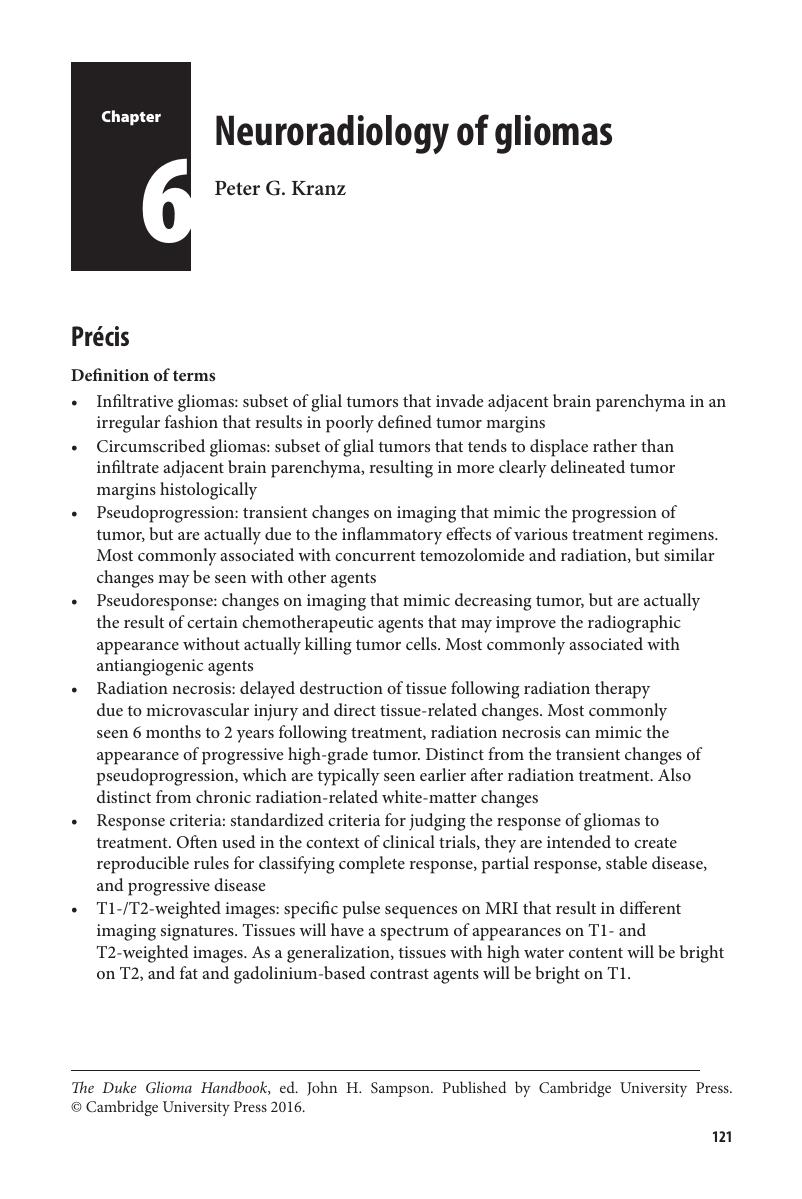
Image of the first page of this content. For PDF version, please use the ‘Save PDF’ preceeding this image.'
Type
Chapter
Information
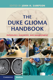
The Duke Glioma Handbook
Pathology, Diagnosis, and Management
, pp. 121 - 145
Publisher: Cambridge University Press
Print publication year: 2016

Access options

Get access to the full version of this content by using one of the access options below. (Log in options will check for institutional or personal access. Content may require purchase if you do not have access.)

References

Albert, FK, Forsting, M, Sartor, K, Adams, HP, Kunze, S. Early postoperative magnetic resonance imaging after resection of malignant glioma: objective evaluation of residual tumor and its influence on regrowth and prognosis. Neurosurgery, 1994;34(1):4560; discussion 61.Google ScholarPubMed
Atlas, SW, Grossman, RI, Gomori, JM, Hackney, DB, Goldberg, HI, Zimmerman, RA, et al. Hemorrhagic intracranial malignant neoplasms: spin-echo MR imaging. Radiology, 1987;164(1):71–7.CrossRefGoogle ScholarPubMed
Barajas, RF, Chang, JS, Segal, MR, Parsa, AT, McDermott, MW, Berger, MS, et al. Differentiation of recurrent glioblastoma multiforme from radiation necrosis after external beam radiation therapy with dynamic susceptibility-weighted contrast-enhanced perfusion MR imaging. Radiology, 2009;253(2):486–96.CrossRefGoogle ScholarPubMed
Basu, S, Alavi, A. Molecular imaging (PET) of brain tumors. Neuroimaging Clinics of North America, 2009;19(4):625–46.CrossRefGoogle ScholarPubMed
Bell, D, Chitnavis, BP, Al-Sarraj, S, Connor, S, Sharr, MM, Gullan, RW. Pilocytic astrocytoma of the adult – clinical features, radiological features and management. British Journal of Neurosurgery, 2004;18(6):613–16.CrossRefGoogle ScholarPubMed
Bisdas, S, Kirkpatrick, M, Giglio, P, Welsh, C, Spampinato, MV, Rumboldt, Z. Cerebral blood volume measurements by perfusion-weighted MR imaging in gliomas: ready for prime time in predicting short-term outcome and recurrent disease? AJNR American Journal of Neuroradiology, 2009;30(4):681–8.CrossRefGoogle ScholarPubMed
Brandsma, D, van den Bent, MJ. Pseudoprogression and pseudoresponse in the treatment of gliomas. Current Opinion in Neurology, 2009;22(6):633–8.CrossRefGoogle ScholarPubMed
Brodbelt, A. Clinical applications of imaging biomarkers. Part 2. The neurosurgeon’s perspective. British Journal of Radiology, 2012;84(Special Issue 2):S205–S8.Google Scholar
Chinot, OL, Macdonald, DR, Abrey, LE, Zahlmann, G, Kerloëguen, Y, Cloughesy, TF. Response assessment criteria for glioblastoma: practical adaptation and implementation in clinical trials of antiangiogenic therapy. Current Neurology and Neuroscience Reports, 2013;13(5):347.CrossRefGoogle ScholarPubMed
da Cruz, LCH, Rodriguez, I, Domingues, RC, Gasparetto, EL, Sorensen, AG. Pseudoprogression and pseudoresponse: imaging challenges in the assessment of posttreatment glioma. AJNR American Journal of Neuroradiology, 2011;32(11):1978–85.Google Scholar
Eisenhauer, EA, Therasse, P, Bogaerts, J, Schwartz, LH, Sargent, D, Ford, R, et al. New response evaluation criteria in solid tumours: revised RECIST guideline (version 1.1). European Journal of Cancer, 2009;45(2):228–47.CrossRefGoogle Scholar
Fatterpekar, GM, Galheigo, D, Narayana, A, Johnson, G, Knopp, E. Treatment-related change versus tumor recurrence in high-grade gliomas: a diagnostic conundrum – use of dynamic susceptibility contrast-enhanced (DSC) perfusion MRI. American Journal of Roentgenology, 2012;198(1):1926.CrossRefGoogle ScholarPubMed
Fuller, GN. The WHO classification of tumours of the central nervous system, 4th edition. Archives of Pathology and Laboratory Medicine, 2008;132(6):906.CrossRefGoogle ScholarPubMed
Gempt, J, Krieg, SM, Hüttinger, S, Buchmann, N, Ryang, Y-M, Shiban, E, et al. Postoperative ischemic changes after glioma resection identified by diffusion-weighted magnetic resonance imaging and their association with intraoperative motor evoked potentials. Journal of Neurosurgery, 2013;119(4):829–36.Google ScholarPubMed
Henson, JW, Gaviani, P, Gonzalez, RG. MRI in treatment of adult gliomas. The Lancet Oncology, 2005;6(3):167–75.CrossRefGoogle ScholarPubMed
Henson, JW, Ulmer, S, Harris, GJ. Brain tumor imaging in clinical trials. American Journal of Neuroradiology, 2008;29(3):419–24.CrossRefGoogle ScholarPubMed
Hilario, A, Ramos, A, Perez-Nunez, A, Salvador, E, Millan, JM, Lagares, A, et al. The added value of apparent diffusion coefficient to cerebral blood volume in the preoperative grading of diffuse gliomas. American Journal of Neuroradiology, 2012;33(4):701–7.CrossRefGoogle ScholarPubMed
Jain, R. Perfusion CT imaging of brain tumors: an overview. American Journal of Neuroradiology, 2011;32(9):1570–7.CrossRefGoogle ScholarPubMed
Johnson, DR, Brown, PD, Galanis, E, Hammack, JE. Pilocytic astrocytoma survival in adults: analysis of the Surveillance, Epidemiology, and End Results Program of the National Cancer Institute. Journal of Neuro-Oncology, 2012;108(1):187–93.CrossRefGoogle ScholarPubMed
Khalid, L, Carone, M, Dumrongpisutikul, N, Intrapiromkul, J, Bonekamp, D, Barker, PB, et al. Imaging characteristics of oligodendrogliomas that predict grade. American Journal of Neuroradiology, 2012;33(5):852–7.CrossRefGoogle ScholarPubMed
Koeller, KK, Rushing, EJ. From the archives of the AFIP: oligodendroglioma and its variants: radiologic–pathologic correlation. Radiographics, 2005;25(6):1669–88.CrossRefGoogle ScholarPubMed
Louis, DN, Ohgaki, H, Wiestler, OD, Cavenee, WK, Burger, PC, Jouvet, A, et al. The 2007 WHO classification of tumours of the central nervous system. Acta Neuropathologica, 2007;114(2):97109.CrossRefGoogle ScholarPubMed
Macdonald, DR, Cascino, TL, Schold, SC, Cairncross, JG. Response criteria for phase II studies of supratentorial malignant glioma. Journal of Clinical Oncology, 1990;8(7):1277–80.CrossRefGoogle ScholarPubMed
McKnight, TR. Proton magnetic resonance spectroscopic evaluation of brain tumor metabolism. Seminars in Oncology, 2004;31(5):605–17.CrossRefGoogle ScholarPubMed
Parisi, JE, Miller, DV, Boyer, PJ, Brat, DJ, Cochran, EJ, Cohen, ML, et al. Protocol for the examination of specimens from patients with tumors of the brain/spinal cord. Archives of Pathology and Laboratory Medicine, 2008;132(6):907–12.CrossRefGoogle Scholar
Pope, WB, Young, JR, Ellingson, BM. Advances in MRI assessment of gliomas and response to anti-VEGF therapy. Current Neurology and Neuroscience Reports, 2011;11(3):336–44.CrossRefGoogle ScholarPubMed
Price, SJ, Gillard, JH. Imaging biomarkers of brain tumour margin and tumour invasion. British Journal of Radiology, 2011;84 Spec No 2:S159–67.CrossRefGoogle ScholarPubMed
Rahmathulla, G, Marko, NF, Weil, RJ. Cerebral radiation necrosis: a review of the pathobiology, diagnosis and management considerations. Journal of Clinical Neuroscience, 2013;20(4):485502.CrossRefGoogle ScholarPubMed
Rees, JH, Smirniotopoulos, JG, Jones, RV, Wong, K. Glioblastoma multiforme: radiologic–pathologic correlation. Radiographics, 1996;16(6):1413–38; quiz 62–3.CrossRefGoogle ScholarPubMed
SaNai, N, Chang, S, Berger, MS. Low-grade gliomas in adults. Journal of Neurosurgery, 2011;115(5):948–65.CrossRefGoogle ScholarPubMed
Sanghera, P, Rampling, R, Haylock, B, Jefferies, S, McBain, C, Rees, JH, et al. The concepts, diagnosis and management of early imaging changes after therapy for glioblastomas. Clinical Oncology, 2012;24(3):216–27.CrossRefGoogle ScholarPubMed
Siu, A, Wind, JJ, Iorgulescu, JB, Chan, TA, Yamada, Y, Sherman, JH. Radiation necrosis following treatment of high grade glioma – a review of the literature and current understanding. Acta Neurochirurgica, 2011;154(2):191201.CrossRefGoogle ScholarPubMed
Valk, PE, Dillon, WP. Radiation injury of the brain. AJNR American Journal of Neuroradiology, 1991;12(1):4562.Google ScholarPubMed
Walker, C, Baborie, A, Crooks, D, Wilkins, S, Jenkinson, MD. Biology, genetics and imaging of glial cell tumours. British Journal of Radiology, 2011;84 Spec No 2:S90106.CrossRefGoogle ScholarPubMed
Wen, PY, Macdonald, DR, Reardon, DA, Cloughesy, TF, Sorensen, AG, Galanis, E, et al. Updated response assessment criteria for high-grade gliomas: response assessment in Neuro-Oncology Working Group. Journal of Clinical Oncology, 2010;28(11):1963–72.CrossRefGoogle ScholarPubMed
White, ML, Zhang, Y, Kirby, P, Ryken, TC. Can tumor contrast enhancement be used as a criterion for differentiating tumor grades of oligodendrogliomas? AJNR American Journal of Neuroradiology, 2005;26(4):784–90.Google ScholarPubMed

Save book to Kindle

To save this book to your Kindle, first ensure coreplatform@cambridge.org is added to your Approved Personal Document E-mail List under your Personal Document Settings on the Manage Your Content and Devices page of your Amazon account. Then enter the ‘name’ part of your Kindle email address below. Find out more about saving to your Kindle.

Note you can select to save to either the @free.kindle.com or @kindle.com variations. ‘@free.kindle.com’ emails are free but can only be saved to your device when it is connected to wi-fi. ‘@kindle.com’ emails can be delivered even when you are not connected to wi-fi, but note that service fees apply.

Find out more about the Kindle Personal Document Service.

Available formats
×

Save book to Dropbox

To save content items to your account, please confirm that you agree to abide by our usage policies. If this is the first time you use this feature, you will be asked to authorise Cambridge Core to connect with your account. Find out more about saving content to Dropbox.

Available formats
×

Save book to Google Drive

To save content items to your account, please confirm that you agree to abide by our usage policies. If this is the first time you use this feature, you will be asked to authorise Cambridge Core to connect with your account. Find out more about saving content to Google Drive.

Available formats
×