Skip to main content Accessibility help
×
Hostname: page-component-586b7cd67f-2brh9 Total loading time: 0 Render date: 2024-11-24T06:26:41.677Z Has data issue: false hasContentIssue false

23 - Breast Screen Reader Assessment Strategy (BREAST): A Research Infrastructure with a Translational Objective

from Part IV - Clinical Performance Assessment

Published online by Cambridge University Press:  20 December 2018
Ehsan Samei
Affiliation:
Duke University Medical Center, Durham
Elizabeth A. Krupinski
Affiliation:
Emory University, Atlanta
Get access

Summary

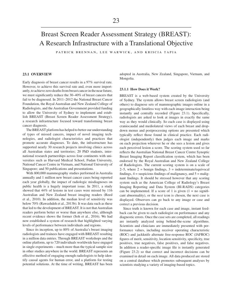
Image of the first page of this content. For PDF version, please use the ‘Save PDF’ preceeding this image.'
Type
Chapter
Information
Publisher: Cambridge University Press
Print publication year: 2018

Access options

Get access to the full version of this content by using one of the access options below. (Log in options will check for institutional or personal access. Content may require purchase if you do not have access.)

References

Al Mousa, D.S., Brennan, P.C., Ryan, E.A., Lee, W.B., Tan, J., Mello-Thoms, C. (2014a). How mammographic breast density affects radiologists’ visual search patterns. Acad Radiol, 21(11), 13861393.Google Scholar
Al Mousa, D.S., Mello-Thoms, C., Ryan, E.A., Lee, W.B., Pietrzyk, M.W., Reed, W.M., et al. (2014b). Mammographic density and cancer detection: does digital imaging challenge our current understanding? Acad Radiol, 21(11), 13771385.Google Scholar
Alakhras, M.M., Brennan, P.C., Rickard, M., Bourne, R., Mello-Thoms, C. (2015). Effect of radiologists’ experience on breast cancer detection and localization using digital breast tomosynthesis. Eur Radiol, 25(2), 402409.Google Scholar
Australian Government. (2009). BreastScreen Australia Evaluation. Screening Monograph No. 1/2009. Canberra, Australia: Australian Government.Google Scholar
Baker, J.A., Rosen, E.L., Lo, J.Y., Gimenez, E.I., Walsh, R., Soo, M.S. (2003). Computer-aided detection (CAD) in screening mammography: sensitivity of commercial CAD systems for detecting architectural distortion. Am J Roentgenol, 181(4), 10831088.Google Scholar
Bargallo, X., Velasco, M., Santamaria, G., Del Amo, M., Arguis, P., Sanchez Gomez, S. (2013). Role of computer-aided detection in very small screening detected invasive breast cancers. J Digit Imag, 26(3), 572577.CrossRefGoogle ScholarPubMed
Barlow, W.E., Chi, C., Carney, P.A., Taplin, S.H., D’Orsi, C., Cutter, G., et al. (2004). Accuracy of screening mammography interpretation by characteristics of radiologists. J Natl Cancer Inst, 96(24), 18401850.Google Scholar
Bassett, L.W. (1997). Diagnosis of Diseases of the Breast. Philadelphia, PA: W.B. Saunders.Google Scholar
Bird, R.E., Wallace, T.W., Yankaskas, B.C. (1992). Analysis of cancers missed at screening mammography. Radiology, 184(3), 613617.CrossRefGoogle ScholarPubMed
Boyd, N.F., Byng, J.W., Jong, R.A., Fishell, E.K., Little, L.E., Miller, A.B., et al. (1995). Quantitative classification of mammographic densities and breast cancer risk: results from the Canadian National Breast Screening Study. J Natl Cancer Inst, 87(9), 670675.Google Scholar
Boyd, N.F., Lockwood, G.A., Martin, L.J., Knight, J.A., Byng, J.W., Yaffe, M.J., Tritchler, D.L. (1998). Mammographic densities and breast cancer risk. Breast Dis, 10, 113126.CrossRefGoogle ScholarPubMed
Brem, R.F., Baum, J., Lechner, M., Kaplan, S., Souders, S., Naul, L.G., Hoffmeister, J. (2003). Improvement in sensitivity of screening mammography with computer-aided detection: a multiinstitutional trial. Am J Roentgenol, 181(3), 687693.CrossRefGoogle ScholarPubMed
Broeders, M.J., Onland-Moret, N.C., Rijken, H.J., Hendriks, J.H., Verbeek, A.L., Holland, R. (2003). Use of previous screening mammograms to identify features indicating cases that would have a possible gain in prognosis following earlier detection. Eur J Cancer, 39(12), 17701775.Google Scholar
Buist, D.S.M., Porter, P.L., Lehman, C., Taplin, S.H., White, E. (2004). Factors contributing to mammography failure in women aged 40–49 years. J Natl Cancer Inst, 96(19), 14321440.Google Scholar
Burrell, H.C., Sibbering, D.M., Wilson, A.R., Pinder, S.E., Evans, A.J., Yeoman, L.J., et al. (1996). Screening interval breast cancers: mammographic features and prognosis factors. Radiology, 199(3), 811817.Google Scholar
Burrell, H.C., Evans, A.J., Wilson, A.R., Pinder, S.E. (2001). False-negative breast screening assessment: what lessons can we learn? Clin Radiol, 56(5), 385388.Google Scholar
Carney, P.A., Miglioretti, D.L., Yankaskas, B.C., Kerlikowske, K., Rosenberg, R., Rutter, C. M., et al. (2003). Individual and combined effects of age, breast density, and hormone replacement therapy use on the accuracy of screening mammography. Ann Intern Med, 138(3), 168175.Google Scholar
Cawson, J.N., Nickson, C., Amos, A., Hill, G., Whan, A.B., Kavanagh, A.M. (2009). Invasive breast cancers detected by screening mammography: a detailed comparison of computer-aided detection-assisted single reading and double reading. J Med Imag Radiat Oncol, 53(5), 442449.Google Scholar
Chiarelli, A.M., Kirsh, V.A., Klar, N.S., Shumak, R., Jong, R., Fishell, E., et al. (2006). Influence of patterns of hormone replacement therapy use and mammographic density on breast cancer detection. Cancer Epidemiol Biomarkers Prev, 15(10), 18561862.Google Scholar
Ciatto, S., Visioli, C., Paci, E., Zappa, M. (2004). Breast density as a determinant of interval cancer at mammographic screening. Br J Cancer, 90(2), 393396.Google Scholar
Ciatto, S., Houssami, N., Bernardi, D., Caumo, F., Pellegrini, M., Brunelli, S., et al. (2013). Integration of 3D digital mammography with tomosynthesis for population breast-cancer screening (STORM): a prospective comparison study. Lancet Oncol, 14(7), 583589.Google Scholar
Committee. (1997). Quality Assurance Guidelines for Radiologists. Sheffield, England: NHSBSP Publications.Google Scholar
Digabel-Chabay, C., Allioux, C., Labbe-Devilliers, C., Meingan, P., Ricaud Couprie, M. (2004). Architectural distortion and diagnostic difficulties. J Radiol, 85, 20992106.CrossRefGoogle ScholarPubMed
Doi, K., MacMahon, H., Katsuragawa, S., Nishikawa, R.M., Jiang, Y. (1999). Computer-aided diagnosis in radiology: potential and pitfalls. Eur J Radiol, 31(2), 97109.Google Scholar
D’Orsi, C.J., Kopans, D.B. (1993). Mammographic feature analysis. Semin Roentgenol, 28(3), 204230.Google Scholar
D’Orsi, C.J.B., Feig, S.A., et al. (1998). Illustrated Breast Imaging Reporting and Data System, Illustrated BI-RADS, 3rd edn. Reston, VA: American College of Radiology.Google Scholar
Elmore, J.G., Jackson, S.L., Abraham, L., Miglioretti, D.L., Carney, P.A., Geller, B.M., et al. (2009). Variability in interpretive performance at screening mammography and radiologists’ characteristics associated with accuracy. Radiology, 253(3), 641651.Google Scholar
Giger, M.L. (2000). Computer-aided diagnosis of breast lesions in medical images. Comput Sci Eng, 2(5), 3945.Google Scholar
Giger, M.L., Karssemeijer, N., Armato, S.G. (2001). Computer-aided diagnosis in medical imaging. IEEE Trans Med Imag, 20(12), 12051208.Google Scholar
Gilbert, F.J., Tucker, L., Gillan, M.G., Willsher, P., Cooke, J., Duncan, K.A., et al. (2015). Accuracy of digital breast tomosynthesis for depicting breast cancer subgroups in a UK retrospective reading study (TOMMY trial). Radiology, 277(3), 697706.Google Scholar
Goergen, S.K., Evans, J., Cohen, G.P., MacMillan, J.H. (1997). Characteristics of breast carcinomas missed by screening radiologists. Radiology, 204(1), 131135.Google Scholar
Homer, M.J.S. (1997). Mammographic Interpretation: A Practical Approach,. New York, NY: McGraw-Hill.Google Scholar
Huynh, P.T., Jarolimek, A.M., Daye, S. (1998). The false-negative mammogram. Radiographics, 18(5), 11371154; quiz 12431134.Google Scholar
Jain, A. (2000). Artificial intelligence techniques in breast cancer diagnosis and prognosis. Paper presented at the World Scientific, Singapore.CrossRefGoogle Scholar
Jiang, Y.N., Nishikawa, R.M., Papaioannou, J. (1998). Requirement of microcalcifications detection for computerized classification of malignant and benign clustered microcalcifications. Proc SPIE Med Imag, 3338.Google Scholar
Kan, L., Olivotto, I.A., Warren Burhenne, L.J., Sickles, E.A., Coldman, A.J. (2000). Standardized abnormal interpretation and cancer detection ratios to assess reading volume and reader performance in a breast screening program. Radiology, 215(2), 563567.Google Scholar
Knutzen, A.M., Gisvold, J.J. (1993). Likelihood of malignant disease for various categories of mammographically detected, nonpalpable breast lesions. Mayo Clin Proc, 68(5), 454460.CrossRefGoogle ScholarPubMed
Kolb, T.M., Lichy, J., Newhouse, J.H. (2002). Comparison of the performance of screening mammography, physical examination, and breast US and evaluation of factors that influence them: an analysis of 27,825 patient evaluations. Radiology, 225(1), 165175.Google Scholar
Kundel, H.L., Nodine, C.F., Carmody, D. (1978). Visual scanning, pattern recognition and decision-making in pulmonary nodule detection. Invest Radiol, 13(3), 175181.CrossRefGoogle ScholarPubMed
Lang, K., Andersson, I., Rosso, A., Tingberg, A., Timberg, P., Zackrisson, S. (2016). Performance of one-view breast tomosynthesis as a stand-alone breast cancer screening modality: results from the Malmo Breast Tomosynthesis Screening Trial, a population-based study. Eur Radiol, 26(1), 184190.Google Scholar
Majid, A.S., de Paredes, E.S., Doherty, R.D., Sharma, N.R., Salvador, X. (2003). Missed breast carcinoma: pitfalls and pearls. Radiographics, 23(4), 881895.Google Scholar
Mandelson, M.T., Oestreicher, N., Porter, P.L., White, D., Finder, C.A., Taplin, S.H., White, E. (2000). Breast density as a predictor of mammographic detection: comparison of interval- and screen-detected cancers. J Natl Cancer Inst, 92(13), 10811087.CrossRefGoogle ScholarPubMed
Miglioretti, D.L., Smith-Bindman, R., Abraham, L., Brenner, R.J., Carney, P.A., Bowles, E.J., et al. (2007). Radiologist characteristics associated with interpretive performance of diagnostic mammography. J Natl Cancer Inst, 99(24), 18541863.Google Scholar
National Accreditation Committee. (1994). National Program for the Early Detection of Breast Cancer – National Accreditation Requirements. Canberra, Australia: National Accreditation Committee.Google Scholar
Pohlman, S., Powell, K.A., Obuchowski, N.A., Chilcote, W.A., Grundfest-Broniatowski, S. (1996). Quantitative classification of breast tumors in digitized mammograms. Med Phys, 23(8), 13371345.CrossRefGoogle ScholarPubMed
Rangayyan, R.M., Mudigonda, N.R., Desautels, J.E. (2000). Boundary modelling and shape analysis methods for classification of mammographic masses. Med Biol Eng Comput, 38(5), 487496.Google Scholar
Rangayyan, R.M., Banik, S., Desautels, J.E. (2010). Computer-aided detection of architectural distortion in prior mammograms of interval cancer. J Digit Imag, 23(5), 611631.Google Scholar
Rawashdeh, M.A., Bourne, R.M., Ryan, E.A., Lee, W.B., Pietrzyk, M.W., Reed, W.M., et al. (2013a). Quantitative measures confirm the inverse relationship between lesion spiculation and detection of breast masses. Acad Radiol, 20(5), 576580.Google Scholar
Rawashdeh, M.A., Lee, W.B., Bourne, R.M., Ryan, E.A., Pietrzyk, M.W., Reed, W.M., et al. (2013b). Markers of good performance in mammography depend on number of annual readings. Radiology, 269(1), 6167.Google Scholar
Reed, W.M., Lee, W.B., Cawson, J.N., Brennan, P.C. (2010). Malignancy detection in digital mammograms: important reader characteristics and required case numbers. Acad Radiol, 17(11), 14091413.Google Scholar
Rosenberg, R.D., Hunt, W.C., Williamson, M.R., Gilliland, F.D., Wiest, P.W., Kelsey, C.A., et al. (1998). Effects of age, breast density, ethnicity, and estrogen replacement therapy on screening mammographic sensitivity and cancer stage at diagnosis: review of 183,134 screening mammograms in Albuquerque, New Mexico. Radiology, 209(2), 511518.Google Scholar
Sampat, M.P., Whitman, G.J., Markey, M.K., Bovik, A.C. (2005). Evidence based detection of spiculated masses and architectural distortions. Proc SPIE Med Imag, 5747.CrossRefGoogle Scholar
Scott, H.J., Gale, A.G. (2006). Breast screening: PERFORMS identifies key mammographic training needs. Br J Radiol, 79, S127–S133.CrossRefGoogle ScholarPubMed
Shen, L., Rangayyan, R.M., Desautels, J.L. (1994). Application of shape analysis to mammographic calcifications. IEEE Trans Med Imag, 13(2), 263274.Google Scholar
Shi, J., Sahiner, B., Chan, H.P., Ge, J., Hadjiiski, L., Helvie, M.A., et al. (2008). Characterization of mammographic masses based on level set segmentation with new image features and patient information. Med Phys, 35(1), 280290.CrossRefGoogle ScholarPubMed
Sickles, E.A. (1989). Breast masses: mammographic evaluation. Radiology, 173(2), 297303.Google Scholar
Soh, B.P., Lee, W., McEntee, M.F., Kench, P.L., Reed, W.M., Heard, R., et al. (2013). Screening mammography: test set data can reasonably describe actual clinical reporting. Radiology, 268(1), 4653.Google Scholar
Soh, B.P., Lee, W.B., McEntee, M.F., Kench, P.L., Reed, W.M., Heard, R., et al. (2014). Mammography test sets: reading location and prior images do not affect group performance. Clin Radiol, 69(4), 397402.Google Scholar
Soh, B.P., Lee, W.B., Mello-Thoms, C., Tapia, K., Ryan, J., Hung, W.T., et al. (2015). Certain performance values arising from mammographic test set readings correlate well with clinical audit. J Med Imag Radiat Oncol, 59(4), 403410.Google Scholar
Soh, B.P.L., Lee, W.B., Wong, J., Sim, L., Hillis, S.L., Tapia, K.A., Brennan, P.C. (2016). Varying performance in mammographic interpretation across two countries: do results indicate reader or population variances? Proc SPIE Med Imag, 9787.Google Scholar
Strickland, R.N., Hahn, H. (1996). Wavelet transforms for detecting microcalcifications in mammograms. IEEE Trans Med Imag, 15(2), 218229.Google Scholar
Suleiman, W.I., McEntee, M.F., Lewis, S.J., Rawashdeh, M.A., Georgian-Smith, D., Heard, R., et al. (2016a). In the digital era, architectural distortion remains a challenging radiological task. Clin Radiol, 71(1), e35–e40.Google Scholar
Suleiman, W.I., Rawashdeh, M.A., Lewis, S.J., McEntee, M.F., Lee, W., Tapia, K., Brennan, P.C. (2016b). Impact of breast reader assessment strategy on mammographic radiologists’ test reading performance. J Med Imag Radiat Oncol, 60(3), 352358.Google Scholar
Troxel, D.B. (2006). Medicolegal aspects of error in pathology. Arch Pathol Lab Med, 130(5), 617619.Google Scholar
US Department of Health and Human Services. (1997). An Overview of the Final Regulations Implementing the Mammography Quality Standards Act of 1992. Rockville, MD: US Department of Health and Human Services.Google Scholar
Vyborny, C.J., Doi, T., O’Shaughnessy, K.F., Romsdahl, H.M., Schneider, A.C., Stein, A.A. (2000a). Breast cancer: importance of spiculation in computer-aided detection. Radiology, 215(3), 703707.Google Scholar
Vyborny, C.J., Giger, M.L., Nishikawa, R.M. (2000b). Computer-aided detection and diagnosis of breast cancer. Radiol Clin N Am, 38(4), 725740.Google Scholar
Yankaskas, B.C., Schell, M.J., Bird, R.E., Desrochers, D.A. (2001). Reassessment of breast cancers missed during routine screening mammography: a community-based study. Am J Roentgenol, 177(3), 535541.Google Scholar

Save book to Kindle

To save this book to your Kindle, first ensure coreplatform@cambridge.org is added to your Approved Personal Document E-mail List under your Personal Document Settings on the Manage Your Content and Devices page of your Amazon account. Then enter the ‘name’ part of your Kindle email address below. Find out more about saving to your Kindle.

Note you can select to save to either the @free.kindle.com or @kindle.com variations. ‘@free.kindle.com’ emails are free but can only be saved to your device when it is connected to wi-fi. ‘@kindle.com’ emails can be delivered even when you are not connected to wi-fi, but note that service fees apply.

Find out more about the Kindle Personal Document Service.

Available formats
×

Save book to Dropbox

To save content items to your account, please confirm that you agree to abide by our usage policies. If this is the first time you use this feature, you will be asked to authorise Cambridge Core to connect with your account. Find out more about saving content to Dropbox.

Available formats
×

Save book to Google Drive

To save content items to your account, please confirm that you agree to abide by our usage policies. If this is the first time you use this feature, you will be asked to authorise Cambridge Core to connect with your account. Find out more about saving content to Google Drive.

Available formats
×