Skip to main content Accessibility help
×
Hostname: page-component-77c89778f8-rkxrd Total loading time: 0 Render date: 2024-07-19T10:17:36.347Z Has data issue: false hasContentIssue false

Chapter 20 - The Fetal Arterial and Venous Circulation in Fetal Growth Restriction

from Section 5 - Characteristics of Fetal Growth Restriction

Published online by Cambridge University Press:  23 July 2018

Christoph Lees
Affiliation:
Imperial College London
Gerard H. A. Visser
Affiliation:
Universiteit Utrecht, The Netherlands
Kurt Hecher
Affiliation:
University Medical Centre, Hamburg
Get access

Summary

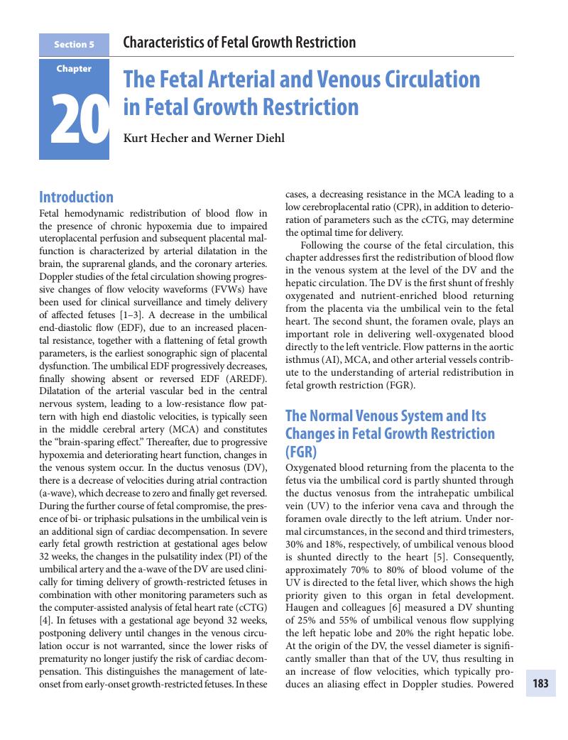
Image of the first page of this content. For PDF version, please use the ‘Save PDF’ preceeding this image.'
Type
Chapter
Information
Publisher: Cambridge University Press
Print publication year: 2018

Access options

Get access to the full version of this content by using one of the access options below. (Log in options will check for institutional or personal access. Content may require purchase if you do not have access.)

References

Hecher, K, Bilardo, CM, Stigter, RH, et al. Monitoring of fetuses with intrauterine growth restriction: A longitudinal study. Ultrasound Obstet Gynecol 2001;18:564–70.CrossRefGoogle ScholarPubMed
Baschat, A, Gembruch, U, Harman, CR. The sequence of changes in Doppler and biophysical parameters as severe fetal growth restriction worsens. Ultrasound Obstet Gynecol 2001;18:571–7.Google Scholar
Ferrazzi, E, Bozzo, M, Rigano, S, et al. Temporal sequence of abnormal Doppler changes in the peripheral and central circulatory systems of the severely growth-restricted fetus. Ultrasound Obstet Gynecol 2002;19:140–6.CrossRefGoogle ScholarPubMed
Bilardo, CM, Wolf, H, Stigter, RH et al. Relationship between monitoring parameters and perinatal outcome in severe, early intrauterine growth restriction. Ultrasound Obstet Gynecol 2004;23:119–25.CrossRefGoogle ScholarPubMed
Kiserud, T, Rasmussen, S, Skulstad, SM. Blood flow and degree of shunting through the ductus venosus in the human fetus. Am J Obstet Gynecol 2000;182:147–53.CrossRefGoogle ScholarPubMed
Haugen, G, Kiserud, T, Godfrey, K, et al. Portal and umbilical venous blood supply to the liver in the human fetus near term. Ultrasound Obstet Gynecol 2004;24:599605.CrossRefGoogle Scholar
Tchirikov, M, Schlabritz-Loutsevitch, H, Hubbard, GB, et al. Structural evidence for mechanisms to redistribute hepatic and ductus venosus blood flows in non-human primate fetuses. Am J Obstet Gynecol 2005;192:1146–52.Google Scholar
Haugen, G, Bollerslev, J, Henriksen, T. Human fetoplacental and fetal liver blood flow after maternal glucose loading: A cross-sectional observational study. Acta Obstet Gynecol Scand 2014;93:778–85.Google Scholar
Tchirikov, M, Schröder, HJ, Hecher, K. Ductus venosus shunting in the fetal venous circulation: Regulatory mechanisms, diagnostic methods and medical importance. Ultrasound Obstet Gynecol 2006;27:452–61.Google Scholar
Kessler, J, Rasmussen, S, Kiserud, T. The fetal portal vein: Normal blood flow development during the second half of human pregnancy. Ultrasound Obstet Gynecol 2007;30:5260.CrossRefGoogle ScholarPubMed
Kiserud, T, Kessler, J, Ebbing, C, et al. Ductus venosus shunting in growth-restricted fetuses and the effect of umbilical circulatory compromise. Ultrasound Obstet Gynecol 2006;28:143–9.CrossRefGoogle ScholarPubMed
Kessler, J, Rasmussen, S, Kiserud, T. The left portal vein as an indicator of watershed in the fetal circulation: Development during the second half of pregnancy and a suggested method of evaluation. Ultrasound Obstet Gynecol 2007;30:757–64.Google Scholar
Kessler, J, Rasmussen, S, Godfrey, K, et al. Longitudinal study of umbilical and portal venous blood flow to the fetal liver: Low pregnancy weight gain is associated with preferential supply to the fetal left liver lobe. Pediatr Res 2008;63:315–20.Google Scholar
Ebbing, C, Rasmussen, S, Godfrey, KM, et al. Redistribution pattern of fetal liver circulation in intrauterine growth restriction. Acta Obstet Gynecol Scand 2009;88:1118–23.Google Scholar
Haugen, G, Hanson, M, Kiserud, T, et al. Fetal liver-sparing cardiovascular adaptations linked to mother’s slimness and diet. Circ Res 2005;96:1214.CrossRefGoogle ScholarPubMed
Godfrey, KM, Haugen, G, Kiserud, T, et al. Fetal liver blood flow distribution: Role in human developmental strategy to prioritize fat deposition versus brain development. PLoS One 2012;7:e41759.CrossRefGoogle ScholarPubMed
Kiserud, T, Ebbing, C, Kessler, J, Rasmussen, S. Fetal cardiac output, distribution to the placenta and impact of placental compromise. Ultrasound Obstet Gynecol 2006;28:126–36.CrossRefGoogle Scholar
Hecher, K, Campbell, S, Snijders, R, et al. Reference ranges for fetal venous and atrioventricular blood flow parameters. Ultrasound Obstet Gynecol 1994; 4:381–90.Google Scholar
Kessler, J, Rasmussen, S, Hanson, M, et al. Longitudinal reference ranges for ductus venosus flow velocities and waveform indices. Ultrasound Obstet Gynecol 2006 Dec;28:890–8.Google Scholar
Kiserud, T, Kilavuz, O, Hellevik, LR. Venous pulsation in the fetal left portal branch: The effect of pulse and flow direction. Ultrasound Obstet Gynecol 2003; 21:359–64.Google Scholar
Hellevik, LR, Stergiopulos, N, Kiserud, T, et al. A mathematical model of umbilical venous pulsation. J Biomech 2000;33:1123–30.CrossRefGoogle ScholarPubMed
Wada, N, Tachibana, D, Kurihara, Y, et al. Alterations of time-intervals of the ductus venosus and atrioventricular flow velocity waveforms in growth restricted fetuses. Ultrasound Obstet Gynecol 2014; doi: 10.1002/uog.14717.CrossRefGoogle Scholar
Rigano, S, Bozzo, M, Ferrazzi, E, et al. Early and persistent reduction in umbilical vein blood flow in the growth-restricted fetus: A longitudinal study. Am J Obstet Gynecol 2001;185:834–8.Google Scholar
Fouron, JC. The unrecognized physiological and clinical significance of the fetal aortic isthmus. Ultrasound Obstet Gynecol 2003;22: 441–7.Google Scholar
Fouron, JC, Gosselin, J, Amiel-Tison, C, et al. Correlation between prenatal velocity waveforms in the aortic isthmus and neurodevelopmental outcome between the ages of 2 and 4 years. Am J Obstet Gynecol 2001;184:630–6.Google Scholar
Figueras, F, Benavides, A, Del Rio, M, et al. Monitoring of fetuses with intrauterine growth restriction: Longitudinal changes in ductus venosus and aortic isthmus flow. Ultrasound Obstet Gynecol 2009;33:3943.CrossRefGoogle ScholarPubMed
Chabaneix, J, Fouron, JC, Sosa-Olavarria, A, et al. Profiling left and right ventricular proportional output during fetal life with a novel systolic index in the aortic isthmus. Ultrasound Obstet Gynecol 2014; 44:176–81.Google Scholar
Cruz-Martinez, R, Figueras, F, Oros, D, et al. Cerebral blood perfusion and neurobehavioral performance in full-term small-for-gestational-age fetuses. Am J Obstet Gynecol 2009;201:474.e1–7.Google Scholar
Figueras, F, Eixarch, E, Meler, E, et al. Small-for-gestational-age fetuses with normal umbilical artery Doppler have suboptimal perinatal and neurodevelopmental outcome. Eur J Obstet Gynecol Reprod Biol 2008;136:34–8.Google Scholar
Konje, JC, Bell, SC, Taylor, DJ. Abnormal Doppler velocimetry and blood flow volume in the middle cerebral artery in very severe intrauterine growth restriction: Is the occurrence of reversal of compensatory flow too late? BJOG 2001;108:973–9.Google Scholar
Benavides-Serralde, JA, Hernandez-Andrade, E, Cruz-Martinez, R, et al. Doppler evaluation of the posterior cerebral artery in normally grown and growth restricted fetuses. Prenat Diagn 2014;34:115–20.Google Scholar
Benavides-Serralde, A, Scheier, M, Cruz-Martinez, R, et al. Changes in central and peripheral circulation in intrauterine growth-restricted fetuses at different stages of umbilical artery flow deterioration: New fetal cardiac and brain parameters. Gynecol Obstet Invest 2011;71:274–80.CrossRefGoogle ScholarPubMed
Hernandez-Andrade, E, Serralde, JA, Cruz-Martinez, R. Can anomalies of fetal brain circulation be useful in the management of growth restricted fetuses? Prenat Diagn 2012;32:103–12.Google Scholar
Baschat, AA, Gembruch, U. The cerebroplacental Doppler ratio revisited. Ultrasound Obstet Gynecol 2003;21:124–7.Google Scholar
Hecher, K, Spernol, R, Stettner, H et al. Potential for diagnosing imminent risk to appropriate- and small-for-gestational-age fetuses by Doppler sonographic examination of umbilical and cerebral arterial blood flow. Ultrasound Obstet Gynecol 1992;2:266–71.Google Scholar
Morales-Roselló, J, Khalil, A, Morlando, M, et al. Changes in fetal Doppler indices as a marker of failure to reach growth potential at term. Ultrasound Obstet Gynecol 2014;43:303–10.CrossRefGoogle ScholarPubMed
Morales-Roselló, J, Khalil, A, Morlando, M, et al. Poor neonatal acid-base status in term fetuses with low cerebroplacental ratio. Ultrasound Obstet Gynecol 2015;45:156–61.Google Scholar
Parra-Saavedra, M, Simeone, S, Triunfo, S, et al. Correlation between histological signs of placental underperfusion and perinatal morbidity in late-onset small-for-gestational-age fetuses. Ultrasound Obstet Gynecol 2015;45:149–55.Google Scholar
Figueras, F, Savchev, S, Triunfo, S., et al. An integrated model with classification criteria to predict small-for-gestational-age fetuses at risk of adverse perinatal outcome. Ultrasound Obstet Gynecol 2015;45:279–85.Google Scholar
Cruz-Lemini, M, Crispi, F, Valenzuela-Alcaraz, B, et al. A fetal cardiovascular score to predict infant hypertension and arterial remodeling in intrauterine growth restriction. Am J Obstet Gynecol 2014;210:552.e1–22CrossRefGoogle ScholarPubMed
Baschat, A, Muench, MV, Gembruch, U. Coronary artery blood flow velocities in various fetal conditions. Ultrasound Obstet Gynecol 2003;21:426–9.Google Scholar
Lees, C, Marlow, N, van Wassenaer-Leemhuis, A., et al. and the TRUFFLE Group. 2 year neurodevelopmental and intermediate perinatal outcomes in infants with very preterm fetal growth restriction (TRUFFLE): A randomised trial. Lancet 2015;385:2162–72.CrossRefGoogle ScholarPubMed

Save book to Kindle

To save this book to your Kindle, first ensure coreplatform@cambridge.org is added to your Approved Personal Document E-mail List under your Personal Document Settings on the Manage Your Content and Devices page of your Amazon account. Then enter the ‘name’ part of your Kindle email address below. Find out more about saving to your Kindle.

Note you can select to save to either the @free.kindle.com or @kindle.com variations. ‘@free.kindle.com’ emails are free but can only be saved to your device when it is connected to wi-fi. ‘@kindle.com’ emails can be delivered even when you are not connected to wi-fi, but note that service fees apply.

Find out more about the Kindle Personal Document Service.

Available formats
×

Save book to Dropbox

To save content items to your account, please confirm that you agree to abide by our usage policies. If this is the first time you use this feature, you will be asked to authorise Cambridge Core to connect with your account. Find out more about saving content to Dropbox.

Available formats
×

Save book to Google Drive

To save content items to your account, please confirm that you agree to abide by our usage policies. If this is the first time you use this feature, you will be asked to authorise Cambridge Core to connect with your account. Find out more about saving content to Google Drive.

Available formats
×