

Book contents
- Frontmatter
- Contents
- Preface
- Acknowledgements
- Glossary
- Abbreviations
- 1 Basics of breast MRI
- 2 Imaging-related anatomy and pathology
- 3 Interpreting breast MRI studies
- 4 MRI-guided biopsy techniques
- 5 High-risk screening using breast MRI
- 6 Preoperative staging with breast MRI
- 7 Problem-solving applications of breast MRI
- 8 MRI after breast augmentation
- Answers to multiple choice questions
- Appendices
- Index
- Plate section
- References
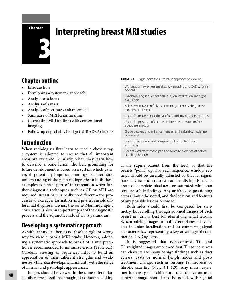
3 - Interpreting breast MRI studies
Published online by Cambridge University Press: 05 March 2012

Book contents
- Frontmatter
- Contents
- Preface
- Acknowledgements
- Glossary
- Abbreviations
- 1 Basics of breast MRI
- 2 Imaging-related anatomy and pathology
- 3 Interpreting breast MRI studies
- 4 MRI-guided biopsy techniques
- 5 High-risk screening using breast MRI
- 6 Preoperative staging with breast MRI
- 7 Problem-solving applications of breast MRI
- 8 MRI after breast augmentation
- Answers to multiple choice questions
- Appendices
- Index
- Plate section
- References
Summary
A summary is not available for this content so a preview has been provided. Please use the Get access link above for information on how to access this content.
- Type
- Chapter
- Information
- Handbook of Breast MRI , pp. 48 - 91Publisher: Cambridge University PressPrint publication year: 2011
References
Concepts for differential diagnosis in breast MR imagingMagn Reson Imaging Clin N Am 2006 14 305CrossRefGoogle ScholarPubMed
Diagnostic architectural and dynamic features at breast MR imaging: multicenter studyRadiology 2006 238 42CrossRefGoogle ScholarPubMed
BI-RADS Breast Imaging Reporting and Data System, Breast Imaging AtlasReston, VAAmerican College of Radiology 2003Google Scholar
Breast Imaging: A Guide for PracticeCamperdown, NSW, AustraliaNational Breast Cancer Center 2002Google Scholar
The Royal College of Radiologists Breast Group breast imaging classificationClin Radiol 2009 64 624CrossRefGoogle ScholarPubMed
The current status of breast MR imaging. Part 1. Choice of technique, image interpretation, diagnostic accuracy, and transfer to clinical practiceRadiology 2007 244 356CrossRefGoogle Scholar
Does size matter? Positive predictive value of MRI-detected breast lesions as a function of lesion sizeAJR Am J Roentgenol 2006 186 426CrossRefGoogle ScholarPubMed
BI-RADS lesion characteristics predict likelihood of malignancy in breast MRI for masses but not for nonmasslike enhancementAJR Am J Roentgenol 2009 193 994CrossRefGoogle Scholar
Characteristics of probably benign breast MRI lesionsAJR Am J Roentgenol 2009 193 861CrossRefGoogle ScholarPubMed
Do T2-weighted pulse sequences help with the differential diagnosis of enhancing lesions in dynamic breast MRIJ Magn Reson Imaging 1999 9 1873.0.CO;2-2>CrossRefGoogle ScholarPubMed
Breast carcinomas with strong high-signal intensity on T2-weighted MR images: pathologic characteristics and differential diagnosisJ Magn Reson Imaging 2007 25 502CrossRefGoogle Scholar
Nonpalpable mammographically occult invasive breast cancers detected by MRAJR Am J Roentgenol 2006 186 865CrossRefGoogle Scholar
Breast MRI: are T2 IR sequences useful in the evaluation of breast lesionsEur J Radiol 2009 71 96CrossRefGoogle ScholarPubMed
MR imaging of mucinous carcinoma of the breastAJR Am J Roentgenol 2002 179 179CrossRefGoogle ScholarPubMed
Triple-negative breast cancer: correlation between MR imaging and pathologic findingsRadiology 2009 250 638CrossRefGoogle ScholarPubMed
MR appearance of metastatic melanotic melanoma in the breastClin Radiol 2000 55 572CrossRefGoogle ScholarPubMed
Screening with magnetic resonance imaging and mammography of a UK population at high familial risk of breast cancer: a prospective multicenter cohort study (MARIBS)Lancet 2005 365 1769Google Scholar
Reading protocol for dynamic contrast-enhanced MR images of the breast: sensitivity and specificity analysisRadiology 2005 236 779CrossRefGoogle ScholarPubMed
Evaluation of a prospective scoring system designed for a multicenter breast MR imaging screening studyRadiology 2006 239 677CrossRefGoogle ScholarPubMed
Positive and negative predictive values of BI-RADS-MRI descriptors for focal breast massesMagn Reson Med Sci 2006 5 7CrossRefGoogle ScholarPubMed
Two different types of ring-like enhancement on dynamic MR imaging in breast cancer: correlation with the histopathologic findingsJ Magn Reson Imaging 2008 28 1435CrossRefGoogle ScholarPubMed
Potential MRI interpretation model: differentiation of benign from malignant breast massesAJR Am J Roentgenol 2005 185 964CrossRefGoogle ScholarPubMed
Typical atypical findings on dynamic MRI of the breastEur J Radiol 2010 76 195CrossRefGoogle ScholarPubMed
Is the “blooming sign” a promising additional tool to determine malignancy in MR mammographyEur Radiol 2004 14 394CrossRefGoogle ScholarPubMed
Morphologic blooming in breast MRI as a characterization of margin for discriminating benign from malignant lesionsAcad Radiol 2006 13 1344CrossRefGoogle ScholarPubMed
The adjacent vessel on dynamic contrast-enhanced breast MRIAJR Am J Roentgenol 2006 187 W147CrossRefGoogle ScholarPubMed
Gadobenate dimeglumine-enhanced MR imaging breast vascular maps: association between invasive cancer and ipsilateral increased vascularityRadiology 2005 235 791CrossRefGoogle ScholarPubMed
Assessment of three different software systems in the evaluation of dynamic MRI of the breastEur J Radiol 2009 69 300CrossRefGoogle ScholarPubMed
MRI-detected suspicious breast lesions: predictive values of kinetic features measured by computer-aided evaluationAJR Am J Roentgenol 2009 193 826CrossRefGoogle ScholarPubMed
Magnetic resonance imaging of intraductal papilloma of the breastMagn Reson Imaging 2003 21 887CrossRefGoogle ScholarPubMed
Mucinous carcinoma of the breast: MRI features of pure and mixed forms with histopathologic correlationAJR Am J Roentgenol 2009 192 W125CrossRefGoogle ScholarPubMed
Unusual malignant tumors of the breast: MRI features and pathologic correlationEur J Radiol 2010 75 178CrossRefGoogle ScholarPubMed
MR imaging features of pure mucinous carcinoma of the breastEur J Radiol 2006 60 405CrossRefGoogle ScholarPubMed
Multimodality imaging of triple receptor-negative tumors with mammography, ultrasound, and MRIAJR Am J Roentgenol 2010 194 1160CrossRefGoogle ScholarPubMed
Mammographic, US, and MR imaging phenotypes of familial breast cancerRadiology 2008 246 58CrossRefGoogle Scholar
Assessment of false-negative cases of breast MR imaging in women with a familial or genetic predispositionBreast Cancer Res Treat 2010 119 399CrossRefGoogle ScholarPubMed
The role of MRI in invasive lobular carcinomaBreast Cancer Res Treat 2004 86 31CrossRefGoogle ScholarPubMed
MRI compared to conventional diagnostic workup in the detection and evaluation of invasive lobular carcinoma of the breast: a review of existing literatureBreast Cancer Res Treat 2008 107 1CrossRefGoogle Scholar
The classic type of invasive lobular carcinoma of the breast is known to be elusive on mammography, but demonstrable when using 3-dimensional automated ultrasoundAm J Clin Oncol 2008 31 513Google Scholar
Preoperative breast MRI in patients with invasive lobular breast cancerEur Radiol 2004 14 1209CrossRefGoogle ScholarPubMed
MR imaging features of infiltrating lobular carcinoma of the breast: histopathologic correlationAJR Am J Roentgenol 2002 178 1227CrossRefGoogle ScholarPubMed
False-positive findings at contrast-enhanced breast MRI: a BI-RADS descriptor studyAJR Am J Roentgenol 2010 194 1658CrossRefGoogle ScholarPubMed
Ductal enhancement on MR imaging of the breastAJR Am J Roentgenol 2003 181 519CrossRefGoogle ScholarPubMed
Pure ductal carcinoma in situ: a range of MRI featuresAJR Am J Roentgenol 2008 191 689CrossRefGoogle ScholarPubMed
Breast MR imaging lexicon updatedMagn Reson Imaging Clin N Am 2006 14 293CrossRefGoogle ScholarPubMed
Determination of the presence and extent of pure ductal carcinoma in situ by mammography and magnetic resonance imagingBreast J 2005 11 382CrossRefGoogle ScholarPubMed
High-spatial-resolution MRI of non-masslike breast lesions: interpretation based on BI-RADS MRI descriptorsAJR Am J Roentgenol 2006 187 330CrossRefGoogle ScholarPubMed
Categorization of non-mass-like breast lesions detected by MRIBreast Cancer 2008 15 241CrossRefGoogle ScholarPubMed
Breast MRI using the VIBE sequence: clustered ring enhancement in the differential diagnosis of lesions showing non-masslike enhancementAJR Am J Roentgenol 2006 187 313CrossRefGoogle ScholarPubMed
Magnetic resonance imaging captures the biology of ductal carcinoma in situJ Clin Oncol 2006 24 4603CrossRefGoogle ScholarPubMed
Biologic significance of false-positive magnetic resonance imaging enhancement in the setting of ductal carcinoma in situAm J Surg 2006 192 520CrossRefGoogle ScholarPubMed
Characterization of pure high-grade DCIS on magnetic resonance imaging using the evolving breast MR lexicon terminology: can it be differentiated from pure invasive disease?Magn Reson Imaging 2005 23 733CrossRefGoogle ScholarPubMed
Breast lesions detected on MR imaging: features and positive predictive valueAJR Am J Roentgenol 2002 179 171CrossRefGoogle ScholarPubMed
Diagnostic usefulness of segmental and linear enhancement in dynamic breast MRIEur Radiol 2005 15 2010CrossRefGoogle ScholarPubMed
Comparison of MDCT and MRI for evaluating the intraductal component of breast cancerAJR Am J Roentgenol 2006 187 322CrossRefGoogle ScholarPubMed
Enhancing area surrounding breast carcinoma on MR mammography: comparison with pathologic examinationEur Radiol 2004 14 1363CrossRefGoogle Scholar
Magnetic resonance evaluation of the presence of an extensive intraductal component in breast cancerActa Radiol 2004 45 721CrossRefGoogle ScholarPubMed
Magnetic resonance imaging of ductal carcinoma in situ: what is its clinical application? A reviewAm J Surg 2009 198 262CrossRefGoogle ScholarPubMed
High grade and non-high grade ductal carcinoma in situ on dynamic MR mammography: characteristic findings for signal increase and morphological pattern of enhancementBr J Radiol 2003 76 3CrossRefGoogle ScholarPubMed
Pure ductal carcinoma in situ: kinetic and morphologic MR characteristics compared with mammographic appearance and nuclear gradeRadiology 2007 245 684CrossRefGoogle ScholarPubMed
Ductal carcinoma in situ: MR imaging–histopathologic correlationRadiology 1995 196 415CrossRefGoogle ScholarPubMed
Science to practice: why do purely intraductal cancers enhance on breast MR imagesRadiology 2009 253 281CrossRefGoogle Scholar
MRI for diagnosis of pure ductal carcinoma in situ: a prospective observational studyLancet 2007 370 485CrossRefGoogle ScholarPubMed
Magnetic resonance imaging characteristics of fibrocystic change of the breastInvest Radiol 2005 40 436CrossRefGoogle ScholarPubMed
Magnetic resonance imaging features of fibrocystic change of the breastMagn Reson Imaging 2008 26 1207CrossRefGoogle ScholarPubMed
The value of MRI compared to mammography in the assessment of tumour extent in invasive lobular carcinoma of the breastEur J Surg Oncol 2008 34 135CrossRefGoogle ScholarPubMed
Clinical indication and patient age predict likelihood of malignancy in suspicious breast MRI lesionsAcad Radiol 2009 16 1282CrossRefGoogle ScholarPubMed
National trends and practices in breast MRIAJR Am J Roentgenol 2008 191 332CrossRefGoogle ScholarPubMed
Targeted ultrasound of the breast in women with abnormal MRI findings for whom biopsy has been recommendedAJR Am J Roentgenol 2009 193 1025CrossRefGoogle ScholarPubMed
Breast lesions detected with MR imaging: utility and histopathologic importance of identification with USRadiology 2003 227 856CrossRefGoogle ScholarPubMed
Utility of targeted sonography for breast lesions that were suspicious on MRIAJR Am J Roentgenol 2009 192 1128CrossRefGoogle ScholarPubMed
US correlation for MRI-detected breast lesions in women with familial risk of breast cancerClin Radiol 2005 60 801CrossRefGoogle ScholarPubMed
Correlation of targeted ultrasound with magnetic resonance imaging abnormalities of the breastAm J Surg 2005 190 592CrossRefGoogle ScholarPubMed
Indeterminate or suspicious breast lesions detected initially with MR imaging: value of MRI-directed breast ultrasoundAcad Radiol 2008 15 618CrossRefGoogle ScholarPubMed
MR-directed (“second-look”) ultrasound examination for breast lesions detected initially on MRI: MR and sonographic findingsAJR Am J Roentgenol 2010 194 370CrossRefGoogle ScholarPubMed
Breast cancers detected with imaging screening in the BRCA population: emphasis on MR imaging with histopathologic correlationRadioGraphics 2007 27 S165CrossRefGoogle ScholarPubMed
Solid breast nodules: use of sonography to distinguish between benign and malignant lesionsRadiology 1995 196 123CrossRefGoogle ScholarPubMed
Ultrasound of solid breast nodules: distinguishing benign from malignantBreast UltrasoundPhiladelphia, PALippincott Williams & Wilkins 2004 445Google Scholar
Pseudocystic lesions and clustered cysts: radiologists bewareAJR Am J Roentgenol 2006 186 A79Google Scholar
Sonographically depicted breast clustered microcysts: is follow-up appropriateAJR Am J Roentgenol 2005 185 952CrossRefGoogle ScholarPubMed
Probably benign lesions at breast magnetic resonance imaging. Preliminary experience in high-risk womenCancer 2003 98 377CrossRefGoogle ScholarPubMed